
🫀 Ischaemic heart disease (IHD), also called coronary heart disease, occurs when the heart muscle does not get enough oxygen for what it is being asked to do. The usual cause is atherosclerosis narrowing the coronary arteries, but spasm, microvascular disease, dissection and high demand can do the same.
The spectrum runs from stable angina (predictable pain on exertion) to acute coronary syndromes (a sudden clot), heart failure and sudden death.
⚠️ Clinical priority: Recognise a blocked coronary artery quickly. Every minute it stays closed, more heart muscle dies.
1.01 What Is IHD?
Ischaemia means that a tissue is receiving too little blood flow for its needs. In the heart this produces two broad clinical pictures:
- Chronic coronary disease: A fixed narrowing limits blood flow. At rest there is enough oxygen, but during exercise demand outstrips supply and the patient gets angina. The muscle recovers when demand falls, so no permanent damage occurs. This is stable angina.
- Acute coronary syndrome (ACS): A plaque suddenly ruptures and a clot forms on it, abruptly cutting off flow. If the blockage lasts long enough, heart muscle dies: this is a myocardial infarction (MI).
The same disease can also cause:
- Ischaemic cardiomyopathy: Heart failure from previous infarcts.
- Arrhythmias: Ischaemic or scarred muscle can trigger dangerous rhythms.
- Sudden cardiac death: Usually from ventricular fibrillation.
- Non-obstructive ischaemia: Vasospastic and microvascular angina, where the large arteries are not significantly narrowed.
💡 Clinical Pearl: Think of IHD as a problem of supply versus demand. Stable angina is a demand problem against a fixed supply; ACS is a sudden collapse in supply.
1.02 Coronary Anatomy
The right and left coronary arteries arise from the aortic root just above the aortic valve. Knowing which artery supplies which territory lets you predict both the ECG changes and the likely complications.
- Left main stem (LMS): Short trunk that divides into the LAD and circumflex. Significant disease here endangers most of the left ventricle.
- Left anterior descending (LAD): Supplies the anterior wall, the front of the septum and the apex. Blockage causes an anterior MI. A proximal LAD occlusion can destroy a large area of muscle, causing heart failure and shock.
- Left circumflex (LCx): Supplies the lateral wall and, in some people, the posterior or inferior wall. Blockage causes a lateral or posterior MI.
- Right coronary artery (RCA): Supplies the right ventricle, the inferior wall and, in most people, the SA and AV nodes. Blockage causes an inferior MI, often with bradycardia or heart block. A proximal occlusion can also cause RV infarction.
- Dominance: The artery that gives rise to the posterior descending artery is "dominant" — the RCA in about 85% of people.
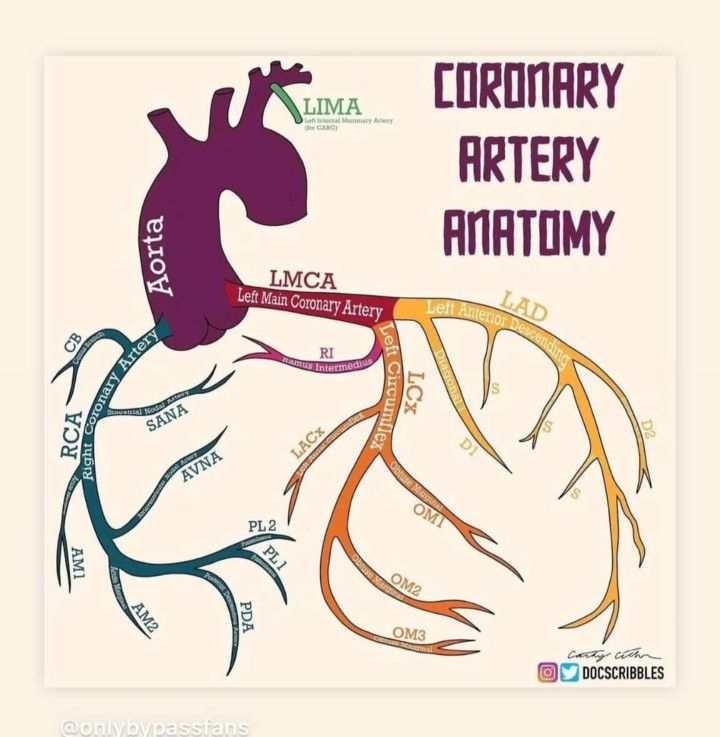
💡 Clinical Pearl: Inferior MI → think bradycardia, heart block and RV infarction (RCA supplies the nodes and RV). Large anterior MI → think heart failure, shock and VT (big LV territory).
1.03 Atherosclerosis and Plaques
Mechanism: Atherosclerosis is a slow, inflammatory disease of the artery wall that begins in childhood and progresses over decades. It develops in stages:
- Endothelial damage: Smoking, high blood pressure, diabetes and high cholesterol damage the inner lining (endothelium), making it "leaky" and sticky.
- LDL entry: LDL cholesterol particles pass into the vessel wall and become oxidised.
- Inflammation: Monocytes enter the wall, become macrophages and engulf the oxidised LDL, turning into lipid-laden foam cells.
- Fatty streak: Collections of foam cells form the earliest visible lesion, already present in many teenagers.
- Mature plaque: Smooth muscle cells migrate in and lay down collagen, forming a fibrous cap over a soft, lipid-rich necrotic core.
- Advanced plaque: The plaque may calcify, bleed internally, ulcerate or rupture.
Stable versus vulnerable plaque. Plaques behave very differently depending on their structure:
- Stable plaque: Thick fibrous cap and small lipid core, often calcified. It narrows the artery gradually and causes exertional angina, but rarely ruptures.
- Vulnerable plaque: Thin cap, large lipid core and active inflammation. It may cause little narrowing but can rupture, exposing its highly thrombogenic contents to blood. Platelets stick, a clot forms and the artery blocks, causing ACS.
- Plaque erosion: The endothelium over a plaque is lost without the cap rupturing. This can also trigger a clot and is relatively more common in younger patients and women.
💡 Important Concept: Heart attacks often arise from plaques that were not severely narrowing the artery beforehand. This is why stenting a stable narrowing does not reliably prevent MI, whereas statins (which stabilise all plaques) do.
1.04 Oxygen Supply and Demand
Ischaemia occurs whenever oxygen delivery falls below what the heart muscle needs. Treatments for angina work by either reducing demand or improving supply.
- Supply depends on: Coronary blood flow and perfusion pressure, haemoglobin, oxygen saturation, and the length of diastole.
- Demand depends on: Heart rate, contractility, wall tension, afterload (blood pressure) and preload (ventricular filling).
- Why diastole matters: The left ventricle is mainly perfused during diastole, because in systole the contracting muscle squeezes its own small vessels shut.
- Why tachycardia is harmful: A fast heart rate both increases demand and shortens diastole, reducing supply. This is why beta blockers and rate control are so effective.
- Non-coronary causes of ischaemia: Severe aortic stenosis, hypertrophic cardiomyopathy, severe anaemia, hypoxia, fast arrhythmias, shock, coronary spasm, coronary embolism, spontaneous coronary artery dissection and vasculitis (e.g. Kawasaki disease).
💡 Clinical Pearl: The subendocardium (inner layer) is the most vulnerable to ischaemia. It is furthest from the epicardial arteries and under the highest pressure during systole. This is why demand ischaemia typically causes ST depression.
1.05 Risk Factors and Risk Assessment
Non-modifiable risk factors:
- Increasing age.
- Male sex (risk in women rises after the menopause).
- Family history of premature coronary disease (a first-degree male relative under 55 or female under 65).
- Familial hypercholesterolaemia.
- South Asian ancestry.
Modifiable risk factors:
- Smoking (including passive smoking).
- Raised LDL cholesterol.
- Hypertension.
- Diabetes.
- Obesity, especially central.
- Physical inactivity.
- Diet high in saturated fat.
- Chronic kidney disease.
- Excess alcohol.
- Psychosocial stress and deprivation.
Risk-enhancing factors:
- Raised lipoprotein(a).
- Chronic inflammatory disease (e.g. rheumatoid arthritis, SLE).
- Previous pre-eclampsia or gestational diabetes.
- Persistent albuminuria.
- Obstructive sleep apnoea.
- Severe mental illness.
UK risk assessment (NICE NG238):
- QRISK3: Estimates 10-year cardiovascular risk in people without known cardiovascular disease.
- Primary prevention: If QRISK3 is 10% or more, offer atorvastatin 20 mg daily after a shared discussion.
- Secondary prevention: People who already have IHD do not need a QRISK score; they all qualify for high-intensity statin therapy.
- Lipids: Measure total cholesterol, HDL, non-HDL cholesterol, LDL and triglycerides.
- Familial hypercholesterolaemia: Suspect it with total cholesterol above 7.5 mmol/L and a personal or family history of premature coronary disease.
💡 Clinical Pearl: There is no "safe" cholesterol threshold — risk rises continuously with lifetime exposure to LDL. Light drinking should not be recommended for heart protection; the apparent benefit in observational studies is due to confounding.
1.06 Stable Angina
Mechanism: A fixed coronary narrowing (usually more than 70% of the lumen) allows enough flow at rest but not during exertion. When demand rises, the muscle beyond the narrowing becomes ischaemic and causes pain. When demand falls, the ischaemia resolves without lasting damage — so troponin does not rise.
Clinical pattern: NICE describes three characteristic features of anginal pain:
- 1. Constricting discomfort in the front of the chest, neck, jaw, shoulders or arms.
- 2. Brought on by physical exertion.
- 3. Relieved by rest or GTN within about 5 minutes.
The number of features present defines the type of pain:
- Typical angina: All three features.
- Atypical angina: Two features.
- Non-anginal chest pain: One or none.
Pain is often predictable, occurring at the same level of activity, and is worse in cold weather, after heavy meals or with emotional stress. Some patients, especially older people and those with diabetes, report breathlessness or fatigue instead of pain ("angina equivalent").
Examination: Often normal. Look for causes and contributors:
- Hypertension.
- Aortic stenosis (ejection systolic murmur).
- Anaemia.
- Heart failure.
- Signs of hyperlipidaemia (xanthelasma, tendon xanthomata, corneal arcus in the young).
💡 Teaching Pearl: Angina that is new, increasing in frequency, occurring at rest or lasting more than 15 minutes is no longer stable. Treat it as possible ACS.
1.07 Investigating Stable Angina
Investigations aim to (1) confirm that the symptoms are due to coronary disease, (2) find contributing conditions and (3) identify patients who would benefit from revascularisation.
- Baseline tests: A 12-lead ECG, which is often normal between episodes, though Q waves suggest a previous MI. Bloods: FBC (anaemia), U&E, HbA1c, lipid profile and thyroid function if indicated.
- Echocardiogram: If there is a murmur, suspected heart failure or a previous MI.
- CT coronary angiography (CTCA): NICE's first-line test when typical or atypical angina cannot be excluded clinically. It shows plaques, calcium and narrowings non-invasively, and a normal CTCA effectively rules out obstructive disease. Heavy calcification reduces accuracy.
- Functional (ischaemia) testing: Used when CTCA shows a narrowing of uncertain significance, or is non-diagnostic. Options include stress echocardiography, stress perfusion cardiac MRI, myocardial perfusion scintigraphy (SPECT) and CT-derived fractional flow reserve (FFR-CT).
- Invasive coronary angiography: For patients whose symptoms persist despite medical therapy, or whose non-invasive tests suggest high-risk anatomy. It allows PCI at the same sitting. Pressure-wire measurements (FFR or iFR) tell whether an intermediate narrowing actually limits flow.
💡 Clinical Pearl: Anatomy is not the same as physiology. A narrowing that looks moderate may cause significant ischaemia, while one that looks severe may not limit flow. Pressure-wire testing settles the question.
1.08 Drug Treatment of Stable Angina
Treatment has two separate goals, and every patient needs both:
- 1. Relieve symptoms: Reduce oxygen demand or improve supply.
- 2. Prevent MI and death: Use antiplatelet, statin and risk-factor treatment.
Step 1 — Sublingual GTN for every patient.
- How it works: GTN dilates veins, reducing preload and therefore oxygen demand.
- Dose: 400 micrograms (one spray) sublingually. Repeat after 5 minutes if needed.
- When to call 999: If pain persists 5 minutes after the second dose.
- Before exertion: It can be taken before activities known to cause angina.
- Side effects: Headache, flushing and dizziness. Sit down when taking it.
- Contraindication: Do not give within 24–48 hours of a PDE-5 inhibitor (e.g. sildenafil) because of the risk of profound hypotension.
Step 2 — First-line: a beta blocker OR a calcium channel blocker, chosen according to comorbidities.
- Beta blockers slow the heart rate and reduce contractility, lowering demand and lengthening diastole.
- Bisoprolol: 2.5–5 mg once daily initially, usually 5–10 mg once daily (max 20 mg).
- Atenolol: 100 mg daily (once daily or 50 mg twice daily).
- Metoprolol: 50–100 mg two to three times daily.
- Target: Resting heart rate of 55–60 bpm.
- Avoid or use caution: Asthma, heart block, severe bradycardia and decompensated heart failure.
- Calcium channel blockers dilate arteries, lowering afterload and improving coronary flow; the rate-limiting types also slow the heart.
- Amlodipine (dihydropyridine): 5 mg once daily, increased to 10 mg if needed. Safe to combine with a beta blocker.
- Diltiazem (rate-limiting): standard-release 60 mg three times daily (60 mg twice daily in older people), max 360 mg/day. Modified-release preparations differ, so prescribe by brand.
- Verapamil (rate-limiting): 80–120 mg three times daily.
- Avoid diltiazem and verapamil in LV systolic dysfunction or conduction disease.
Step 3 — If symptoms are not controlled: Switch to the other class, or combine a beta blocker with a dihydropyridine CCB (e.g. amlodipine).
⚠️ Dangerous combination: Never combine a beta blocker with verapamil (risk of complete heart block and asystole). Diltiazem with a beta blocker should only be used with caution and monitoring.
Step 4 — Add-on drugs if two drugs fail or are not tolerated. NICE advises adding a third anti-anginal only while awaiting revascularisation, or when revascularisation is not appropriate.
- Long-acting nitrates: Reduce preload. Tolerance develops with continuous exposure, so leave a nitrate-free period each day.
- Isosorbide mononitrate 10 mg twice daily initially, usually 20–40 mg twice daily, given asymmetrically (e.g. 8 am and 2 pm).
- Or modified-release 25–60 mg once daily in the morning (max 120 mg daily).
- GTN patch 5–15 mg/24 h with an 8–12-hour patch-free period.
- Contraindicated with PDE-5 inhibitors.
- Ivabradine: Slows the sinus node only, without lowering blood pressure. Only works in sinus rhythm.
- 5 mg twice daily (2.5 mg twice daily if aged 75 or over), increased after 3–4 weeks to a max of 7.5 mg twice daily.
- Start only if resting heart rate is 70 bpm or more.
- Reduce the dose if the heart rate stays below 50 bpm; stop if there is no improvement after 3 months.
- Contraindicated in AF. Do not combine with diltiazem or verapamil.
- Nicorandil: Potassium-channel opener with nitrate-like effects.
- 10 mg twice daily (5 mg twice daily if prone to headache), usually 10–20 mg twice daily, max 30 mg twice daily.
- Can cause ulceration of the mouth, gut, skin or eyes — stop if this develops (MHRA warning).
- Contraindicated with PDE-5 inhibitors.
- Ranolazine: Blocks the late sodium current, improving symptoms with little effect on heart rate or blood pressure.
- 375 mg twice daily for 2–4 weeks, then 500 mg twice daily, max 750 mg twice daily.
- Avoid with strong CYP3A4 inhibitors or a creatinine clearance below 30 mL/min.
- Prolongs the QT interval.
Preventing MI and death (all patients):
- Aspirin: 75 mg daily (consider, balancing bleeding risk).
- High-intensity statin: Atorvastatin 80 mg daily.
- ACE inhibitor: If there is diabetes, hypertension, CKD or LV dysfunction.
- Risk-factor control: Treat blood pressure and diabetes; stop smoking; exercise regularly; eat a healthy diet.
💡 Clinical Pearl: Anti-anginal drugs make patients feel better but do not prolong life. Aspirin and statins prolong life but do not relieve pain. Make sure every patient gets both.
1.09 Revascularisation: PCI and CABG
Revascularisation restores blood flow past a narrowing. In stable disease it is mainly used for symptoms that persist despite optimal medical therapy.
- PCI (percutaneous coronary intervention):
- What it involves: A catheter, usually via the radial artery, is passed into the coronary artery. The narrowing is opened with a balloon and held open with a drug-eluting stent.
- Recovery: Quick, often same-day discharge.
- After the procedure: In stable disease, patients need aspirin plus clopidogrel (dual antiplatelet therapy) for about 6 months, to prevent clot forming on the stent.
- Benefit: Improves angina and quality of life, but in stable disease does not reduce death or MI compared with good medical therapy (COURAGE and ISCHEMIA trials).
- CABG (coronary artery bypass grafting):
- What it involves: Open-heart surgery that routes blood around blockages using grafts. The left internal mammary artery (LIMA) is usually grafted to the LAD and has excellent long-term patency; vein grafts from the leg are also used.
- Who benefits: Generally favoured for significant left main disease, complex multivessel disease and multivessel disease with diabetes. It can improve survival in these groups and in some patients with impaired LV function.
- Choosing between them: A multidisciplinary heart team (cardiologist and cardiac surgeon) decides, considering the anatomy, LV function, diabetes, frailty and the patient's preferences.
💡 Clinical Pearl: In stable angina, a stent is a symptom treatment. Its prognostic value depends on anatomy and LV function; it is not automatically life-saving. In ACS, by contrast, urgent PCI clearly saves lives.
1.10 Vasospastic Angina
Mechanism: A coronary artery goes into sudden, intense spasm, temporarily reducing or stopping blood flow. The artery is often otherwise normal or only mildly diseased. It is also called variant or Prinzmetal angina.
Clinical pattern: Chest pain at rest, typically at night or in the early morning, sometimes in clusters. It is not reliably brought on by exercise. Smoking, cocaine, amphetamines, triptans and cold exposure can trigger it.
Diagnosis: An ECG during pain may show transient ST elevation that resolves with the spasm. Angiography shows no fixed blockage. Specialist provocation testing (e.g. intracoronary acetylcholine) can confirm spasm.
Management:
- Prevention: Calcium channel blockers are first-line (e.g. diltiazem, verapamil or amlodipine).
- Add-on: Long-acting nitrates; sublingual GTN for attacks.
- Triggers: Stop smoking and avoid cocaine and other triggers.
- Avoid non-selective beta blockers (e.g. propranolol) because unopposed alpha-receptor activity can worsen spasm.
💡 Clinical Pearl: Recurrent chest pain at rest in the early morning, with transient ST elevation and normal coronaries, is vasospastic angina. Treat with a calcium channel blocker, not a beta blocker.
1.11 Microvascular Angina (INOCA)
Mechanism: INOCA (Ischaemia with Non-Obstructive Coronary Arteries) describes genuine ischaemia without any significant narrowing of the large coronary arteries. The problem lies in the tiny vessels (the microcirculation), which fail to dilate normally or go into spasm.
Clinical pattern:
- Typical angina, on exertion or at rest, with a "normal" angiogram.
- More often recognised in women.
- It is not benign: quality of life is often poor and cardiovascular risk is increased.
Diagnosis:
- Exclude obstructive coronary disease first.
- Stress perfusion imaging may show ischaemia.
- Specialist invasive testing measures coronary flow reserve and microvascular resistance, and tests for spasm.
Management:
- Control risk factors and give a statin.
- Anti-anginal drugs matched to the mechanism: beta blockers, calcium channel blockers or ranolazine. CCBs are best when spasm predominates.
- Specialist review for persistent symptoms.
💡 Clinical Pearl: A normal angiogram does not mean the chest pain is "not cardiac". Microvascular dysfunction and vasospasm are real causes of angina.
1.12 What Is ACS?
An acute coronary syndrome is a sudden reduction in coronary blood flow, nearly always caused by a clot forming on a ruptured or eroded plaque.
The sequence of events:
- 1. A vulnerable plaque ruptures or erodes.
- 2. Collagen and lipid from inside the plaque are exposed to the blood.
- 3. Platelets stick to the exposed surface, become activated and clump together. This is why antiplatelet drugs are central to treatment.
- 4. The clotting cascade adds fibrin, building a thrombus. This is why anticoagulants are also used.
- 5. Blood flow is reduced (partial occlusion) or stopped (complete occlusion).
- 6. Muscle downstream becomes ischaemic. If this persists, it dies, beginning within about 20–30 minutes and spreading from the inner layer outwards over several hours.
What determines infarct size: How long the artery stays blocked, how much muscle it supplies, and whether collateral vessels provide some backup flow. Early reperfusion limits the damage.
🚨 Time is muscle: Opening the artery in the first hours saves the most heart muscle. After about 12 hours, most of the territory has already died.
1.13 Unstable Angina, NSTEMI and STEMI
ACS is divided into three types according to the ECG and the troponin. This division matters because it decides the immediate treatment.
| Type | ECG, troponin and what it means |
|---|---|
| Unstable angina | Ischaemic pain (new, worsening or at rest). ECG normal or ST depression / T inversion. No troponin rise. Ischaemia without muscle death. |
| NSTEMI | Ischaemic pain. ECG normal or ST depression / T inversion. Troponin rises. Muscle death, usually from a partially blocked artery. |
| STEMI | Ischaemic pain with persistent ST elevation (or an equivalent pattern). Usually a completely blocked artery. Needs immediate reperfusion. |
Key points:
- In STEMI, troponin will rise but may still be normal at first. Never wait for the troponin before treating a STEMI.
- A normal ECG does not exclude NSTEMI.
- Unstable angina has become less common because high-sensitivity troponin assays now detect small amounts of muscle damage that older tests missed.
💡 Clinical Pearl: STEMI and NSTEMI are ECG labels, not measures of how big the infarct is. Some NSTEMIs are large, and some completely blocked arteries (especially the circumflex) do not produce ST elevation.
1.14 Presentation and Examination
Typical symptoms:
- Central, heavy, crushing or tight chest pain.
- Pain radiating to the left arm, both arms, the neck, jaw or back.
- Lasting more than 15–20 minutes, often at rest and not relieved by GTN.
- Associated sweating, nausea, vomiting and breathlessness.
Atypical presentations — common, and frequently missed:
- Older people: Breathlessness, collapse, delirium, falls or sudden functional decline, often without pain.
- People with diabetes: Reduced pain perception from autonomic neuropathy ("silent" MI).
- Women: More likely to report breathlessness, fatigue, nausea or back or jaw pain, although central chest pain remains the commonest symptom.
- Other presentations: Epigastric pain, palpitations, syncope or cardiac arrest.
Examination helps assess severity and look for complications and alternative diagnoses:
- General: Pale, sweaty, distressed; reduced consciousness suggests shock.
- Pulse: Tachycardia, bradycardia (inferior MI) or an irregular rhythm.
- Blood pressure: Hypotension suggests shock, RV infarction or a mechanical complication. Check both arms if dissection is possible.
- JVP and lungs: Crackles mean pulmonary oedema; a raised JVP with clear lungs suggests RV infarction.
- Heart sounds: A new murmur suggests mitral regurgitation or ventricular septal rupture.
- Perfusion: Cold peripheries, slow capillary refill and low urine output mean low cardiac output.
💡 Clinical Pearl: In an older patient with sudden breathlessness, delirium or collapse, always do an ECG and troponin, even without chest pain.
1.15 The ECG in ACS
The 12-lead ECG is the most important immediate test. Record it within 10 minutes of first contact and repeat it if the pain persists or changes.
STEMI criteria: New ST elevation at the J point in two contiguous leads — at least 1 mm in limb leads, and at least 2 mm in V2–V3 for men aged 40 or over (2.5 mm under 40; 1.5 mm in women) and 1 mm in other chest leads.
Localising the infarct:
| Territory | ECG leads → usual artery and what to watch for |
|---|---|
| Anterior / septal | ST elevation V1–V4 → LAD. Large territory: risk of heart failure, shock and VT. |
| Lateral | ST elevation I, aVL, V5–V6 → LCx or a diagonal branch of the LAD. |
| Inferior | ST elevation II, III, aVF (reciprocal depression in I and aVL) → RCA (sometimes LCx). Watch for bradycardia, AV block and RV infarction. |
| Posterior | ST depression V1–V3 with tall R waves and upright T waves; ST elevation in posterior leads V7–V9 → LCx or RCA. Treat as STEMI. |
| Right ventricle | ST elevation in right-sided lead V4R, with an inferior STEMI → proximal RCA. |
Other important ECG findings:
- Hyperacute T waves: Tall, broad T waves that may come before ST elevation — the earliest sign of occlusion.
- ST depression: Subendocardial ischaemia (NSTEMI) or a reciprocal change mirroring a STEMI elsewhere.
- T-wave inversion: Ischaemia, or an evolving or reperfused infarct.
- Pathological Q waves: An established infarct (dead muscle); may persist permanently.
- Left bundle branch block: Hides ST changes. Use clinical judgement and the modified Sgarbossa criteria; get urgent senior or cardiology advice if the patient is unstable or has ongoing ischaemia.


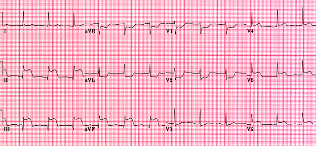
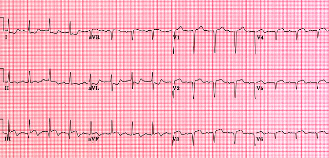
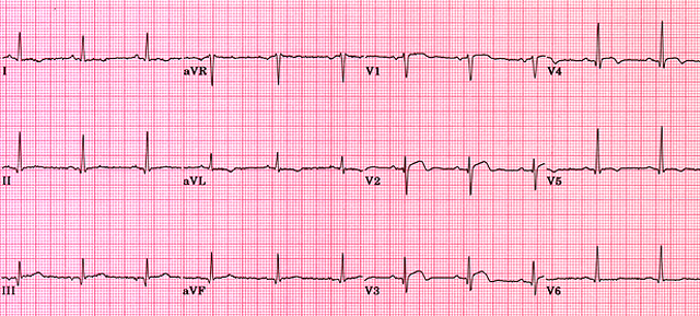
⚠️ ECG Safety Pearl: A normal first ECG does not exclude ACS — repeat it. ST depression in V1–V3 may be a posterior STEMI: request posterior leads. With every inferior STEMI, record right-sided leads.
1.16 Troponin
What it is: Troponin I and T are proteins in the contractile apparatus of heart muscle. They leak into the blood when heart muscle cells are damaged, so a raised troponin means myocardial injury.
How to use it:
- High-sensitivity assays: Detect injury within 1–3 hours of symptom onset.
- Abnormal threshold: Above the 99th percentile of the normal population for that particular assay.
- Serial testing: A rise or fall between samples shows an acute process. A stable raised level suggests chronic injury (e.g. CKD, heart failure). Use the local validated pathway, commonly 0/1-hour or 0/3-hour sampling.
- Very early samples: A sample taken soon after pain onset may be normal even in an evolving MI.
Causes of a raised troponin other than ACS:
- Myocarditis.
- Heart failure.
- Pulmonary embolism.
- Sepsis and critical illness.
- Renal failure.
- Fast arrhythmias.
- Severe hypertension.
- Takotsubo syndrome.
- Cardiac contusion.
- Aortic dissection.
- Stroke and subarachnoid haemorrhage.
- Strenuous endurance exercise.
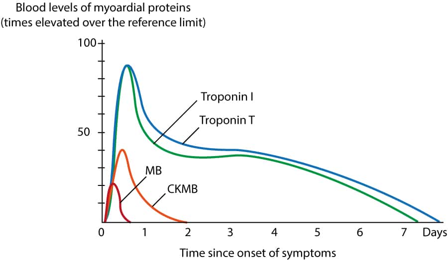
💡 Essential Teaching Point: Troponin tells you the heart muscle is injured, not why. A septic patient with a raised troponin does not usually need ACS treatment; they need treatment for sepsis.
1.17 Definition and Types of MI
Universal definition: MI requires a rise and/or fall of troponin (at least one value above the 99th percentile) plus evidence of ischaemia — at least one of:
- Ischaemic symptoms.
- New ischaemic ECG changes.
- New pathological Q waves.
- New loss of viable muscle or a regional wall-motion abnormality on imaging.
- Coronary thrombus on angiography or at autopsy.
Raised troponin without evidence of ischaemia is called myocardial injury, not infarction.
Types of MI:
- Type 1: Plaque rupture or erosion with coronary thrombosis — the "classic" heart attack. Treat with antiplatelets, anticoagulation and angiography.
- Type 2: A supply–demand mismatch without a new clot, e.g. severe anaemia, fast AF, hypoxia, hypotension or spasm. Treat the underlying cause; routine ACS antithrombotic treatment and urgent angiography are not automatically appropriate.
- Type 3: Sudden cardiac death with features of ischaemia before troponin could be measured.
- Type 4: Related to PCI, including stent thrombosis.
- Type 5: Related to CABG.
💡 Clinical Pearl: Type 2 MI is very common in older inpatients (sepsis, anaemia, AF). Giving them dual antiplatelets and anticoagulation adds bleeding risk without treating the cause.
1.18 Differential Diagnosis of Chest Pain
Life-threatening alternatives — consider these before giving antithrombotics:
- Aortic dissection: Sudden, severe, tearing pain radiating to the back. Look for unequal arm pulses or blood pressures, a new aortic regurgitation murmur and neurological signs. Antiplatelets, anticoagulants and especially fibrinolysis can be fatal.
- Pulmonary embolism: Pleuritic pain, breathlessness, tachycardia, hypoxia and risk factors for VTE.
- Tension pneumothorax: Acute distress, absent breath sounds on one side, hypotension.
- Oesophageal rupture (Boerhaave): Severe pain after vomiting, surgical emphysema and sepsis.
Other diagnoses:
- Pericarditis: Sharp pain, worse lying flat and better sitting forward; widespread saddle-shaped ST elevation with PR depression.
- Myocarditis: Chest pain and raised troponin after a viral illness, often in a young person.
- Gastro-oesophageal reflux: Burning pain related to meals or lying down.
- Musculoskeletal pain: Reproducible on palpation or movement.
- Pneumonia: Fever, cough, pleuritic pain and focal chest signs.
⚠️ Safety Pearl: Before thrombolysis or anticoagulation, always ask: "Could this be a dissection?" Chest pain with syncope, neurological signs, unequal pulses or a new diastolic murmur needs urgent CT aortography first.
1.19 Immediate Management
Use an ABCDE approach while working out which ACS pathway the patient needs.
First 10 minutes:
- ABCDE assessment with heart rate, BP, oxygen saturations and respiratory rate.
- 12-lead ECG immediately — if STEMI, activate the primary PCI pathway now.
- IV access and cardiac monitoring.
- Look for shock, pulmonary oedema and dangerous arrhythmias.
Bloods and imaging:
- High-sensitivity troponin per local protocol.
- FBC, U&E and creatinine (affects drug choice and doses).
- Glucose and HbA1c, lipid profile.
- Clotting if anticoagulation or procedures are planned.
- Chest X-ray if pulmonary oedema or an alternative diagnosis is suspected.
- Echo if unstable or a mechanical complication is suspected.
Initial drugs:
- Aspirin 300 mg orally (chewed or dispersed) as soon as ACS is suspected, unless there is true allergy or suspected dissection.
- GTN 400 micrograms sublingually for pain if systolic BP is above 90 mmHg. Avoid in RV infarction and after recent PDE-5 inhibitor use.
- Analgesia: IV morphine 2.5–5 mg slowly, titrated to effect, with an antiemetic (e.g. metoclopramide 10 mg IV). Opioids can delay absorption of oral P2Y12 inhibitors, so use the lowest effective dose.
- Oxygen: Only if hypoxic. Target saturations of 94–98%, or 88–92% if at risk of hypercapnia. Routine oxygen in normoxic patients gives no benefit.
- Further antithrombotic treatment: A second antiplatelet and an anticoagulant are chosen according to the ACS type, bleeding risk and planned procedure (see the sections that follow).
⚠️ Clinical Pearl: Do not let blood tests delay treatment of a clear STEMI. Equally, do not reflexively give the full ACS drug package to every chest pain — think about dissection first.
1.20 STEMI: Primary PCI
The aim in STEMI is to reopen the blocked artery as fast as possible. The preferred method is primary PCI: emergency angiography, followed by removing the clot and stenting the culprit artery.
NICE NG185 timing:
- Within 12 hours of symptom onset: Offer primary PCI if it can be delivered within 120 minutes of the time fibrinolysis could have been given. In practice, paramedics transfer patients directly to a heart attack centre.
- More than 12 hours: Consider angiography if there is ongoing ischaemia or cardiogenic shock.
Procedure points:
- Radial access is preferred (fewer bleeding complications); drug-eluting stents are used.
- Antiplatelet: Prasugrel with aspirin is preferred if the patient is not on an oral anticoagulant.
- Anticoagulation: Unfractionated heparin during the procedure.
- Multivessel disease without shock: Complete revascularisation of other significant narrowings is usually offered, during the same admission or soon after.
🚨 Time-Critical Principle: Every 30-minute delay to reperfusion increases mortality. The ECG and the story make the diagnosis — not the troponin.
1.21 STEMI: Fibrinolysis
Fibrinolytic ("clot-busting") drugs activate plasminogen to plasmin, which dissolves fibrin in the clot. They are less effective than PCI and carry a risk of bleeding (including intracranial haemorrhage in about 1%), but they can be given anywhere.
Indications:
- STEMI with symptom onset under 12 hours.
- Primary PCI cannot be delivered within 120 minutes of the time fibrinolysis could be given (NICE NG185).
- No contraindications.
- Give as early as possible — ideally within 10 minutes of the diagnostic ECG, pre-hospital if available.
Fibrinolytic doses:
- Tenecteplase (preferred): a single IV bolus over 5–10 seconds, weight-based. A half dose should be considered in patients aged 75 or over to reduce intracranial haemorrhage.
| Weight | Tenecteplase dose |
|---|---|
| <60 kg | 30 mg |
| 60–69 kg | 35 mg |
| 70–79 kg | 40 mg |
| 80–89 kg | 45 mg |
| ≥90 kg | 50 mg |
- Reteplase: 10 units IV bolus, repeated once after 30 minutes.
- Alteplase (accelerated 90-minute regimen): 15 mg IV bolus, then 0.75 mg/kg over 30 minutes (max 50 mg), then 0.5 mg/kg over 60 minutes (max 35 mg); total max 100 mg.
Drugs given with the fibrinolytic:
- Aspirin: 300 mg loading, then 75 mg daily.
- Clopidogrel: 300 mg loading if aged 75 or under; 75 mg (no loading) if over 75; then 75 mg daily. Ticagrelor and prasugrel are not used with fibrinolysis.
- Enoxaparin (preferred anticoagulant), given at the same time as the fibrinolytic:
- Under 75: 30 mg IV bolus, then 15 minutes later 1 mg/kg SC twice daily (max 100 mg for the first two SC doses).
- 75 or over: No IV bolus; 0.75 mg/kg SC twice daily (max 75 mg for the first two doses).
- Creatinine clearance below 30 mL/min: 1 mg/kg SC once daily.
- Duration: Until revascularisation or for up to 8 days.
- Unfractionated heparin (alternative): 60 units/kg IV bolus (max 4,000 units), then 12 units/kg/hour (max 1,000 units/hour), adjusted to an aPTT ratio of 1.5–2.0, for 24–48 hours.
Absolute contraindications — the risk of catastrophic bleeding is too high:
- Previous intracranial haemorrhage, or stroke of unknown cause, at any time.
- Ischaemic stroke within 6 months.
- CNS tumour, damage or arteriovenous malformation.
- Major trauma, surgery or head injury within 1 month.
- GI bleeding within 1 month.
- Known bleeding disorder (excluding menses).
- Suspected aortic dissection.
- Non-compressible punctures in the past 24 hours (e.g. liver biopsy, lumbar puncture).
Relative contraindications:
- TIA within 6 months.
- Oral anticoagulant therapy.
- Pregnancy or within 1 week postpartum.
- Refractory hypertension (systolic above 180 or diastolic above 110 mmHg).
- Advanced liver disease.
- Infective endocarditis.
- Active peptic ulcer.
- Prolonged or traumatic CPR.
After fibrinolysis:
- Transfer to a PCI-capable centre.
- Repeat the ECG at 60–90 minutes. Less than 50% ST resolution, ongoing pain or instability means the drug has failed: arrange urgent rescue PCI, not a second dose.
- If reperfusion succeeds, perform coronary angiography within 2–24 hours.
- Watch for bleeding. New headache, falling GCS or focal neurology suggests intracranial haemorrhage: stop anticoagulation, arrange urgent CT and seek haematology and neurosurgical advice.
💡 Clinical Pearl: Fibrinolysis is a bridge, not the end of treatment. Every patient should still go on to angiography.
1.22 NSTEMI and Unstable Angina
In NSTEMI the artery is usually only partly blocked, so there is time to assess risk before deciding on angiography.
Initial treatment:
- Aspirin 300 mg loading, then 75 mg daily.
- Fondaparinux 2.5 mg SC daily for patients without high bleeding risk who are not having immediate angiography (NICE). Use unfractionated heparin instead if creatinine is above 265 µmol/L.
- A second antiplatelet (P2Y12 inhibitor) — see Antiplatelet Therapy.
- Anti-ischaemic treatment (GTN, beta blocker if no heart failure or shock) and pain relief.
- High-intensity statin (atorvastatin 80 mg).
- Serial ECGs and troponin; continuous monitoring if pain continues or the patient is unstable.
Risk stratification: Use the GRACE score, which combines age, heart rate, systolic BP, creatinine, Killip class (heart failure), cardiac arrest, ST changes and troponin. Assess bleeding risk at the same time.
Timing of angiography (NICE NG185):
- Immediate: If clinically unstable — ongoing pain, haemodynamic instability, life-threatening arrhythmia or acute heart failure. These patients are managed like STEMI.
- Within 72 hours: If the predicted 6-month mortality is above 3% (intermediate or higher risk).
- Lower risk: Consider conservative management, with further testing guided by symptoms and evidence of ischaemia.
Revascularisation: PCI for suitable lesions; CABG for complex multivessel or left main disease; medical therapy alone may be best for some, especially frail patients.
💡 Clinical Pearl: NSTEMI is not a "small" heart attack. Long-term mortality is similar to STEMI because these patients are often older, with more widespread disease.
1.23 Antiplatelet Therapy
Platelets drive coronary thrombosis, so blocking them is central to ACS treatment. Two pathways are targeted, and blocking both is more effective than blocking either alone. This is dual antiplatelet therapy (DAPT).
Aspirin irreversibly blocks cyclooxygenase-1, stopping thromboxane A₂ production and platelet aggregation.
- Dose: 300 mg loading, then 75 mg daily for life.
P2Y12 inhibitors block the ADP receptor on platelets:
- Clopidogrel: An irreversible prodrug needing liver activation, so its response varies.
- Dose: 300 mg loading (600 mg before PCI), then 75 mg daily.
- Used: With fibrinolysis, in high bleeding risk, and when the patient also needs an oral anticoagulant.
- Ticagrelor: Reversible and direct-acting, with a faster, stronger effect.
- Dose: 180 mg loading, then 90 mg twice daily for 12 months.
- Beyond 12 months: 60 mg twice daily may be continued for up to 3 years in selected high-risk patients.
- Side effects: Breathlessness and bradyarrhythmias.
- Prasugrel: Potent and irreversible.
- Dose: 60 mg loading, then 10 mg daily (5 mg daily if under 60 kg or aged 75 or over — generally avoid at 75 or over).
- Contraindicated after previous stroke or TIA.
Which P2Y12 inhibitor? (NICE NG185):
- STEMI with primary PCI: Prasugrel with aspirin if not on an oral anticoagulant; ticagrelor or clopidogrel as alternatives depending on bleeding risk.
- NSTEMI or unstable angina having angiography: Prasugrel or ticagrelor. Give prasugrel only once the anatomy is known and PCI is planned.
- Conservative management or high bleeding risk: Ticagrelor or clopidogrel.
- Patient on an oral anticoagulant (e.g. for AF): Clopidogrel. Keep triple therapy (anticoagulant + aspirin + clopidogrel) as short as possible, often up to 1 month, then anticoagulant plus clopidogrel.
Duration: DAPT usually continues for 12 months after ACS (shorter if bleeding risk is high), then aspirin alone for life. Stopping DAPT early after a stent can cause stent thrombosis, a frequently fatal event. Consider a PPI for patients at risk of GI bleeding.
💡 Clinical Pearl: After ACS a patient takes aspirin plus one P2Y12 inhibitor — never two P2Y12 inhibitors together. Every antithrombotic added increases bleeding, especially in older, frail patients and those with CKD.
1.24 Anticoagulation
Anticoagulants stop the coagulation cascade from building fibrin into the clot. They are used for the first few days of ACS and during PCI.
- Fondaparinux (indirect factor Xa inhibitor):
- Dose: 2.5 mg SC once daily for up to 8 days or until discharge.
- Use: NICE's choice for NSTEMI/unstable angina without high bleeding risk when immediate angiography is not planned.
- Avoid if creatinine clearance is below 20 mL/min.
- During PCI: Extra unfractionated heparin must be given, because fondaparinux alone allows clots to form on catheters.
- Unfractionated heparin (UFH): Acts via antithrombin. It is short-acting, easily adjusted and reversible with protamine, which makes it ideal around procedures and in renal failure.
- Medical ACS: 60–70 units/kg IV bolus (max 5,000 units), then 12–15 units/kg/hour (max 1,000 units/hour), adjusted to an aPTT ratio of 1.5–2.5.
- During PCI: 70–100 units/kg IV, guided by the ACT (50–70 units/kg if a GP IIb/IIIa inhibitor is used).
- Enoxaparin (low-molecular-weight heparin):
- NSTEMI/unstable angina: 1 mg/kg SC twice daily for 2–8 days.
- STEMI under 75: 30 mg IV bolus plus 1 mg/kg SC, then 1 mg/kg SC twice daily (max 100 mg for the first two SC doses).
- STEMI aged 75 or over: No bolus; 0.75 mg/kg SC twice daily (max 75 mg for the first two doses).
- Creatinine clearance 15–30 mL/min: 1 mg/kg SC once daily.
Before anticoagulating, check:
- Active bleeding and platelet count.
- Renal function and weight.
- Whether the patient already takes warfarin or a DOAC.
- That combinations of antiplatelets and anticoagulants are not continued for longer than needed.
1.25 MINOCA, SCAD and Coronary Embolism
MINOCA (Myocardial Infarction with Non-Obstructive Coronary Arteries):
- What it is: The patient meets the criteria for MI, but angiography shows no significant narrowing. It is a working diagnosis, not a final one.
- Possible causes: Plaque disruption without a lasting blockage, coronary spasm, embolism, spontaneous dissection and microvascular dysfunction.
- Mimics: Myocarditis and takotsubo syndrome.
- Key test: Cardiac MRI separates true infarction from myocarditis and takotsubo.
- Treatment: Directed at the underlying cause.
Spontaneous coronary artery dissection (SCAD):
- Mechanism: A tear or bleed within the artery wall compresses the true lumen.
- Who gets it: Typically women under 60 without conventional risk factors, including in late pregnancy and after delivery. Associated with fibromuscular dysplasia and extreme physical or emotional stress.
- Treatment: Usually conservative if stable, because the artery usually heals by itself and PCI risks extending the tear. Beta blockers are commonly used. PCI or CABG is reserved for ongoing ischaemia or instability.
Coronary embolism:
- Sources: AF, prosthetic or diseased valves, infective endocarditis and LV thrombus.
- Management: Find and treat the source. Long-term anticoagulation is usually needed.
💡 Clinical Pearl: A young woman with an MI in the weeks after childbirth has SCAD until proven otherwise.
1.26 Arrhythmias After MI
Ischaemic and dying muscle is electrically unstable, so arrhythmias are commonest in the first 48 hours. Always correct potassium (aim above 4.0 mmol/L) and magnesium, and treat the ischaemia itself.
- Ventricular fibrillation:
- Immediate defibrillation and ALS: adrenaline 1 mg and amiodarone 300 mg after the third shock.
- Urgent angiography after return of circulation.
- VF within the first 48 hours does not by itself require an ICD.
- Ventricular tachycardia:
- Pulseless: ALS with defibrillation.
- Pulse but adverse features (shock, syncope, ischaemia, heart failure): synchronised DC cardioversion (up to 3 attempts), then amiodarone 300 mg IV over 10–20 minutes.
- Stable: Amiodarone 300 mg IV over 20–60 minutes.
- Arrange urgent revascularisation.
- Sinus bradycardia: Common and usually vagal in inferior MI. Treat only with adverse features: atropine 500 micrograms IV, repeated to a maximum of 3 mg. Review rate-limiting drugs.
- AV block — inferior MI: Usually at the AV node and transient. Atropine if compromised; monitor.
- AV block — anterior MI: Mobitz II or complete block means extensive damage below the node and a poor prognosis. Temporary pacing (transcutaneous, then transvenous), with adrenaline or isoprenaline as a bridge. Permanent pacing if the block persists.
- Asystole: CPR and the non-shockable ALS pathway, with adrenaline 1 mg IV as soon as possible and every 3–5 minutes; treat the 4 Hs and 4 Ts. Consider pacing if P waves are present (ventricular standstill).
💡 Clinical Pearl: Heart block in inferior MI usually resolves; heart block in anterior MI signals a large infarct and usually needs pacing.
1.27 Heart Failure and Cardiogenic Shock
Acute LV failure and pulmonary oedema:
- Sit the patient upright; give oxygen to target saturations.
- IV furosemide 40–80 mg (higher if already on a loop diuretic).
- IV nitrate infusion if systolic BP allows.
- CPAP if hypoxic or distressed.
- Urgent echo and revascularisation.
- Avoid routine opiates.
Cardiogenic shock occurs when the heart cannot pump enough blood to perfuse the organs. It usually follows a large anterior MI, and mortality is around 40–50%.
- Features:
- Systolic BP below 90 mmHg despite adequate filling, or the need for vasopressors.
- Cold, clammy peripheries.
- Confusion.
- Oliguria.
- Raised lactate.
- Pulmonary oedema (the lungs may be clear if the RV is mainly affected).
- Management:
- Senior cardiology and critical care input immediately.
- Emergency PCI of the culprit artery.
- Bedside echo to exclude mechanical complications.
- Noradrenaline for hypotension, with or without dobutamine for low output, under invasive monitoring.
- Consider mechanical circulatory support (e.g. Impella, ECMO) in specialist centres.
- Assess fluid needs individually rather than giving large volumes.
- Avoid beta blockers and other negative inotropes.
⚠️ Clinical Pearl: Hypotension after MI is not always LV failure. Think RV infarction, papillary muscle rupture, septal rupture, tamponade and arrhythmia — echo distinguishes them.
1.28 Right Ventricular Infarction
Mechanism: A proximal RCA occlusion can infarct the right ventricle as well as the inferior LV wall. A damaged RV cannot push enough blood through the lungs to fill the left ventricle, so LV output depends on adequate venous return (it is preload-dependent).
Clinical pattern: Inferior STEMI with the triad of hypotension, raised JVP and clear lungs, often with bradycardia or AV block.
Diagnosis: ST elevation in right-sided lead V4R; echo shows a dilated, poorly moving RV.
Management:
- Urgent reperfusion.
- Maintain preload: Cautious IV fluid boluses.
- Avoid nitrates, diuretics and opiates — they reduce preload and can cause collapse.
- Inotropes if hypotension persists despite fluid.
- Pacing for AV block (keeping atrial and ventricular contraction in sequence helps the RV).
💡 Clinical Pearl: Inferior MI + low BP + high JVP + clear lungs = RV infarction. GTN may cause profound hypotension.
1.29 Mechanical Complications
Dead muscle is soft and weakest about 3–5 days after MI, before scar forms. Mechanical complications are now less common thanks to early PCI, but they are frequently fatal. Suspect them with sudden deterioration, a new murmur or shock.
- Papillary muscle rupture:
- Presentation: Sudden severe mitral regurgitation, flash pulmonary oedema and a new pansystolic murmur (which may be quiet), usually within the first week.
- Management: Urgent echo; diuretics and afterload reduction (IV nitrates if BP allows); intra-aortic balloon pump or other mechanical support as a bridge; emergency mitral valve surgery.
- Ventricular septal rupture:
- Presentation: A hole in the septum creates a left-to-right shunt, with a new harsh pansystolic murmur and shock.
- Management: Urgent echo; immediate cardiothoracic referral; haemodynamic support (diuretics, vasodilators if BP allows, IABP or Impella) as a bridge to surgical repair; percutaneous closure in selected patients.
- Free-wall rupture:
- Presentation: Blood fills the pericardium, causing tamponade (hypotension, raised JVP, PEA arrest).
- Management: Emergency echo; pericardiocentesis only as a temporary measure; immediate cardiac surgery. Often fatal.
🚨 Clinical Pearl: Sudden deterioration several days after MI with pulmonary oedema, shock or a new murmur needs an immediate echo and cardiothoracic referral.
1.30 Pericarditis and Late Complications
Pericarditis:
- Early post-MI pericarditis (days 1–3): Inflammation over the infarct, causing pleuritic pain and a friction rub. Usually self-limiting.
- Treat with high-dose aspirin; colchicine may be added.
- Avoid other NSAIDs and corticosteroids, which impair scar healing.
- Exclude re-infarction and check for an effusion.
- Dressler syndrome (weeks later): Immune-mediated pericarditis with fever and effusion.
- High-dose aspirin plus colchicine 500 micrograms twice daily (once daily if under 70 kg) for about 3 months.
- Echo to exclude effusion and tamponade.
- Corticosteroids only if refractory.
- Review anticoagulation if there is an effusion.
Late complications:
- Ischaemic cardiomyopathy and heart failure:
- Full heart failure therapy for reduced EF: ACE inhibitor/ARB or sacubitril/valsartan, beta blocker, MRA and SGLT2 inhibitor, plus diuretics for congestion.
- Cardiac rehabilitation.
- Consider an ICD or CRT if the EF remains 35% or less despite optimal therapy (reassess at least 40 days after MI).
- LV aneurysm:
- Suspect if ST elevation persists weeks after MI; confirm with echo or cardiac MRI.
- Causes heart failure, thrombus and VT.
- Heart failure therapy; anticoagulate if thrombus is present; surgery for refractory heart failure, angina or VT.
- LV thrombus (risk of embolic stroke):
- Anticoagulation (warfarin or DOAC) for 3–6 months with repeat imaging.
- Keep triple therapy with antiplatelets as short as possible and give a PPI.
- Recurrent ventricular arrhythmias: Optimise beta blockade, correct electrolytes and treat residual ischaemia; amiodarone if needed; electrophysiology referral and ablation; ICD for secondary prevention.
- Recurrent MI: Treat as a new ACS, considering stent thrombosis; check adherence to DAPT.
1.31 Secondary Prevention
Atherosclerosis affects all the arteries, so an MI marks a patient at high risk of another event. Secondary prevention reduces that risk and should be started before discharge. A simple way to remember the drugs is "5 drugs for life after MI", adjusted to the individual.
- Antiplatelets:
- Aspirin 75 mg daily for life.
- Plus a P2Y12 inhibitor, usually for 12 months.
- High-intensity statin:
- Dose: Atorvastatin 80 mg daily; lower doses if there are interactions or intolerance.
- Target (NICE NG238): LDL cholesterol 2.0 mmol/L or less, or non-HDL cholesterol 2.6 mmol/L or less.
- Monitoring: Recheck lipids at 2–3 months.
- If the target is not met: Add ezetimibe 10 mg daily, then consider other NICE-approved options (e.g. inclisiran, PCSK9 inhibitors).
- ACE inhibitor:
- Dose: Ramipril 2.5 mg twice daily once stable, titrated to 5 mg twice daily.
- Monitoring: BP, renal function and potassium.
- If not tolerated: Use an ARB.
- Beta blocker:
- When to start: Once haemodynamically stable.
- Reduced EF: Continue indefinitely.
- Preserved EF: NICE advises about 12 months; recent trials have questioned the benefit, so review continuation individually.
- MRA (eplerenone):
- Who: Patients with EF 40% or less plus heart failure or diabetes.
- When: Start 3–14 days after MI.
- Dose: 25 mg daily, increasing to 50 mg daily.
- Monitoring: Potassium.
- SGLT2 inhibitor: If there is HFrEF, diabetes or CKD.
- Before discharge: Assess LV function by echo in all patients.
💡 Clinical Pearl: Revascularisation fixes one artery; secondary prevention protects all of them. Both are needed.
1.32 Rehabilitation and Lifestyle
- Cardiac rehabilitation: Offer to every patient. Structured exercise and education reduce cardiac death and readmission, but uptake is poor, so encourage it actively.
- Smoking cessation: The single most effective lifestyle change — it substantially reduces the risk of death and further MI.
- Exercise: Regular graded aerobic exercise, increasing gradually.
- Diet: Mediterranean-style, low in saturated fat, high in fibre, fruit and vegetables.
- Risk factors: Control blood pressure and diabetes, and optimise weight.
- Psychological health: Depression and anxiety are common after MI and worsen outcomes — screen and support.
- Driving: Group 1 (car) licence holders usually stop driving for 1 week after successful PCI, or 4 weeks otherwise; they do not need to notify the DVLA unless other conditions apply. Group 2 (bus and lorry) drivers must stop for at least 6 weeks and notify the DVLA.
- Returning to normal life: Most people can return to work within weeks, depending on the job. Sexual activity can usually resume about 4 weeks after an uncomplicated MI.
1.33 Drug Reference Table
🫀 Common adult doses (UK). Doses vary with indication, renal function, blood pressure, bleeding risk and local protocols. Always check the BNF.
| Drug | Usual adult dose |
|---|---|
| Aspirin | ACS: 300 mg loading, then 75 mg daily for life. |
| Clopidogrel | 300–600 mg loading (600 mg before PCI), then 75 mg daily. With fibrinolysis: 300 mg if ≤75 years, 75 mg if >75. |
| Ticagrelor | 180 mg loading, then 90 mg twice daily for 12 months; 60 mg twice daily thereafter in selected patients. |
| Prasugrel | 60 mg loading, then 10 mg daily (5 mg if <60 kg or ≥75 years). Contraindicated after stroke/TIA. |
| Fondaparinux | 2.5 mg SC daily up to 8 days. Avoid if CrCl <20 mL/min. Extra UFH needed during PCI. |
| Enoxaparin | NSTEMI: 1 mg/kg SC twice daily. STEMI: see Anticoagulation for age- and renal-adjusted regimens. |
| Unfractionated heparin | Medical ACS: 60–70 units/kg IV bolus, then 12–15 units/kg/h (aPTT 1.5–2.5). PCI: 70–100 units/kg guided by ACT. |
| Tenecteplase | Single IV bolus 30–50 mg by weight band; consider half dose if ≥75 years. |
| GTN spray | 400 micrograms sublingually; repeat after 5 minutes. Call 999 if pain persists 5 minutes after the second dose. |
| Morphine | 2.5–5 mg IV slowly, titrated, with an antiemetic. |
| Bisoprolol | Angina: 2.5–5 mg daily, usually 5–10 mg (max 20 mg). Target heart rate 55–60 bpm. |
| Amlodipine | 5 mg daily, increase to 10 mg if needed. |
| Diltiazem | 60 mg three times daily (twice daily in older people), max 360 mg/day. Prescribe MR by brand. |
| Verapamil | 80–120 mg three times daily. Never with a beta blocker. |
| Isosorbide mononitrate | 20–40 mg twice daily (asymmetric), or MR 25–60 mg each morning (max 120 mg). |
| Ivabradine | 5 mg twice daily (2.5 mg if ≥75), max 7.5 mg twice daily. Sinus rhythm, HR ≥70. |
| Nicorandil | 10 mg twice daily, usually 10–20 mg twice daily (max 30 mg twice daily). Watch for ulceration. |
| Ranolazine | 375 mg twice daily, then 500 mg, max 750 mg twice daily. Check QT and interactions. |
| Atorvastatin | Secondary prevention: 80 mg daily. Primary prevention: 20 mg daily. |
| Ezetimibe | 10 mg daily when further LDL lowering is needed. |
| Ramipril | Post-MI: 2.5 mg twice daily once stable, titrated to 5 mg twice daily. |
| Eplerenone | Post-MI LV dysfunction: 25 mg daily, increasing to 50 mg daily. |
⚠️ Prescribing Pearls: Aspirin plus one P2Y12 inhibitor after ACS. Prasugrel is contraindicated after stroke or TIA. Never give nitrates with PDE-5 inhibitors or in RV infarction. Monitor potassium and creatinine with ACE inhibitors and eplerenone. Do not start beta blockers in shock or heart block.
1.34 Spot the Diagnosis
| Clue | Think of |
|---|---|
| Predictable exertional chest tightness relieved by rest | Stable angina |
| New or worsening angina at rest, normal troponin | Unstable angina |
| ST elevation II, III, aVF with bradycardia | Inferior STEMI (RCA) |
| Inferior STEMI, low BP, high JVP, clear lungs | RV infarction |
| ST depression V1–V3 with tall R waves | Posterior STEMI |
| Early-morning rest pain, transient ST elevation, normal coronaries | Vasospastic angina |
| Angina in a woman, normal angiogram, positive stress test | Microvascular angina (INOCA) |
| Young woman, MI weeks after childbirth | SCAD |
| Raised troponin in sepsis with fast AF | Type 2 MI / myocardial injury |
| Day 4 post-MI, flash pulmonary oedema, new murmur | Papillary muscle rupture |
| Day 4 post-MI, shock, harsh pansystolic murmur | Ventricular septal rupture |
| Sudden PEA arrest days after MI | Free-wall rupture with tamponade |
| Persistent ST elevation weeks after MI | LV aneurysm |
| Fever, pleuritic pain and effusion weeks after MI | Dressler syndrome |
| Tearing pain to back, unequal arm BPs | Aortic dissection |
1.35 Exam-style Scenarios
Decide your answer before reading on.
- 1. A 58-year-old man has 40 minutes of central chest pain. ECG shows ST elevation in V1–V4. The nearest PCI centre is 30 minutes away. Answer: Anterior STEMI. Give aspirin 300 mg and analgesia, and transfer for primary PCI (prasugrel loading in the cath lab if not on an anticoagulant). Fibrinolysis is not needed because PCI is available within 120 minutes.
- 2. A 72-year-old woman on a remote island has an inferior STEMI; the nearest PCI is 4 hours away. She had an ischaemic stroke 3 months ago. Answer: Ischaemic stroke within 6 months is an absolute contraindication to fibrinolysis. Arrange urgent transfer for primary PCI despite the delay.
- 3. Ninety minutes after tenecteplase, a patient still has pain and less than 50% ST resolution. Answer: Failed reperfusion. Arrange urgent rescue PCI; do not repeat the fibrinolytic.
- 4. A patient with an inferior STEMI becomes hypotensive after GTN. JVP is raised; lungs are clear. Answer: RV infarction. Stop nitrates, give IV fluid boluses, record V4R, and arrange urgent reperfusion.
- 5. An 80-year-old woman admitted with pneumonia and fast AF has a troponin rising from 60 to 90 ng/L and no chest pain. Answer: Probable type 2 MI or myocardial injury. Treat the sepsis and control the AF; do not start routine ACS DAPT and anticoagulation.
- 6. Four days after an anterior MI, a man suddenly becomes breathless and hypotensive with a new loud pansystolic murmur. Answer: Ventricular septal rupture (or papillary muscle rupture). Immediate echo and cardiothoracic referral, with mechanical support as a bridge to surgery.
- 7. A patient with stable angina on bisoprolol still has symptoms. Which drug should be added? Answer: A dihydropyridine CCB such as amlodipine — not verapamil, because of the risk of heart block.
- 8. A 34-year-old woman 3 weeks postpartum has chest pain and a raised troponin. Angiography shows a long, smooth narrowing of the LAD. Answer: SCAD. Treat conservatively if stable, with a beta blocker; avoid PCI unless there is ongoing ischaemia.
1.36 Key Points
- 1. IHD is a mismatch between oxygen supply and demand; atherosclerosis is the usual cause.
- 2. Stable angina is reversible ischaemia; troponin does not rise.
- 3. ACS is plaque rupture plus clot; antiplatelets and anticoagulants target the clot.
- 4. MI often arises from plaques that were not severely narrowing the artery.
- 5. A normal ECG and a normal first troponin do not exclude ACS — repeat both.
- 6. STEMI needs reperfusion now; primary PCI within 120 minutes, otherwise fibrinolysis.
- 7. ST depression in V1–V3 may be a posterior STEMI.
- 8. Inferior MI: watch for heart block and RV infarction; avoid nitrates if the RV is involved.
- 9. Troponin means injury, not necessarily ACS; distinguish type 1 from type 2 MI.
- 10. Give oxygen only if hypoxic.
- 11. Aspirin plus one P2Y12 inhibitor for 12 months; never stop DAPT early after a stent.
- 12. A new murmur or shock days after MI needs an immediate echo.
- 13. Never combine a beta blocker with verapamil.
- 14. Secondary prevention: aspirin, P2Y12 inhibitor, atorvastatin 80 mg, ACE inhibitor and beta blocker, plus cardiac rehabilitation.
- 15. Exclude aortic dissection before giving antithrombotics or fibrinolysis.
1.37 NICE and European Guidelines
- 📄 NICE NG185: Acute Coronary Syndromes — STEMI, NSTEMI, unstable angina and secondary prevention.
- 📄 NICE CG95: Recent-Onset Chest Pain — assessment and investigation of suspected angina.
- 📄 NICE CG126: Stable Angina — anti-anginal therapy and revascularisation.
- 📄 NICE NG238: Cardiovascular Risk and Lipid Modification — statins and prevention.
- 📄 NICE NG106: Chronic Heart Failure — LV dysfunction after MI.
- 📄 ESC: Acute Coronary Syndromes
- 📄 ESC: Chronic Coronary Syndromes