Related Subjects:
|Assessing Breathlessness
|Assessing Chest Pain
|Pericardial Effusion and Tamponade
|Constrictive Pericarditis
|Colchicine
|Chest X Ray Interpretation
📖 About
- Constrictive pericarditis = rigid pericardium → heart trapped in a “small rigid box.”
- ❌ Restricts normal diastolic filling → impaired cardiac output.
- JVP: prominent x descent; y descent prominent (lost in tamponade).
- Key haemodynamics: dip & plateau / “square root sign” on pressure tracings.
🦠 Causes
- Idiopathic / Post-viral 🌐 → most common in developed countries.
- Tuberculosis 🦠 → commonest cause worldwide, esp. developing countries.
- Post-surgical or Post-radiation 🏥 → scarring after cardiac surgery or mediastinal radiotherapy.
- Inflammatory / Autoimmune 🤲 → RA, SLE, systemic autoimmune disease.
- Metabolic 💉 → Uraemia, trauma.
- Malignancy 🎗️ → primary pericardial tumours or secondary invasion.
- Drugs 💊 → hydralazine, procainamide, methysergide.
🧬 Pathophysiology
- Ventricular filling impaired in mid/late diastole.
- All diastolic pressures equalise (RA, RV, LA, LV).
- Dip and plateau waveform = rapid early filling then abrupt halt ⏹.
🩺 Clinical Features
- 📉 Symptoms: fatigue, weakness, ascites, breathlessness, orthopnoea.
- 🫀 Signs:
- Soft heart sounds, impalpable apex beat.
- Signs of right heart failure: hepatomegaly, ascites, pleural effusion.
- Raised JVP with both x & y descents (Friedreich’s sign).
- Kussmaul’s sign → JVP rises with inspiration (normally falls).
- Pericardial knock → early diastolic sound (mimics S3).
- Pulsus paradoxus (more classic in tamponade, but can occur here).
- ❌ Pulmonary oedema suggests another diagnosis (think restrictive cardiomyopathy).
 💡 Key exam pearl: differentiate from restrictive cardiomyopathy (myocardial disease) and cardiac tamponade (fluid compression). This may need multimodal imaging and haemodynamics.
🔎 Investigations
- 🧪 Bloods: FBC, U&E, CRP → look for TB, uraemia, inflammation.
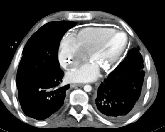
- 📷 CXR: pericardial calcification, pleural effusions.
- 📉 ECG: low QRS voltage, widespread T wave flattening/inversion, P mitrale.
- 🩻 Echo: thickened, bright pericardium ± septal bounce.
- 🧭 Cardiac catheterisation: square root sign (early dip + plateau).
- 🧲 CT / MRI: confirm thickened or calcified pericardium, assess extent.
- 🔬 Pericardial biopsy if unclear (TB, malignancy).
🛠️ Management
- Medical 💊:
- Diuretics → relieve fluid overload (symptomatic only).
- NSAIDs or steroids if active inflammation present.
- Surgical 🔪:
- Pericardiectomy = definitive treatment.
- High-risk surgery but often curative in severe symptomatic cases.
📚 References
Cases - Constrictive Pericarditis
- Case 1 - Post-Tuberculosis Pericarditis:
A 45-year-old man from South Asia presents with progressive breathlessness, abdominal distension, and leg swelling. On exam: raised JVP with prominent y descent, hepatomegaly, ascites, and peripheral oedema. Heart sounds are quiet, and a pericardial knock is heard in early diastole. CXR: pericardial calcification. Echocardiography: septal bounce and ventricular interdependence.
Diagnosis: Constrictive pericarditis secondary to TB infection.
Management: Optimise with diuretics for fluid overload; definitive treatment is pericardiectomy in suitable patients.
- Case 2 - Post-Cardiac Surgery Patient:
A 62-year-old woman presents 1 year after CABG surgery with exertional dyspnoea, fatigue, and ankle oedema. Examination: raised JVP with Kussmaul’s sign, hepatomegaly, ascites. ECG shows low-voltage QRS. Echocardiography: thickened pericardium with abnormal ventricular filling. Cardiac catheterisation: equalisation of diastolic pressures in all four chambers.
Diagnosis: Constrictive pericarditis post-cardiac surgery.
Management: Symptomatic relief with diuretics initially; surgical pericardiectomy is the only curative therapy.
Teaching Commentary ❤️
Constrictive pericarditis occurs when the pericardium becomes fibrosed and rigid, impairing diastolic filling and mimicking right heart failure. Causes include tuberculosis, previous cardiac surgery, radiation, and idiopathic/viral. Classic findings: raised JVP with rapid y descent, Kussmaul’s sign, ascites, hepatomegaly, peripheral oedema, and a pericardial knock. Echo and CT/MRI can confirm thickened, calcified pericardium.
Management is mainly supportive with diuretics, but definitive treatment is pericardiectomy in patients with severe, persistent symptoms.
