
| Download the amazing global Makindo app: ✅ Means NICE/National Guidelines 2026 compliant Android | Apple | |
|---|---|
| MEDICAL DISCLAIMER: Educational use only. Not for diagnosis or management. See below for full disclaimer. |
Idiopathic Fascicular Left Ventricular Tachycardia
Related Subjects: |Classical Ventricular Tachycardia |Idiopathic Ventricular Tachycardia |Right Ventricular Outflow Tract Tachycardia |Idiopathic Fascicular Left Ventricular Tachycardia |Left Ventricular Outflow Tract Tachycardia |Ventricular Fibrillation |Resuscitation - Adult Tachycardia Algorithm |Resuscitation - Advanced Life Support |Automatic Implantable Cardioverter Defibrillator (AICD)
⚡ Idiopathic ventricular tachycardia (IVT) in patients with an anatomically normal heart is a distinct entity whose management and prognosis differs from ventricular tachycardia associated with structural heart disease. This is a specialist diagnosis and should not be confused with VT in the setting of cardiomyopathy. ✅
📖 About
- A form of Idiopathic Ventricular Tachycardia (IVT).
- 💓 The most common idiopathic VT of the left ventricle, often arising from the Purkinje system.
🧬 Aetiology
- Absence of structural heart disease 🫀.
- Due to a re-entrant arrhythmia mechanism involving the fascicles of the left bundle branch.
- Mainly affects males (60–80%), young to middle-aged (15–40 years). 👨
🔎 Forms (Subtypes)
- Posterior fascicular VT (most common, ⬇️ axis).
- Anterior fascicular VT (less common, ⬆️ axis).
- Upper septal fascicular VT (rare, narrowest QRS).
✅ Diagnostic Criteria
- No structural heart disease (confirmed by Echo/CMR).
- No metabolic or electrolyte abnormalities (exclude hypoK/Mg, etc.).
- No inherited arrhythmia syndromes (e.g., Long QT, Brugada, CPVT).
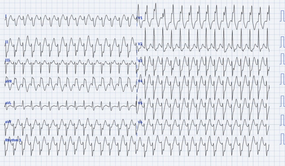
📊 Fascicular VT ECG Example
🩺 Classical VT is still the most common cause of wide-complex regular tachycardia. ❗ There is no perfectly reliable ECG method to distinguish classical VT from idiopathic VT or SVT with aberrancy. 👉 Always treat as VT until proven otherwise – follow the Adult Tachycardia (ALS) algorithm.
👩⚕️ Clinical Features
- Typically young male adults (15–40 years).
- Symptoms: palpitations 💓, dizziness, presyncope/syncope 😵, often triggered by exertion, febrile illness, or stress.
- Usually well tolerated, unlike scar-related VT, but still causes functional limitation.
🧪 Investigations
- 🧾 Bloods: Normal (exclude electrolytes, thyroid).
- 🫀 Echocardiogram: Normal LV size and function.
- 📷 CXR: Normal.
- 🩺 12-lead ECG (baseline): QT normal, no structural abnormality.
- 📉 Acute ECG during VT: RBBB morphology, axis depending on subtype; QRS narrower (100–140 ms) compared to other VT.
- 💡 Cardiac MRI may be used to exclude subtle myocarditis, sarcoid or ARVC.
💊 Management
- 🔑 First principle: If in doubt, treat as classical VT and involve cardiology early.
- 🛑 ABC approach. If unstable ➝ DC cardioversion (per ALS). ⚡
- 👨⚕️ In stable, proven fascicular VT with normal LV function ➝ IV Verapamil 10 mg over 3–5 min under senior cardiology supervision (be ready to DC convert). ❗ NEVER use verapamil if diagnosis uncertain as it may be fatal in scar-related VT.
- For recurrent but moderate symptoms ➝ Oral Verapamil (120–480 mg/day).
- Radiofrequency catheter ablation 🔥 offers curative therapy in >90% of patients with symptomatic or drug-refractory VT.
- 💡 Unlike scar VT, idiopathic fascicular VT has an excellent long-term prognosis with low risk of sudden death once properly diagnosed and managed.
📚 References
| The content on this website is provided for educational and informational purposes only to support exam preparation (e.g., MLA, MRCP, USMLE) and learning. This is NOT medical advice, diagnosis, treatment, or professional guidance. It does not replace consultation with a qualified healthcare professional, official guidelines (e.g., NICE, GMC, BNF), or supervised clinical practice. Always verify information with current, authoritative sources. Makindo and its contributors accept no liability for any reliance on this content, including errors, omissions, or any resulting harm, loss, or consequences. By using this site, you agree to these terms. |
|
|
Categories
- About
- Acute Medicine
- Anaesthetics and Critical Care
- Anatomy
- Anatomy and Physiology
- Biochemistry
- Book
- Cardiology
- Collections
- CompSci
- Crib Sheets
- Critical care
- Dental
- Dermatology
- Differentials
- Drugs
- ENT
- Electrocardiogram
- Embryology
- Emergency Medicine
- Endocrinology
- Ethics
- Foundation Doctors
- GCSE
- Gastroenterology
- General Practice
- Genetics
- Geriatric Medicine
- Geriatrics
- Guidelines
- Haematology
- Hepatology
- Immunology
- Infectious Diseases
- Infographic
- Investigations
- Lists
- MRCP
- Mandatory Training
- Medical Students
- Microbiology
- Nephrology
- Neurology
- Neurosurgery
- Nutrition
- OSCE
- Obstetrics Gynaecology
- Oncology
- Ophthalmology
- Oral Medicine and Dentistry
- Orthopaedics
- Paediatrics
- Palliative
- Palliative Care
- Pathology
- Pharmacology
- Physiology
- Procedures
- Psychiatry
- Public Health
- Radiology
- Respiratory
- Resuscitation
- Revision Topics
- Rheumatology
- Statistics and Research
- Stroke
- Surgery
- Toxicology
- Trauma and Orthopaedics
- USMLE
- Urology
- Vascular Surgery