Pantothenate Kinase–Associated Neurodegeneration
🐯🧠 Eye of the Tiger Sign see below is a classic MRI appearance strongly associated with PKAN (Pantothenate Kinase–Associated Neurodegeneration), one of the NBIA disorders (Neurodegeneration with Brain Iron Accumulation).
It reflects pathological iron deposition in the basal ganglia and is a high-yield radiology clue in patients with progressive dystonia/parkinsonism.
Definition
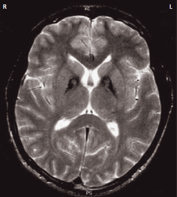
- 🐯 The “eye of the tiger” sign describes a characteristic pattern in the globus pallidus on MRI.
- 🧲 It is most classically seen on T2-weighted (and often T2*/SWI) imaging.
- 🧠 It is strongly linked to PKAN, an autosomal recessive NBIA disorder caused by mutations in PANK2.
Where is it seen?
- 🧠 Basal ganglia, specifically the globus pallidus.
- 🎯 This region is vulnerable because iron handling and mitochondrial metabolism are tightly linked to neuronal survival here.
Imaging Appearance (How to Describe It)
- 🧲 T2 MRI: central area of high signal in the globus pallidus.
- ⬛ Surrounding rim of low signal due to iron deposition (paramagnetic effect).
- 🧠 The result resembles a “tiger’s eye”: bright centre + dark surrounding ring.
- 🧪 SWI/T2* often highlights the iron much more clearly (marked blooming).
- 🚫 Typically no enhancement and no mass effect (this is degeneration, not tumour).
Pathophysiology (Why the Sign Happens)
- 🧬 PANK2 mutation disrupts coenzyme A metabolism in mitochondria.
- ⚙️ Neurons become vulnerable to oxidative stress and abnormal lipid metabolism.
- ⬛ Iron accumulates in the globus pallidus → produces low T2 signal.
- ✨ The central bright region represents gliosis, vacuolisation, and tissue degeneration.
- 🔥 Iron drives oxidative injury → progressive neurodegeneration (a vicious cycle).
Clinical Presentation (When to Suspect It)
- 👶 Classic PKAN: onset in childhood, often with rapid progression.
- 🧑 Atypical PKAN: later onset (teen/adult), slower progression.
- 🌀 Dystonia (often generalised), painful spasms, abnormal posturing.
- 🧍 Parkinsonism (bradykinesia, rigidity, postural instability).
- 🗣️ Dysarthria and swallowing difficulty.
- 🧠 Cognitive change and psychiatric features (impulsivity, mood symptoms) may occur.
- 🚶 Gait deterioration, falls, increasing care needs.
Key Exam / Bedside Clues
- 🎯 Progressive dystonia in a child or teenager is a major red flag.
- 🧠 Mixed movement disorder (dystonia + parkinsonism) suggests basal ganglia disease.
- ⚠️ Look for bulbar dysfunction (dysarthria/dysphagia) as this drives morbidity.
Differential Diagnosis (Important)
- 🧲 Other NBIA disorders can mimic aspects of the imaging pattern:
- PLA2G6-associated neurodegeneration (PLAN)
- Neuroferritinopathy
- Aceruloplasminemia
- 🧠 Wilson disease (different pattern; look for hepatic disease + KF rings).
- 🩸 Manganese deposition (e.g., chronic liver disease) can affect basal ganglia.
- 💊 Drug/toxin-related basal ganglia injury (CO poisoning, methanol) - usually different clinical context and imaging distribution.
How Reliable is the Sign?
- ✅ Strongly associated with PKAN, especially in classic childhood cases.
- ⚠️ Not absolutely exclusive - rare “eye-of-the-tiger–like” appearances can occur in other disorders.
- 🧬 Diagnosis should be confirmed with PANK2 genetic testing.
🔎 Investigations
- 🧲 MRI brain with T2, T2*/SWI sequences to assess iron deposition.
- 🧬 Genetic testing: PANK2 sequencing (and NBIA panels if uncertain).
- 🧪 Bloods to consider depending on differential:
- Caeruloplasmin and copper studies (exclude Wilson)
- Ferritin and iron studies (selected cases)
- Liver function tests (metabolic/toxic causes)
- 🧑⚕️ Specialist neurology assessment with movement disorder expertise.
Management (Principles)
- 🎯 Multidisciplinary care is essential: neurology, physiotherapy, speech and language therapy, dietetics, neurodisability, palliative care support as needed.
- 💊 Symptomatic treatment (often the mainstay):
- Antispasmodics for dystonia (e.g., baclofen)
- Botulinum toxin injections for focal dystonia
- Trial of dopaminergic therapy for parkinsonian features in selected patients
- Management of pain, spasticity, and sleep disturbance
- 🧠 Deep brain stimulation (DBS) may benefit severe dystonia in selected cases.
- 🗣️ Swallow assessment and aspiration prevention strategies are critical in progressive disease.
Prognosis
- ⏳ Variable: classic childhood PKAN is often progressive with increasing disability.
- 🧑 Adult-onset forms may progress more slowly.
- 🚨 Complications often relate to immobility, aspiration, and malnutrition.
Teaching Points (High Yield)
- 🐯 “Eye of the tiger” = think PKAN (PANK2).
- ⬛ Dark rim = iron deposition; ✨ bright centre = gliosis/vacuolisation.
- 🌀 Progressive dystonia/parkinsonism in young person → request MRI with SWI/T2*.
- 🧬 Imaging suggests the diagnosis, but genetics confirms it.
Mini Case (Spot Diagnosis Style)
- 👦 A 12-year-old develops progressive dystonia and falls with worsening dysarthria.
- 🧲 MRI shows bilateral globus pallidus hypointensity with central hyperintensity on T2 (“eye of the tiger”).
- 🧬 Most likely diagnosis: PKAN (NBIA disorder).
- 🎯 Next step: genetic testing (PANK2) + MDT management for movement disorder symptoms.