
| Download the amazing global Makindo app: ✅ Means NICE/National Guidelines 2026 compliant Android | Apple | |
|---|---|
| MEDICAL DISCLAIMER: Educational use only. Not for diagnosis or management. See below for full disclaimer. |
Abdominal X Ray Collections
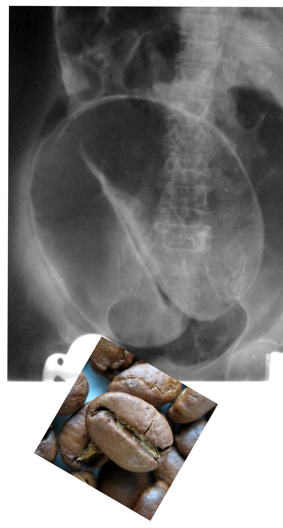
🌀 Expected X-ray findings:
• Massively dilated sigmoid colon arising from the pelvis.
• Classic “coffee-bean” sign with the apex pointing to the right upper quadrant.
• Loss of normal haustral markings in the distended loop.
• Little or no gas in the rectum.
⚠️ If prolonged → risk of ischaemia and perforation (may coexist with free air).
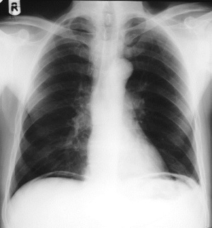
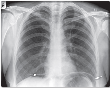
Free Air (Pneumoperitoneum)
💥 Expected X-ray findings:
• Free gas under the diaphragm on erect chest X-ray (right side most sensitive).
• Rigler’s sign: both sides of the bowel wall visible.
• Football sign with large volumes of intraperitoneal air.
🚨 Indicates perforated viscus until proven otherwise → immediate surgical review.
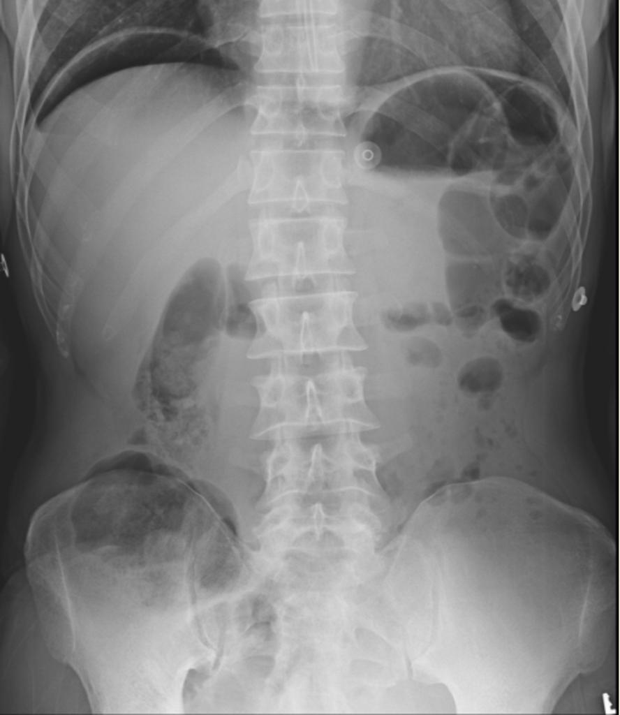
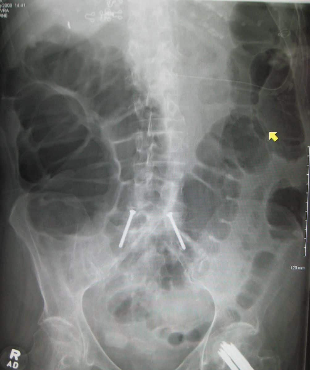
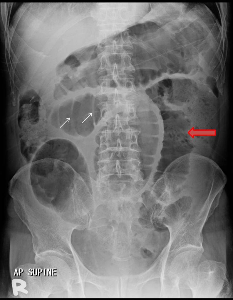
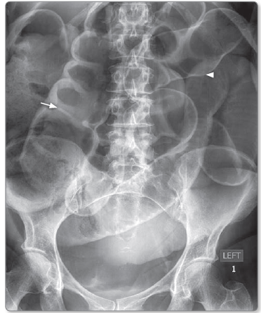
Large Bowel Obstruction (LBO)
🚧 Expected X-ray findings:
• Dilated colon proximal to obstruction (>6 cm; caecum >9 cm).
• Visible haustra that do not extend fully across the bowel lumen.
• Little or no gas in the distal colon or rectum.
• If ileocaecal valve competent → minimal small bowel dilatation.
⚠️ Caecal diameter >12 cm suggests imminent perforation.
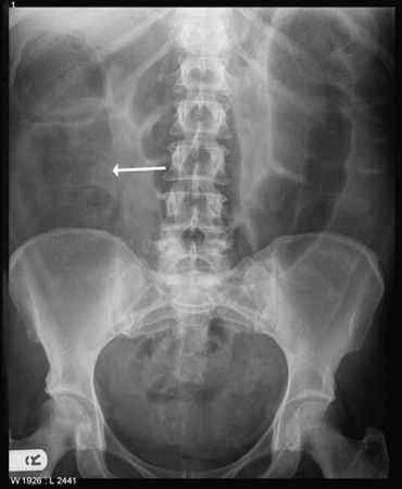
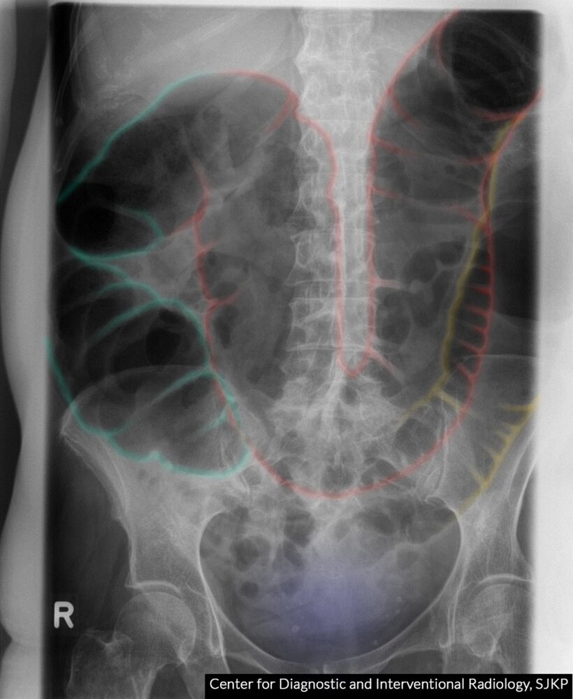
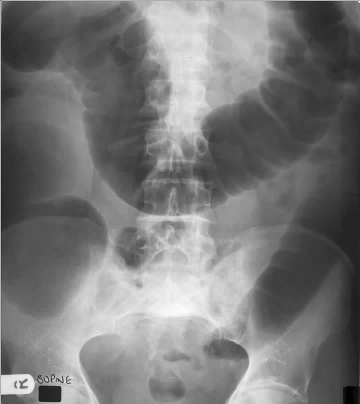
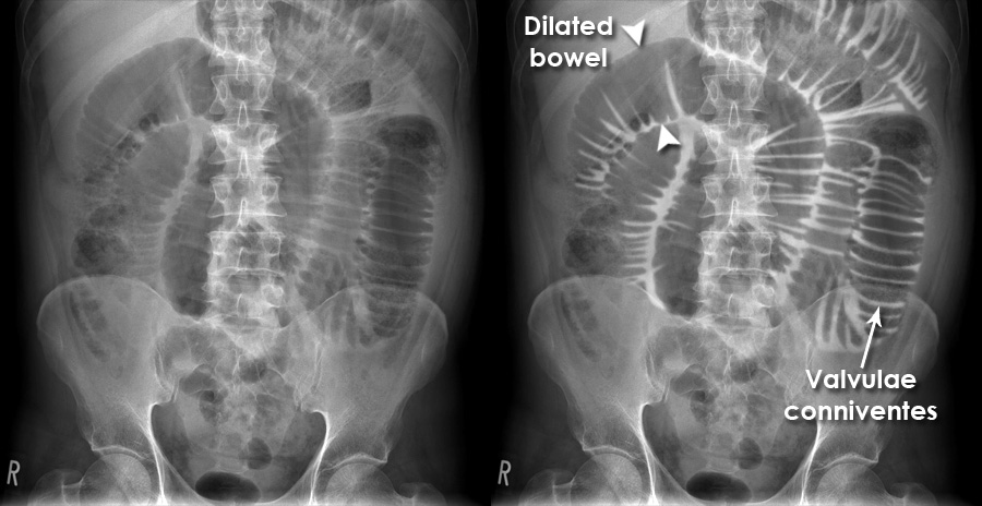
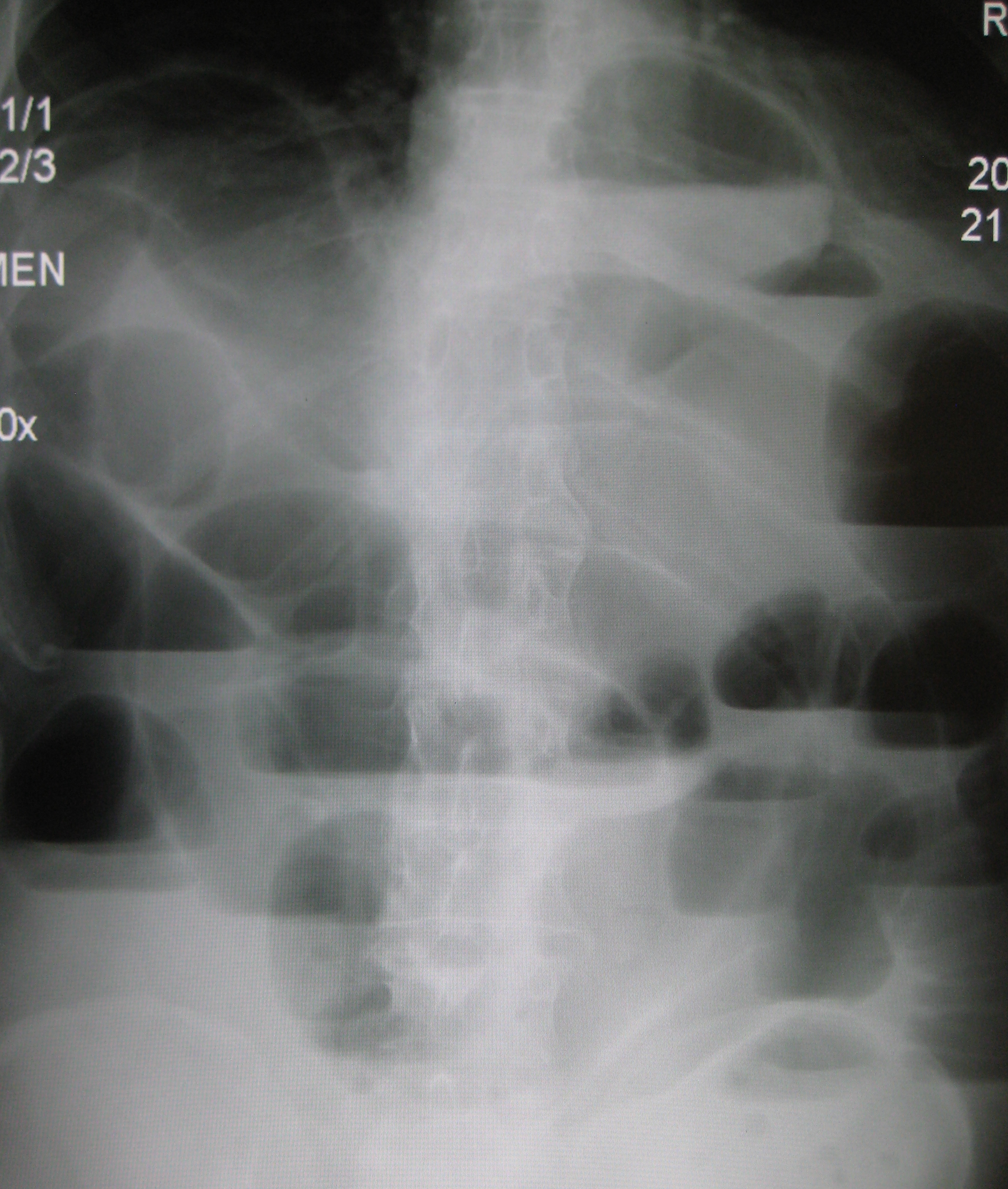
Small Bowel Obstruction (SBO)
🔄 Expected X-ray findings:
• Dilated central small bowel loops (>3 cm diameter).
• Valvulae conniventes visible across the full width (“stacked coins”).
• Multiple air–fluid levels on erect films (step-ladder pattern).
• Relative absence of gas in the colon.
⚠️ Early SBO may have a near-normal X-ray - CT abdomen is definitive.
🧠 Rapid Pattern Recognition (Exam & On-Call Pearl)
- 📍 Central loops + valvulae across full width → SBO
- 📍 Peripheral colon + haustra → LBO
- 📍 Single huge loop with direction → Volvulus
- 📍 Gas under diaphragm → Perforation until proven otherwise
| The content on this website is provided for educational and informational purposes only to support exam preparation (e.g., MLA, MRCP, USMLE) and learning. This is NOT medical advice, diagnosis, treatment, or professional guidance. It does not replace consultation with a qualified healthcare professional, official guidelines (e.g., NICE, GMC, BNF), or supervised clinical practice. Always verify information with current, authoritative sources. Makindo and its contributors accept no liability for any reliance on this content, including errors, omissions, or any resulting harm, loss, or consequences. By using this site, you agree to these terms. |
|
|
Categories
- About
- Acute Medicine
- Anaesthetics and Critical Care
- Anatomy
- Anatomy and Physiology
- Biochemistry
- Book
- Cardiology
- Collections
- CompSci
- Crib Sheets
- Critical care
- Dental
- Dermatology
- Differentials
- Drugs
- ENT
- Electrocardiogram
- Embryology
- Emergency Medicine
- Endocrinology
- Ethics
- Foundation Doctors
- GCSE
- Gastroenterology
- General Practice
- Genetics
- Geriatric Medicine
- Geriatrics
- Guidelines
- Haematology
- Hepatology
- Immunology
- Infectious Diseases
- Infographic
- Investigations
- Lists
- MRCP
- Mandatory Training
- Medical Students
- Microbiology
- Nephrology
- Neurology
- Neurosurgery
- Nutrition
- OSCE
- Obstetrics Gynaecology
- Oncology
- Ophthalmology
- Oral Medicine and Dentistry
- Orthopaedics
- Paediatrics
- Palliative
- Palliative Care
- Pathology
- Pharmacology
- Physiology
- Procedures
- Psychiatry
- Public Health
- Radiology
- Respiratory
- Resuscitation
- Revision Topics
- Rheumatology
- Statistics and Research
- Stroke
- Surgery
- Toxicology
- Trauma and Orthopaedics
- USMLE
- Urology
- Vascular Surgery