
| Download the amazing global Makindo app: ✅ Means NICE/National Guidelines 2026 compliant Android | Apple | |
|---|---|
| MEDICAL DISCLAIMER: Educational use only. Not for diagnosis or management. See below for full disclaimer. |
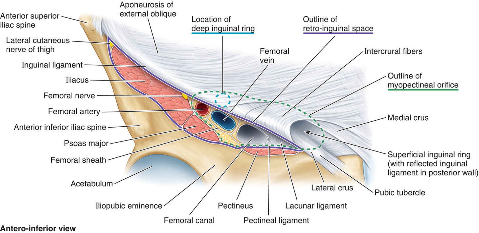
Anatomy of the Inguinal and Femoral canal
🦴 Groin Anatomy - Femoral Canal vs Inguinal Canal
| Item | Femoral Canal | Inguinal Canal |
|---|---|---|
| 📍 Location | Most medial compartment of the femoral sheath, below the inguinal ligament, medial to femoral vein. | Oblique passage in the lower anterior abdominal wall, running from deep inguinal ring to superficial ring, above the inguinal ligament. |
| 🧱 Boundaries (walls) |
Anterior (roof): Inguinal ligament
Posterior (floor): Pectineal (Cooper’s) ligament over pectineus fascia Medial: Lacunar ligament Lateral: Femoral vein |
Anterior wall: External oblique aponeurosis (+ internal oblique laterally)
Posterior wall: Transversalis fascia, reinforced medially by conjoint tendon Roof: Arching fibres of internal oblique & transversus abdominis Floor: Inguinal ligament (medially reinforced by lacunar ligament) |
| 🎒 Contents | Lymphatics & areolar tissue only (incl. deep inguinal node of Cloquet) + fat. ➜ Allows femoral vein to expand. (Femoral artery/vein/nerve are not in the canal; artery/vein lie in adjacent femoral sheath compartments; nerve is outside the sheath.) |
Both sexes: Ilioinguinal nerve (sensory; runs in part of the canal), genital branch of genitofemoral nerve (within cord in males).
Males: Spermatic cord and contents. Females: Round ligament of the uterus. |
| 🚨 Clinical | Femoral hernia: passes through femoral canal, inferolateral to pubic tubercle, below inguinal ligament, medial to femoral vein. Higher risk of strangulation (rigid borders). | Inguinal hernia: above inguinal ligament; Indirect via deep ring (lateral to inferior epigastrics), Direct through Hesselbach’s triangle (medial to inferior epigastrics). |
| 🧭 Surface anatomy tips | Femoral pulse just below inguinal ligament, midway ASIS→pubic tubercle; canal is medial to this (medial to vein). | Superficial ring superolateral to pubic tubercle; deep ring ≈ 1.5–2 cm above midpoint of inguinal ligament. |
🧠 Common Exam Pitfalls
- Femoral canal ≠ triangle: boundaries you listed for adductor longus/sartorius belong to the femoral triangle, not the canal.
- Femoral canal contents are lymphatics (Cloquet node), not artery/vein/nerve.
- Ilioinguinal nerve travels through part of the inguinal canal but does not enter via the deep ring.
| The content on this website is provided for educational and informational purposes only to support exam preparation (e.g., MLA, MRCP, USMLE) and learning. This is NOT medical advice, diagnosis, treatment, or professional guidance. It does not replace consultation with a qualified healthcare professional, official guidelines (e.g., NICE, GMC, BNF), or supervised clinical practice. Always verify information with current, authoritative sources. Makindo and its contributors accept no liability for any reliance on this content, including errors, omissions, or any resulting harm, loss, or consequences. By using this site, you agree to these terms. |
|
|
Categories
- About
- Acute Medicine
- Anaesthetics and Critical Care
- Anatomy
- Anatomy and Physiology
- Biochemistry
- Book
- Cardiology
- Collections
- CompSci
- Crib Sheets
- Critical care
- Dental
- Dermatology
- Differentials
- Drugs
- ENT
- Electrocardiogram
- Embryology
- Emergency Medicine
- Endocrinology
- Ethics
- Foundation Doctors
- GCSE
- Gastroenterology
- General Practice
- Genetics
- Geriatric Medicine
- Geriatrics
- Guidelines
- Haematology
- Hepatology
- Immunology
- Infectious Diseases
- Infographic
- Investigations
- Lists
- MRCP
- Mandatory Training
- Medical Students
- Microbiology
- Nephrology
- Neurology
- Neurosurgery
- Nutrition
- OSCE
- Obstetrics Gynaecology
- Oncology
- Ophthalmology
- Oral Medicine and Dentistry
- Orthopaedics
- Paediatrics
- Palliative
- Palliative Care
- Pathology
- Pharmacology
- Physiology
- Procedures
- Psychiatry
- Public Health
- Radiology
- Respiratory
- Resuscitation
- Revision Topics
- Rheumatology
- Statistics and Research
- Stroke
- Surgery
- Toxicology
- Trauma and Orthopaedics
- USMLE
- Urology
- Vascular Surgery