
| Download the amazing global Makindo app: ✅ Means NICE/National Guidelines 2026 compliant Android | Apple | |
|---|---|
| MEDICAL DISCLAIMER: Educational use only. Not for diagnosis or management. See below for full disclaimer. |
Anatomy of the Muscles
🔬Anatomy
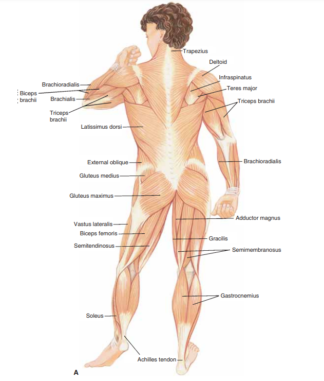
Posterior
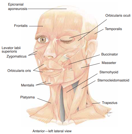
Face and neck
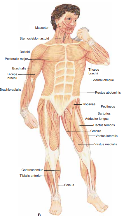
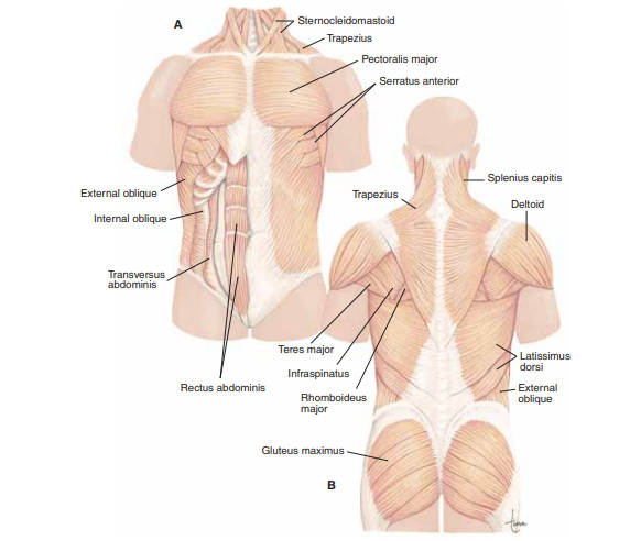
Chest and abdomen
Arms
Legs
📊 Muscles of the Head & Neck – Comprehensive Atlas Table
| Region | Muscle | Origin | Insertion | Action | Innervation | Artery | Notes |
|---|---|---|---|---|---|---|---|
| Facial Expression | Orbicularis Oculi | Medial orbital margin, palpebral ligament | Skin around orbit, eyelids | Closes eyelids (blink, squint) | Facial n. (VII) | Facial, ophthalmic aa. | Key in blink reflex; weakness → exposure keratitis |
| Orbicularis Oris | Maxilla & mandible (around mouth) | Skin/mucosa of lips | Closes, protrudes lips | Facial n. | Labial branches of facial a. | “Kissing muscle” | |
| Buccinator | Pterygomandibular raphe, alveolar margins | Orbicularis oris (corner of mouth) | Compresses cheek, aids mastication | Facial n. | Buccal a. | Pierced by parotid duct | |
| Frontalis (Occipitofrontalis, frontal belly) | Epicranial aponeurosis | Skin of forehead, eyebrows | Raises eyebrows, wrinkles forehead | Facial n. | Superficial temporal a. | Communicates emotion | |
| Platysma | Fascia over clavicle/pectoralis | Mandible, skin of lower face | Tenses skin of neck, depresses mandible | Facial n. | Submental a. | Superficial, thin sheet in anterior neck | |
| Mastication | Masseter | Zygomatic arch | Angle & ramus of mandible | Elevates mandible | Mandibular n. (V3) | Masseteric a. | Powerful jaw closer |
| Temporalis | Temporal fossa | Coronoid process of mandible | Elevates, retracts mandible | V3 (deep temporal nn.) | Deep temporal aa. | Main retractor of mandible | |
| Medial Pterygoid | Medial surface lateral pterygoid plate | Medial mandibular ramus | Elevates, protrudes mandible | V3 | Pterygoid branches maxillary a. | Forms “pterygoid sling” with masseter | |
| Lateral Pterygoid | Lateral surface lateral pterygoid plate | Neck of mandible, TMJ capsule | Protrudes, depresses mandible; side-to-side | V3 | Pterygoid branches maxillary a. | Opens mouth (protraction + depression) | |
| Tongue | Genioglossus | Mental spine of mandible | Tongue & hyoid | Protrudes tongue | Hypoglossal n. (XII) | Lingual a. | Main tongue protruder; keeps airway patent |
| Hyoglossus | Hyoid bone | Side of tongue | Depresses tongue | Hypoglossal n. | Lingual a. | Lingual a. runs deep, hypoglossal n. superficial | |
| Styloglossus | Styloid process | Side of tongue | Retracts, elevates tongue | Hypoglossal n. | Ascending pharyngeal a. | Works with genioglossus in swallowing | |
| Palatoglossus | Palatine aponeurosis | Side of tongue | Elevates posterior tongue | Vagus (X, via pharyngeal plexus) | Ascending palatine a. | Only tongue muscle not by XII | |
| Pharynx & Soft Palate | Superior / Middle / Inferior Pharyngeal Constrictors | Pterygoid hamulus, hyoid, thyroid/cricoid cartilage | Median pharyngeal raphe | Constrict pharynx (swallowing) | Vagus (X, pharyngeal plexus) | Ascending pharyngeal a. | Sequential contraction propels bolus |
| Levator Veli Palatini | Petrous temporal bone, auditory tube | Palatine aponeurosis | Elevates soft palate | Vagus (X) | Ascending palatine a. | Blocks nasopharynx in swallowing | |
| Tensor Veli Palatini | Scaphoid fossa, auditory tube | Palatine aponeurosis | Tenses soft palate, opens auditory tube | Mandibular n. (V3) | Ascending palatine a. | Hooks around pterygoid hamulus | |
| Larynx | Posterior Cricoarytenoid | Posterior cricoid | Arytenoid cartilage | Abducts vocal cords (opens airway) | Recurrent laryngeal n. (X) | Superior & inferior laryngeal aa. | Only abductor of cords → breathing |
| Lateral Cricoarytenoid | Lateral cricoid | Arytenoid cartilage | Adducts vocal cords | Recurrent laryngeal n. | Laryngeal branches | Closes rima glottidis | |
| Cricothyroid | Cricoid cartilage | Inferior thyroid cartilage | Tenses cords (raises pitch) | External branch superior laryngeal n. | Cricothyroid a. | Only laryngeal muscle not by recurrent laryngeal | |
| Neck | Sternocleidomastoid | Manubrium & clavicle | Mastoid process | Rotates head opposite side, flexes neck | Spinal accessory n. (XI), C2–C3 proprioception | Occipital a. | Clinical landmark for triangles of neck |
| Scalenes (Ant, Mid, Post) | Cervical TPs | 1st & 2nd ribs | Elevate ribs (inspiration), flex neck | Cervical spinal nn. | Ascending cervical a. | Brachial plexus passes between ant & mid | |
| Sternohyoid | Manubrium, clavicle | Hyoid | Depresses hyoid | Ansa cervicalis (C1–C3) | Superior thyroid a. | Infrahyoid “strap” muscle | |
| Sternothyroid | Manubrium | Thyroid cartilage | Depresses larynx | Ansa cervicalis | Superior thyroid a. | Deep to sternohyoid | |
| Omohyoid | Superior scapula | Hyoid | Depresses hyoid | Ansa cervicalis | Inferior thyroid a. | Has two bellies with intermediate tendon | |
| Mylohyoid | Mylohyoid line (mandible) | Hyoid, midline raphe | Elevates floor of mouth | Mylohyoid n. (V3) | Submental a. | Forms oral diaphragm |
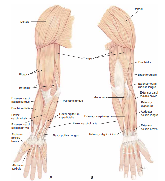
📊 Muscles of the Upper Limb – Comprehensive Atlas Table
| Region | Muscle | Origin | Insertion | Action | Innervation | Artery | Notes |
|---|---|---|---|---|---|---|---|
| Shoulder / Scapular | Deltoid | Clavicle, acromion, spine of scapula | Deltoid tuberosity (humerus) | Abducts (15–90°), flex/IR (ant.), extend/ER (post.) | Axillary n. (C5–C6) | Posterior circumflex humeral a. | Main shoulder abductor; fails in axillary n. injury |
| Supraspinatus | Supraspinous fossa | Greater tubercle (superior facet) | Initiates abduction (0–15°) | Suprascapular n. | Suprascapular a. | Commonly torn rotator cuff muscle | |
| Infraspinatus | Infraspinous fossa | Greater tubercle (middle facet) | Lateral rotation | Suprascapular n. | Suprascapular a. | Part of rotator cuff | |
| Teres Minor | Lateral border of scapula | Greater tubercle (inferior facet) | Lateral rotation | Axillary n. | Circumflex scapular a. | Rotator cuff muscle | |
| Subscapularis | Subscapular fossa | Lesser tubercle of humerus | Medial rotation, adduction | Upper & lower subscapular nn. | Subscapular a. | Only rotator cuff muscle inserting on lesser tubercle | |
| Teres Major | Inferior angle of scapula | Medial lip of intertubercular sulcus | Adduction, medial rotation | Lower subscapular n. | Circumflex scapular a. | “Lat’s little helper” | |
| Serratus Anterior | Ribs 1–8 | Medial scapular border | Protracts, upwardly rotates scapula | Long thoracic n. | Lateral thoracic a. | Winged scapula if paralysed | |
| Arm – Anterior | Biceps Brachii | Short: coracoid; Long: supraglenoid tubercle | Radial tuberosity, bicipital aponeurosis | Flexes elbow, supinates forearm | Musculocutaneous n. (C5–C6) | Brachial a. | Supination most powerful when elbow flexed |
| Brachialis | Anterior humeral shaft | Coronoid process of ulna | Main elbow flexor | Musculocutaneous n. | Brachial a. | Deep to biceps | |
| Coracobrachialis | Coracoid process | Medial mid-humerus | Flexes, adducts arm | Musculocutaneous n. | Brachial a. | Musculocutaneous n. pierces it | |
| Arm – Posterior | Triceps Brachii | Long: infraglenoid tubercle; Lat/Med: posterior humerus | Olecranon process | Main elbow extensor | Radial n. (C6–C8) | Deep brachial a. | Long head crosses shoulder joint |
| Forearm – Anterior
Superficial |
Pronator Teres | Medial epicondyle, coronoid process | Lateral mid-radius | Pronation, weak elbow flexion | Median n. | Ulnar a. | Median n. passes between 2 heads |
| Flexor Carpi Radialis | Medial epicondyle | Base of 2nd/3rd metacarpal | Flexes, abducts wrist | Median n. | Radial a. | Pierces flexor retinaculum | |
| Palmaris Longus | Medial epicondyle | Palmar aponeurosis | Flexes wrist | Median n. | Ulnar a. | Absent in ~15% population | |
| Flexor Carpi Ulnaris | Medial epicondyle, olecranon | Pisiform, hook of hamate, 5th metacarpal | Flexes, adducts wrist | Ulnar n. | Ulnar a. | Ulnar n. runs between its heads | |
| Flexor Digitorum Superficialis | Medial epicondyle, radius | Middle phalanges digits 2–5 | Flexes PIP & MCP joints | Median n. | Ulnar a. | Forms intermediate layer | |
| Forearm – Anterior
Deep |
Flexor Digitorum Profundus | Ulna, interosseous membrane | Distal phalanges 2–5 | Flexes DIP joints | Ulnar n. (medial ½), Median n. (lateral ½) | Anterior interosseous a. | Dual innervation |
| Flexor Pollicis Longus | Radius, interosseous membrane | Distal phalanx of thumb | Flexes thumb | Median n. (ant. interosseous) | Anterior interosseous a. | Key for grip strength | |
| Pronator Quadratus | Distal ulna | Distal radius | Main pronator | Median n. (ant. interosseous) | Anterior interosseous a. | Deepest anterior forearm muscle | |
| Forearm – Posterior
Superficial |
Brachioradialis | Lateral supracondylar ridge | Styloid process of radius | Flexes elbow (mid-pronation) | Radial n. | Radial recurrent a. | “Drinking muscle” |
| Extensor Carpi Radialis Longus | Lateral supracondylar ridge | Base of 2nd metacarpal | Extends, abducts wrist | Radial n. | Radial a. | Part of “mobile wad” | |
| Extensor Carpi Radialis Brevis | Lateral epicondyle | Base of 3rd metacarpal | Extends, abducts wrist | Deep branch radial n. | Radial a. | Tennis elbow origin | |
| Extensor Digitorum | Lateral epicondyle | Extensor expansions digits 2–5 | Extends fingers | Posterior interosseous n. | Posterior interosseous a. | Main finger extensor | |
| Extensor Digiti Minimi | Lateral epicondyle | Extensor expansion 5th digit | Extends little finger | Posterior interosseous n. | Posterior interosseous a. | Specialises finger 5 | |
| Extensor Carpi Ulnaris | Lateral epicondyle, ulna | Base of 5th metacarpal | Extends, adducts wrist | Posterior interosseous n. | Ulnar a. | Active in gripping | |
| Forearm – Posterior
Deep |
Supinator | Lateral epicondyle, supinator crest of ulna | Proximal radius | Supinates forearm | Deep branch radial n. | Radial recurrent a. | Radial n. passes through |
| Abductor Pollicis Longus | Posterior ulna & radius, interosseous membrane | Base of 1st metacarpal | Abducts thumb | Posterior interosseous n. | Posterior interosseous a. | Forms lateral snuffbox border | |
| Extensor Pollicis Brevis | Posterior radius | Base of proximal phalanx of thumb | Extends thumb (MCP) | Posterior interosseous n. | Posterior interosseous a. | Snuffbox border | |
| Extensor Pollicis Longus | Posterior ulna | Base distal phalanx of thumb | Extends thumb (IP) | Posterior interosseous n. | Posterior interosseous a. | Forms medial snuffbox border | |
| Extensor Indicis | Posterior ulna | Extensor expansion index finger | Extends index | Posterior interosseous n. | Posterior interosseous a. | Gives independence to index finger | |
| Hand – Thenar | Abductor Pollicis Brevis | Flexor retinaculum, scaphoid, trapezium | Proximal phalanx thumb (base) | Abducts thumb | Median n. (recurrent) | Superficial palmar br. radial a. | Superficial thenar muscle |
| Flexor Pollicis Brevis | Flexor retinaculum, trapezium | Base proximal phalanx thumb | Flexes thumb | Median n. (superficial head), Ulnar n. (deep head) | Superficial palmar br. radial a. | Dual innervation | |
| Opponens Pollicis | Flexor retinaculum, trapezium | Lateral 1st metacarpal | Opposes thumb | Median n. (recurrent) | Radial a. | Key for precision grip | |
| Hand – Hypothenar | Abductor Digiti Minimi | Pisiform | Base proximal phalanx 5th digit | Abducts little finger | Ulnar n. | Ulnar a. | Most medial hypothenar muscle |
| Flexor Digiti Minimi Brevis | Hook of hamate, flexor retinaculum | Base proximal phalanx 5th digit | Flexes little finger | Ulnar n. | Ulnar a. | Absent in some individuals | |
| Opponens Digiti Minimi | Hook of hamate, flexor retinaculum | Medial border 5th metacarpal | Opposition of little finger | Ulnar n. | Ulnar a. | Deepest hypothenar muscle | |
| Hand – Central | Adductor Pollicis | Oblique head: metacarpals 2–3; Transverse head: 3rd metacarpal | Base proximal phalanx thumb | Adducts thumb | Ulnar n. (deep branch) | Deep palmar arch | Key pinch grip muscle |
| Lumbricals (1–4) | Tendons of FDP | Extensor expansions digits 2–5 | Flex MCP, extend IP joints | 1–2: Median n.; 3–4: Ulnar n. | Superficial & deep palmar arches | “Writing muscle” | |
| Interossei (Palmar, Dorsal) | Metacarpals | Bases proximal phalanges, extensor expansions | PAD = ADduct; DAB = ABduct fingers | Ulnar n. (deep branch) | Palmar & dorsal metacarpal aa. | Critical for finger spreading & closing |
📊 Muscles of the Chest, Abdomen & Pelvis – Comprehensive Atlas Table
| Region | Muscle | Origin | Insertion | Action | Innervation | Artery | Notes |
|---|---|---|---|---|---|---|---|
| Thorax | Diaphragm | Xiphoid, lower 6 ribs, L1–L3 bodies (crura) | Central tendon | Main muscle of inspiration | Phrenic n. (C3–C5) | Phrenic, musculophrenic, inf. phrenic aa. | Openings: T8 (IVC), T10 (oesophagus), T12 (aorta) |
| External Intercostals | Inferior border of rib | Superior border of rib below | Elevate ribs (inspiration) | Intercostal nn. (T1–T11) | Intercostal aa. | Fibres run down & forward (“hands in pockets”) | |
| Internal Intercostals | Inferior border of rib | Superior border of rib below | Depress ribs (forced expiration) | Intercostal nn. | Intercostal aa. | Fibres run down & back (opposite to external) | |
| Innermost Intercostals | Inferior rib margin | Superior rib below | Assist respiration | Intercostal nn. | Intercostal aa. | Deepest layer; neurovascular bundle lies superficial | |
| Transversus Thoracis | Posterior sternum | Costal cartilages 2–6 | Depresses ribs (forced expiration) | Intercostal nn. | Internal thoracic aa. | Part of innermost layer; variable fibres | |
| Subcostals | Inner rib surface near angle | 2–3 ribs below | Assist in depressing ribs | Intercostal nn. | Intercostal aa. | Best seen posteriorly | |
| Abdomen | External Oblique | Lower 8 ribs | Linea alba, iliac crest, pubic tubercle | Flexes, rotates trunk; compresses viscera | T7–T12, iliohypogastric, ilioinguinal nn. | Lower intercostal, superior/inferior epigastric aa. | Forms inguinal ligament; gives spermatic fascia |
| Internal Oblique | Iliac crest, thoracolumbar fascia | Lower ribs, linea alba | Flexes, rotates trunk; compresses viscera | T7–T12, iliohypogastric, ilioinguinal nn. | Same as external | Forms cremaster muscle in males | |
| Transversus Abdominis | Costal cartilages 7–12, iliac crest | Linea alba | Compresses abdominal contents | T7–T12, L1 | Deep circumflex iliac, inferior epigastric aa. | Main “corset” muscle; deep abdominal stabiliser | |
| Rectus Abdominis | Pubic symphysis & crest | Xiphoid & 5th–7th costal cartilages | Flexes trunk; stabilises pelvis | T7–T12 intercostal nn. | Superior & inferior epigastric aa. | Interrupted by tendinous intersections (“six-pack”) | |
| Pyramidalis | Pubis | Linea alba | Tenses linea alba | T12 | Inferior epigastric a. | Absent in ~20% of people | |
| Psoas Major | L1–L5 vertebral bodies | Lesser trochanter (with iliacus) | Hip flexor, trunk flexor | L1–L3 ventral rami | Lumbar aa. | Key in posture & gait | |
| Iliacus | Iliac fossa | Lesser trochanter (with psoas) | Flexes thigh | Femoral n. (L2–L3) | Iliac branch of iliolumbar a. | With psoas = iliopsoas | |
| Pelvis & Perineum | Levator Ani (Puborectalis, Pubococcygeus, Iliococcygeus) | Pubis, ischial spine, tendinous arch | Coccyx, anococcygeal body | Supports pelvic viscera, maintains continence | Pudendal n., S3–S4 | Inferior gluteal a. | Main pelvic diaphragm muscle |
| Coccygeus (Ischiococcygeus) | Ischial spine | Coccyx & sacrum | Supports pelvic floor, pulls coccyx forward | S3–S4 | Inferior gluteal a. | Completes pelvic diaphragm posteriorly | |
| External Anal Sphincter | Perineal body | Coccyx & encircles anal canal | Voluntary anal continence | Inferior rectal branch pudendal n. | Inferior rectal a. | Voluntary, skeletal muscle | |
| Internal Anal Sphincter | Smooth muscle of anal wall | Encircles anal canal | Involuntary continence | Autonomic (S4, pelvic splanchnic) | Middle rectal a. | Involuntary smooth muscle | |
| Bulbospongiosus (M/F) | Perineal body | Clitoris / Corpus spongiosum | Compresses bulb; expels urine/semen | Pudendal n. | Perineal a. | Sexual function, ejaculation, erection | |
| Ischiocavernosus | Ischiopubic ramus | Crus of penis/clitoris | Compresses crus; maintains erection | Pudendal n. | Perineal a. | Sexual function | |
| Superficial & Deep Transverse Perineal | Ischial rami | Perineal body | Stabilise perineal body | Pudendal n. | Perineal a. | Support pelvic floor | |
| Detrusor (Bladder) | Wall of bladder | Wall of bladder | Contracts to void urine | Parasymp. (S2–S4) | Vesical aa. | Smooth muscle; failure → retention |
📊 Muscles of the Back – Summary Table
| Muscle | Origin | Insertion | Action | Innervation | Artery | Notes |
|---|---|---|---|---|---|---|
| 🟢 Erector Spinae Group | ||||||
| Iliocostalis | Iliac crest, sacrum | Angles of ribs | Extends & laterally bends trunk/neck | Dorsal rami C4–S5 | Deep cervical, intercostal, lumbar aa. | Lateral column (lumborum, thoracis, cervicis) |
| Longissimus | Transverse processes (inferior) | Transverse processes (superior), mastoid | Extends & laterally bends trunk/neck/head | Dorsal rami C1–S1 | Deep cervical, intercostal, lumbar aa. | Intermediate column (thoracis, cervicis, capitis) |
| Spinalis | Spinous processes (lower) | Spinous processes (upper), skull base | Extension & lateral flexion | Dorsal rami C2–L3 | Deep cervical, intercostal, lumbar aa. | Most medial erector spinae |
| 🟣 Transversospinalis Group | ||||||
| Multifidus | Sacrum, TP C3–L5 | SP 2–4 levels above | Extension, contralateral rotation | Dorsal rami C1–L5 | Segmental supply | Key stabiliser |
| Semispinalis | TP C7–T12 | Skull (capitis) + SP 4–6 levels above | Extension, contralateral rotation | Dorsal rami C1–T12 | Segmental supply | Superficial to multifidus |
| Rotatores | Transverse processes | SP 1–2 levels above | Contralateral rotation | Dorsal rami C1–L5 | Segmental supply | Likely proprioceptive |
| Interspinales | SP (upper border) | SP above | Extension | Dorsal rami C1–L5 | Segmental supply | Small, insignificant |
| Intertransversarii | TP (upper border) | TP above | Lateral flexion | Dorsal rami C1–L5 | Segmental supply | Small, insignificant |
| 🔵 Suboccipital Muscles | ||||||
| Rectus Capitis Posterior Major | SP of C2 | Inferior nuchal line | Extension, ipsilateral rotation | Suboccipital nerve (C1 DPR) | Occipital a. | Forms border of suboccipital triangle |
| Rectus Capitis Posterior Minor | Posterior tubercle of C1 | Medial inferior nuchal line | Extension | Suboccipital nerve (C1 DPR) | Occipital a. | Deeper & more medial than major |
| Obliquus Capitis Superior | TP of C1 | Occiput above inferior nuchal line | Extension, ipsilateral rotation | Suboccipital nerve (C1 DPR) | Occipital a. | Forms suboccipital triangle |
| Obliquus Capitis Inferior | SP of C2 | TP of C1 | Ipsilateral rotation | Suboccipital nerve (C1 DPR) | Occipital a. | Greater occipital nerve passes above |
| 🟡 Splenius Group | ||||||
| Splenius Capitis | Ligamentum nuchae, SP C7–T6 | Mastoid, sup. nuchal line | Extension, ipsilateral rotation | Dorsal rami C2–C6 | Deep cervical, intercostal aa. | “Bandage” muscle of neck |
| Splenius Cervicis | SP C7–T6 | TP C1–C3 | Extension, ipsilateral rotation | Dorsal rami C2–C6 | Deep cervical, intercostal aa. | More caudal insertion |
💡 Teaching Pearls
✔️ Erector spinae mnemonic: “I Love Spaghetti” (Iliocostalis, Longissimus, Spinalis).
✔️ Transversospinalis mnemonic: “Some Muscles Rotate” (Semispinalis, Multifidus, Rotatores).
✔️ Suboccipital triangle: Borders = RCP Major, OCS, OCI. Contents = Vertebral artery + Suboccipital nerve.
✔️ Splenius = “bandage muscle” (wraps around neck).
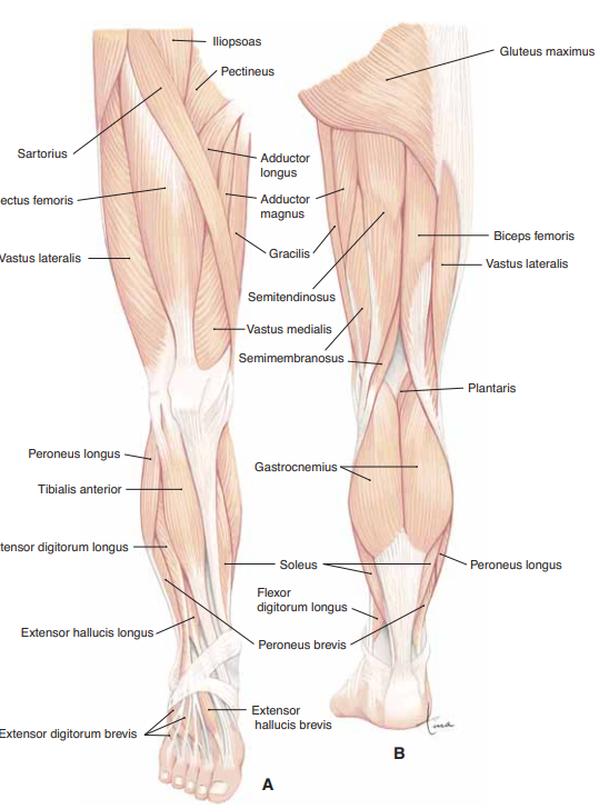
📊 Muscles of the Lower Limb – Comprehensive Master Table
| Region | Muscle | Origin | Insertion | Action | Innervation | Artery | Notes |
|---|---|---|---|---|---|---|---|
| Gluteal | Gluteus Maximus | Ilium (posterior), sacrum, coccyx | Iliotibial tract, gluteal tuberosity | Extends & laterally rotates hip | Inferior gluteal n. | Inferior gluteal a. | Main power extensor; climbing/rising |
| Gluteus Medius | Lateral ilium | Greater trochanter | Abducts, medially rotates thigh | Superior gluteal n. | Superior gluteal a. | Trendelenburg sign | |
| Gluteus Minimus | Lateral ilium | Greater trochanter | Abducts, medially rotates thigh | Superior gluteal n. | Superior gluteal a. | Assists pelvis stabilisation | |
| Piriformis | Anterior sacrum | Greater trochanter | Lateral rotation, abduction (flexed) | N. to piriformis | Inferior gluteal a. | Sciatic nerve exits below | |
| Obturator Internus | Obturator membrane (internal) | Greater trochanter (medial) | Lateral rotation, abduction | N. to obturator internus | Obturator a. | Exits lesser sciatic foramen | |
| Gemelli (Sup & Inf) | Sup: ischial spine; Inf: ischial tuberosity | Greater trochanter (via obturator internus tendon) | Lateral rotation | Sup: n. to obt. internus; Inf: n. to quad. femoris | Inferior gluteal a. | Small twin muscles | |
| Quadratus Femoris | Ischial tuberosity (lat. border) | Intertrochanteric crest | Lateral rotation | N. to quadratus femoris | Inferior gluteal a. | Short quadrangular muscle | |
| Anterior Thigh | Iliopsoas | Iliac fossa & lumbar vertebrae | Lesser trochanter | Main hip flexor | Femoral n. (iliacus); L1–L3 (psoas) | Lumbar aa., femoral a. | Strongest hip flexor |
| Sartorius | ASIS | Pes anserinus | Flex, abduct, lat. rotate hip; flex knee | Femoral n. | Femoral a. | Longest muscle | |
| Rectus Femoris | AIIS & acetabular rim | Tibial tuberosity (via patella) | Knee extension; hip flexion | Femoral n. | Femoral a. | Biarticular quadriceps | |
| Vastus Lateralis | Greater trochanter, linea aspera | Tibial tuberosity | Knee extension | Femoral n. | Lateral circumflex femoral a. | Largest quadriceps | |
| Vastus Medialis | Linea aspera, intertrochanteric line | Tibial tuberosity | Knee extension | Femoral n. | Femoral a. | VMO stabilises patella | |
| Vastus Intermedius | Anterior femur | Tibial tuberosity | Knee extension | Femoral n. | Femoral a. | Deep to rectus femoris | |
| Medial Thigh | Adductor Longus | Pubis | Linea aspera (mid) | Adducts thigh | Obturator n. | Obturator a. | Forms femoral triangle border |
| Adductor Brevis | Inf. pubic ramus | Pectineal line, prox. linea aspera | Adducts thigh | Obturator n. | Obturator a. | Between longus & magnus | |
| Adductor Magnus | Pubis & ischial tuberosity | Linea aspera, adductor tubercle | Adducts; hamstring part extends hip | Obturator n. + Tibial n. | Obturator, deep femoral aa. | Adductor hiatus → popliteal | |
| Gracilis | Inf. pubic ramus | Pes anserinus | Adducts thigh, flexes knee | Obturator n. | Obturator a. | Only medial thigh crossing knee | |
| Pectineus | Pecten pubis | Pectineal line femur | Adducts, flexes hip | Femoral ± Obturator n. | Obturator, medial circumflex femoral | Often dual innervation | |
| Posterior Thigh | Biceps Femoris | Long: ischial tuberosity; Short: linea aspera | Head fibula | Extends hip, flexes knee | Long: Tibial n.; Short: Common fibular n. | Perforating branches | Hamstring prone to injury |
| Semitendinosus | Ischial tuberosity | Pes anserinus | Extends hip, flexes knee | Tibial n. | Deep femoral branches | Long cord-like tendon | |
| Semimembranosus | Ischial tuberosity | Post. medial tibial condyle | Extends hip, flexes knee | Tibial n. | Deep femoral branches | Contributes to oblique popliteal ligament | |
| Anterior Leg | Tibialis Anterior | Lateral tibia, interosseous membrane | Medial cuneiform, 1st metatarsal | Dorsiflexes, inverts foot | Deep fibular n. | Anterior tibial a. | Main dorsiflexor |
| Extensor Digitorum Longus | Lateral tibial condyle, fibula | Extensor expansions toes 2–5 | Extends toes, dorsiflexes | Deep fibular n. | Anterior tibial a. | Anterior compartment syndrome risk | |
| Extensor Hallucis Longus | Ant. fibula, interosseous membrane | Distal phalanx hallux | Extends hallux, dorsiflexes | Deep fibular n. | Anterior tibial a. | Thin, between TA & EDL | |
| Lateral Leg | Fibularis Longus | Upper lat. fibula | 1st metatarsal & medial cuneiform | Plantarflexes, everts | Superficial fibular n. | Fibular a. | Supports transverse arch |
| Fibularis Brevis | Lower lat. fibula | Base 5th metatarsal | Plantarflexes, everts | Superficial fibular n. | Fibular a. | 5th metatarsal stress fractures | |
| Fibularis Tertius | Distal ant. fibula | Dorsum 5th metatarsal | Everts, weak dorsiflexion | Deep fibular n. | Anterior tibial a. | Often absent (~20%) | |
| Posterior Leg
(Superficial) |
Gastrocnemius | Femoral condyles | Calcaneus (Achilles tendon) | Plantarflexes ankle, flexes knee | Tibial n. | Posterior tibial a. | 2-headed calf muscle |
| Soleus | Post. tibia & fibula | Calcaneus | Plantarflexes ankle | Tibial n. | Posterior tibial a. | Postural, slow-twitch | |
| Plantaris | Lat. supracondylar femur | Calcaneus | Weak plantarflexion | Tibial n. | Posterior tibial a. | Absent in 10% | |
| Posterior Leg
(Deep) |
Popliteus | Lateral femoral condyle | Post. tibia | Unlocks knee | Tibial n. | Posterior tibial a. | Rotates femur on tibia |
| Tibialis Posterior | Post. tibia & fibula | Navicular, cuneiforms, MT 2–4 | Inverts, plantarflexes, arch support | Tibial n. | Posterior tibial a. | Main arch supporter | |
| Flexor Digitorum Longus | Post. tibia | Distal phalanges toes 2–5 | Flexes toes, plantarflexes | Tibial n. | Posterior tibial a. | “Dick” of Tom–Dick–Harry | |
| Flexor Hallucis Longus | Post. fibula | Distal phalanx hallux | Flexes great toe, plantarflexes | Tibial n. | Posterior tibial a. | “Harry”; toe-off in gait | |
| Foot Intrinsics | Abductor Hallucis | Calcaneus | Prox. phalanx hallux | Abducts, flexes hallux | Medial plantar n. | Medial plantar a. | Medial sole border |
| Abductor Digiti Minimi | Calcaneus | Prox. phalanx 5th toe | Abducts, flexes little toe | Lateral plantar n. | Lateral plantar a. | Lateral sole border | |
| Flexor Digitorum Brevis | Calcaneus | Middle phalanges 2–5 | Flexes toes 2–5 | Medial plantar n. | Plantar aa. | FDS equivalent | |
| Lumbricals (4) | Tendons of FDL | Extensor expansions 2–5 | Flex MTP, extend IP | 1st: medial plantar n.; others: lateral plantar n. | Plantar metatarsal aa. | “Bye-bye” muscles | |
| Quadratus Plantae | Calcaneus | Tendons of FDL | Assists FDL | Lateral plantar n. | Lateral plantar a. | Straightens FDL pull | |
| Adductor Hallucis | MT 2–4 (oblique), ligaments MTP 3–5 (transverse) | Prox. phalanx hallux | Adducts hallux | Lateral plantar n. (deep) | Lateral plantar a. | Supports transverse arch | |
| Interossei (Plantar 3, Dorsal 4) | MT shafts | Prox. phalanges 2–5 | PAD = ADduct; DAB = ABduct | Lateral plantar n. | Plantar & dorsal metatarsal aa. | Intrinsic stabilisers of toes |
| The content on this website is provided for educational and informational purposes only to support exam preparation (e.g., MLA, MRCP, USMLE) and learning. This is NOT medical advice, diagnosis, treatment, or professional guidance. It does not replace consultation with a qualified healthcare professional, official guidelines (e.g., NICE, GMC, BNF), or supervised clinical practice. Always verify information with current, authoritative sources. Makindo and its contributors accept no liability for any reliance on this content, including errors, omissions, or any resulting harm, loss, or consequences. By using this site, you agree to these terms. |
|
|
Categories
- About
- Acute Medicine
- Anaesthetics and Critical Care
- Anatomy
- Anatomy and Physiology
- Biochemistry
- Book
- Cardiology
- Collections
- CompSci
- Crib Sheets
- Critical care
- Dental
- Dermatology
- Differentials
- Drugs
- ENT
- Electrocardiogram
- Embryology
- Emergency Medicine
- Endocrinology
- Ethics
- Foundation Doctors
- GCSE
- Gastroenterology
- General Practice
- Genetics
- Geriatric Medicine
- Geriatrics
- Guidelines
- Haematology
- Hepatology
- Immunology
- Infectious Diseases
- Infographic
- Investigations
- Lists
- MRCP
- Mandatory Training
- Medical Students
- Microbiology
- Nephrology
- Neurology
- Neurosurgery
- Nutrition
- OSCE
- Obstetrics Gynaecology
- Oncology
- Ophthalmology
- Oral Medicine and Dentistry
- Orthopaedics
- Paediatrics
- Palliative
- Palliative Care
- Pathology
- Pharmacology
- Physiology
- Procedures
- Psychiatry
- Public Health
- Radiology
- Respiratory
- Resuscitation
- Revision Topics
- Rheumatology
- Statistics and Research
- Stroke
- Surgery
- Toxicology
- Trauma and Orthopaedics
- USMLE
- Urology
- Vascular Surgery