| Download the amazing global Makindo app: Android | Apple | |
|---|---|
| MEDICAL DISCLAIMER: Educational use only. Not for diagnosis or management. See below for full disclaimer. |
Spina Bifida
📖 About
- A congenital abnormality of neural tube closure affecting the CNS, most often in the lumbosacral region.
- Results from failure of the spinal column to fuse properly, leaving neural tissue vulnerable.
- May cause severe dysfunction of cauda equina roots or conus medullaris.
🧬 Aetiology
- Folate deficiency: Major modifiable risk factor.
- Genetic factors: Account for ~60–70% of cases.
- Drugs: Anticonvulsants (esp. valproate, carbamazepine) increase risk.
- Maternal conditions: Diabetes mellitus, obesity, hyperthermia.
🧠 Features
- The neural tube normally closes by day 28 post-conception.
- In spina bifida, failure of closure leaves neural elements exposed/damaged.
- Common associations: hydrocephalus, Chiari II malformation, neurogenic bladder, recurrent UTIs.
🔎 Types
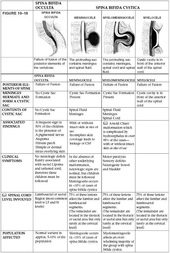
- Myelomeningocele: Most severe. Sac contains spinal cord + damaged nerves → motor, sensory, sphincter deficits.
- Meningocele: Sac contains CSF and meninges only → little/no nerve damage.
- Spina bifida occulta: Mildest form. Small bony defect, often only visible as tuft of hair/dimple. Often asymptomatic.
💡 Classification: Open NTDs (myelomeningocele, meningocele) vs Closed (occulta).
🩺 Clinical Presentation
- At birth: Visible sac or defect in severe forms.
- Occulta: Subtle skin markers (dimple, hair tuft, lipoma).
- Neurological signs: leg weakness, absent reflexes, bladder/bowel incontinence.
🧪 Investigations
- Maternity screening: Raised maternal serum alpha-fetoprotein (AFP) at 15–16 weeks.
- Ultrasound: 18–20 week anomaly scan detects open NTDs.
- Amniotic fluid AChE: Diagnostic if ultrasound is equivocal.
- Postnatal: MRI spine/brain for associated malformations.
🖼️ Diagram

🛡️ Prevention
- Folic acid 400 mcg daily for all women from pre-conception until 12 weeks gestation.
- High risk women: 5 mg daily (epilepsy, diabetes, obesity, family history).
- Avoid teratogenic drugs (esp. valproate) if alternatives exist.
⚕️ Management
- Prenatal: In-utero surgical closure (improves motor outcomes vs postnatal repair) in selected cases; option of termination in severe cases.
- Postnatal: Early neurosurgical closure to prevent infection/meningitis.
- Hydrocephalus: May require ventriculoperitoneal shunt.
- Orthopaedic: Bracing, physiotherapy for mobility.
- Urological: CIC (clean intermittent catheterisation), anticholinergics, bladder augmentation for neurogenic bladder.
- Multidisciplinary care: Specialist nurses, physiotherapy, urology, orthopaedics, neurosurgery, psychology.
🌟 Summary: Spina bifida is a neural tube defect due to failure of closure by day 28. Risk reduced with folate supplementation. Management requires early neurosurgical repair and long-term multidisciplinary support for mobility, bladder, and bowel function.
Cases — Spina Bifida 🧠🦴
- Case 1 — Spina Bifida Occulta (Mildest Form) 🔍: A 12-year-old girl is found to have a small dimple with a tuft of hair over her lumbosacral spine during a routine exam. She has no neurological deficits and normal bladder function. Diagnosis: Spina bifida occulta. Management: Usually benign, but MRI spine to exclude tethered cord; reassure family; surgery only if symptomatic tethering develops.
- Case 2 — Meningocele 🎈: A newborn boy presents with a fluid-filled sac protruding from the lower back, covered by thin skin. Neuro exam: normal limb movements and bladder function. MRI: meningeal sac containing CSF, no spinal cord tissue. Diagnosis: Spina bifida with meningocele. Management: Surgical repair to prevent infection; long-term prognosis good if no cord involvement.
- Case 3 — Myelomeningocele (Most Severe) ⚠️: A term infant is born with an open defect in the lumbosacral region, with exposed spinal cord and meninges. Exam: flaccid paralysis of lower limbs, absent anal tone, urinary incontinence. Hydrocephalus noted on cranial ultrasound. Diagnosis: Spina bifida with myelomeningocele. Management: Urgent neurosurgical closure, antibiotics, ventriculoperitoneal shunt for hydrocephalus, long-term multidisciplinary care (urology, orthopaedics, physiotherapy).
Teaching Commentary 🧠
Spina bifida = failure of neural tube closure (usually lumbar/sacral). - Occulta: Hidden, no neuro deficit, only skin signs (dimple, hair tuft). - Meningocele: Protruding CSF sac, cord intact → usually no neuro deficits. - Myelomeningocele: Protruding sac containing cord + meninges → severe neurological impairment + bladder/bowel dysfunction. Prevention: Maternal folic acid supplementation before conception and in early pregnancy. Associations: Hydrocephalus (Chiari II malformation), orthopaedic deformities, neurogenic bladder.
Categories
- A Level
- About
- Acute Medicine
- Anaesthetics and Critical Care
- Anatomy
- Anatomy and Physiology
- Biochemistry
- Book
- Cardiology
- Collections
- CompSci
- Crib Sheets
- Crib sheets
- Dental
- Dermatology
- Differentials
- Drugs
- ENT
- Education
- Electrocardiogram
- Embryology
- Emergency Medicine
- Endocrinology
- Ethics
- Foundation Doctors
- GCSE
- Gastroenterology
- General Practice
- Genetics
- Geriatric Medicine
- Guidelines
- Gynaecology
- Haematology
- Hepatology
- Immunology
- Infectious Diseases
- Infographic
- Investigations
- Lists
- Mandatory Training
- Medical Students
- Microbiology
- Nephrology
- Neurology
- Neurosurgery
- Nutrition
- OSCE
- OSCEs
- Obstetrics
- Obstetrics Gynaecology
- Oncology
- Ophthalmology
- Oral Medicine and Dentistry
- Orthopaedics
- Paediatrics
- Palliative
- Pathology
- Pharmacology
- Physiology
- Procedures
- Psychiatry
- Public Health
- Radiology
- Renal
- Respiratory
- Resuscitation
- Revision
- Rheumatology
- Statistics and Research
- Stroke
- Surgery
- Toxicology
- Trauma and Orthopaedics
- USMLE
- Urology
- Vascular Surgery
