
| Download the amazing global Makindo app: ✅ Means NICE/National Guidelines 2026 compliant Android | Apple | |
|---|---|
| MEDICAL DISCLAIMER: Educational use only. Not for diagnosis or management. See below for full disclaimer. |
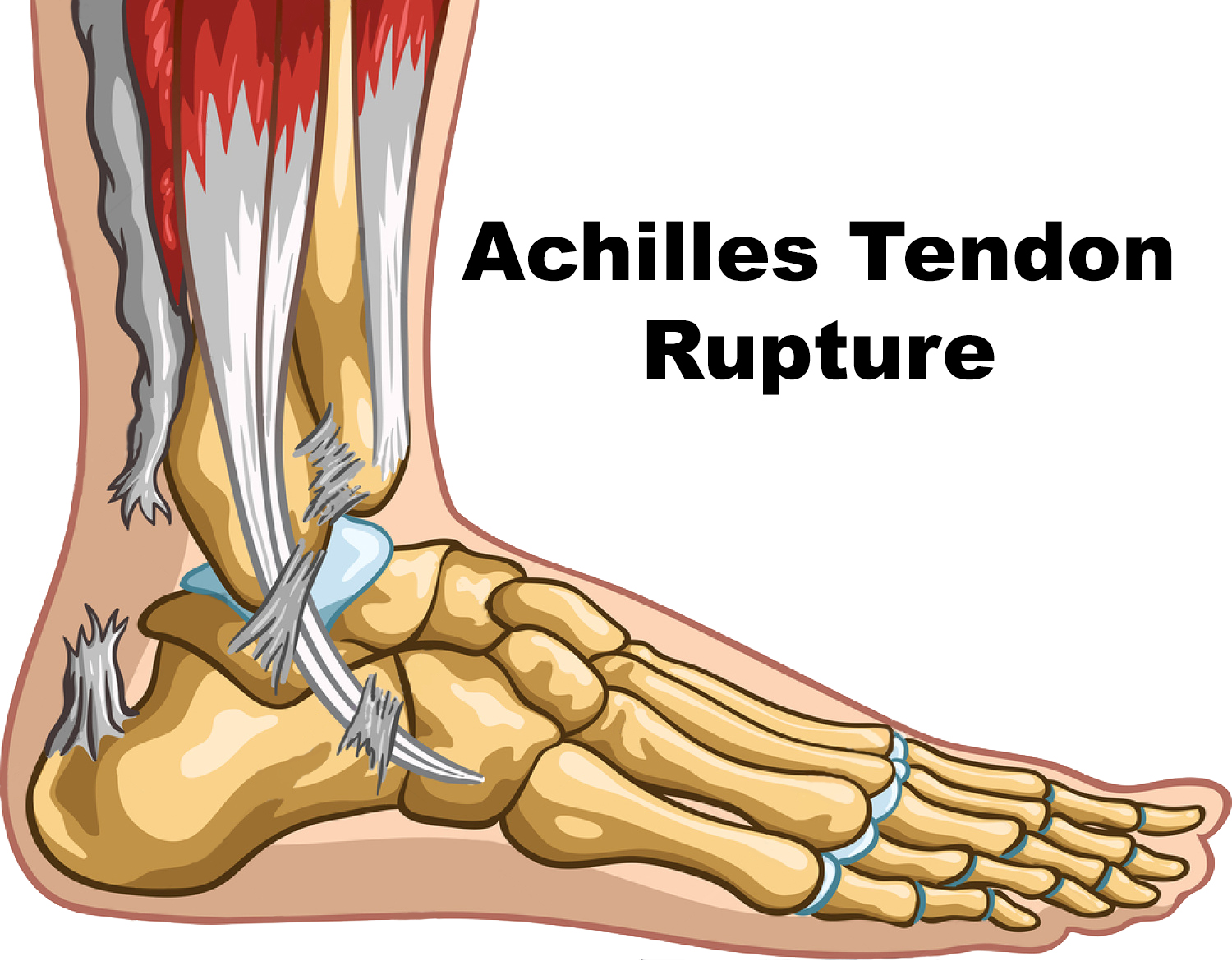
Achilles Tendon rupture
Related Subjects: |Sprained Ankle |Achilles Tendon rupture
🦶 Achilles tendon rupture typically presents as a sudden “snap” or sensation of being kicked at the back of the ankle, most often during sporting activity. 🚫 Key risk factors: fluoroquinolones, corticosteroid injections, diabetes mellitus, obesity, and increasing age. 🔎 Hallmark sign: a positive Thompson calf squeeze test (absence of plantarflexion). ⚖️ Management trade-off: conservative treatment has a higher re‑rupture rate, whereas surgery lowers recurrence but increases wound and thrombotic risk.
📖 About Achilles Tendon Injury
- The Achilles is the strongest tendon in the human body, yet is vulnerable to rupture during sudden eccentric loading (e.g. jumping or sprinting).
- Often referred to as the “weekend warrior injury”, it most commonly affects middle‑aged recreational athletes.
⚠️ Aetiology & Risk Factors
- Sudden acceleration or deceleration during sport.
- Age‑related degenerative changes, exacerbated by diabetes or inflammatory arthritis.
- Medication‑associated: fluoroquinolones (e.g. ciprofloxacin) and repeated corticosteroid injections.
- Additional risks: obesity, poor conditioning, and systemic inflammatory disease.
🩺 Clinical Features
- Sudden sharp pain, audible “snap” or sensation of being kicked in the posterior ankle or calf.
- Weak plantarflexion — patients may still walk, but often cannot run, push off, or stand on tiptoe on the affected side.
- A palpable gap along the Achilles tendon, usually best felt 2–6 cm proximal to the calcaneal insertion.
- Simmonds' triad suggests Achilles tendon rupture:
- 🕳️ Palpable gap in the Achilles tendon.
- 🦶 Increased passive ankle dorsiflexion compared with the other side.
- ❌ Positive calf squeeze / Thompson test — squeezing the calf fails to produce normal plantarflexion.
- Thompson / Simmonds calf squeeze test: with the patient prone and feet hanging over the couch edge, squeezing the calf should cause plantarflexion. Little or no plantarflexion suggests rupture.
- Matles test: with the patient prone and knees flexed to 90°, the affected foot rests in a more dorsiflexed position than the normal side, rather than maintaining slight plantarflexion.
- Bruising, swelling and tenderness may occur around the posterior ankle or calf.
- Partial ruptures can present subtly and may still allow some plantarflexion or limited bilateral tiptoe standing.
📌 Clinical pearl: Ability to walk does not exclude Achilles rupture because other flexor tendons can still produce some plantarflexion. The most useful bedside clues are a palpable tendon gap, a positive calf squeeze test, and the Matles test.
🔍 Investigations
- Ultrasound: first‑line investigation; dynamic and readily identifies rupture location and severity.
- MRI: reserved for indeterminate cases or pre‑operative planning.
💊 Management
- Non‑surgical: functional bracing or boot immobilisation in plantarflexion; appropriate for partial tears or less physically active patients.
- Surgical repair: end‑to‑end tendon suturing; preferred in complete ruptures and younger, active individuals. Enables earlier rehabilitation but carries wound and DVT risk.
- Rehabilitation: physiotherapy is essential for all patients. Gradual strengthening, stretching, and proprioceptive training; return to sport typically takes 6–12 months.
⚡ Complications
- Re‑rupture (higher with conservative management).
- Wound complications, infection, or adhesions following surgery.
- Chronic calf weakness or muscle atrophy.
- Venous thromboembolism related to immobilisation.
🩺 Case 1 – Sudden Sports Injury
A 34‑year‑old man experiences a sudden “pop” in the posterior ankle while playing football, followed by pain and difficulty walking. Examination reveals swelling, a positive Thompson’s test, and inability to stand on tiptoe.
Management: 🏥 Immobilisation in plantarflexion (cast or functional brace), urgent orthopaedic referral. Surgical repair often advised for young, active patients. Early physiotherapy is essential.
Avoid: ❌ Misdiagnosis as a simple ankle sprain; avoid weight‑bearing until stabilised.
🩺 Case 2 – Older Patient on Steroids
A 65‑year‑old man on long‑term prednisolone for COPD presents with sudden ankle pain after stepping off a curb. Examination reveals a palpable tendon gap and weak plantarflexion.
Management: 💊 Non‑surgical functional bracing may be appropriate given comorbidities, with analgesia and physiotherapy. Consider surgery if functionally independent and fit.
Avoid: ❌ Unreviewed continuation of steroids or fluoroquinolones; prolonged immobilisation without VTE prophylaxis.
| The content on this website is provided for educational and informational purposes only to support exam preparation (e.g., MLA, MRCP, USMLE) and learning. This is NOT medical advice, diagnosis, treatment, or professional guidance. It does not replace consultation with a qualified healthcare professional, official guidelines (e.g., NICE, GMC, BNF), or supervised clinical practice. Always verify information with current, authoritative sources. Makindo and its contributors accept no liability for any reliance on this content, including errors, omissions, or any resulting harm, loss, or consequences. By using this site, you agree to these terms. |
|
|
Categories
- About
- Acute Medicine
- Anaesthetics and Critical Care
- Anatomy
- Anatomy and Physiology
- Biochemistry
- Book
- Cardiology
- Collections
- CompSci
- Crib Sheets
- Critical care
- Dental
- Dermatology
- Differentials
- Drugs
- ENT
- Electrocardiogram
- Embryology
- Emergency Medicine
- Endocrinology
- Ethics
- Foundation Doctors
- GCSE
- Gastroenterology
- General Practice
- Genetics
- Geriatric Medicine
- Geriatrics
- Guidelines
- Haematology
- Hepatology
- Immunology
- Infectious Diseases
- Infographic
- Investigations
- Lists
- MRCP
- Mandatory Training
- Medical Students
- Microbiology
- Nephrology
- Neurology
- Neurosurgery
- Nutrition
- OSCE
- Obstetrics Gynaecology
- Oncology
- Ophthalmology
- Oral Medicine and Dentistry
- Orthopaedics
- Paediatrics
- Palliative
- Palliative Care
- Pathology
- Pharmacology
- Physiology
- Procedures
- Psychiatry
- Public Health
- Radiology
- Respiratory
- Resuscitation
- Revision Topics
- Rheumatology
- Statistics and Research
- Stroke
- Surgery
- Toxicology
- Trauma and Orthopaedics
- USMLE
- Urology
- Vascular Surgery