
| Download the amazing global Makindo app: ✅ Means NICE/National Guidelines 2026 compliant Android | Apple | |
|---|---|
| MEDICAL DISCLAIMER: Educational use only. Not for diagnosis or management. See below for full disclaimer. |
Heterochromia Iridium
👁️ Heterochromia - a difference in iris colour between the two eyes or within one eye. Usually benign, but occasionally a sign of underlying systemic or ocular disease. ⚠️ Neuroblastoma (childhood) and melanoma (later life, with dark iris spot) are rare but important causes of acquired heterochromia.
🌈 About
- Heterochromia means different eye colours or multi-toned irises within one eye.
- Usually harmless and may be congenital or acquired.
- Common in animals (e.g. huskies, cats), rare in humans.
🧬 Types
- 🔹 Complete heterochromia: Each eye a different colour (e.g., one blue, one brown).
- 🔸 Sectoral / partial heterochromia: Segment of one iris differs in colour (as below).
- 🔹 Central heterochromia: Inner ring around pupil differs from outer iris colour.
🎨 Split Iris (Sectoral Heterochromia)
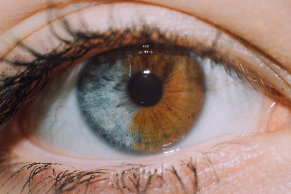
Example of partial heterochromia - a wedge of lighter pigment within one iris.
👶 Heterochromia in Infancy - Causes
- 🌈 Benign (physiological) heterochromia - normal pigment variation.
- 🦋 Horner’s syndrome (congenital) - smaller pupil (miosis), mild ptosis, lighter iris.
- 🧠 Sturge–Weber syndrome - facial port-wine stain, glaucoma risk.
- 🎧 Waardenburg syndrome - white forelock, broad nasal root, deafness.
- 🧬 Piebaldism - patchy albinism of skin and hair.
- 🚼 Hirschsprung disease - may co-occur in syndromic forms (e.g., Waardenburg type IV).
- 🌀 Bloch–Sulzberger syndrome (Incontinentia pigmenti).
- 📘 Neurofibromatosis (von Recklinghausen disease).
- 🍃 Tuberous sclerosis (Bourneville disease).
- 🕊️ Parry–Romberg syndrome - facial hemiatrophy, sometimes with iris pigment loss.
🧑⚕️ Waardenburg Syndrome (example)
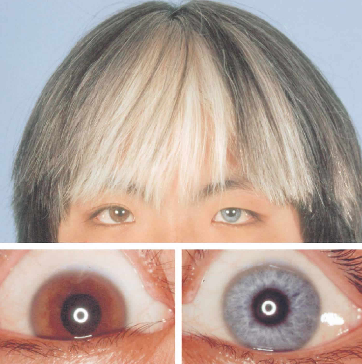
Note iris colour variation, white forelock, and characteristic facial features.
⚡ Acquired Heterochromia - Causes
- 🩸 Eye trauma or post-surgical pigment change.
- 🧠 Neuroblastoma (childhood) - tumour of sympathetic chain; may present with unilateral Horner’s syndrome.
- 🦠 Inflammation: Iritis, uveitis, Fuchs’ heterochromic cyclitis.
- 💊 Medications: Prostaglandin analogues for glaucoma (e.g., latanoprost, bimatoprost, “Latisse”).
- 🌀 Glaucoma or pigment dispersion syndrome.
- 🎯 Melanoma or melanocytoma of the iris - often darkened, irregular patch.
- 💉 Bleeding (hyphema) or iron deposition post-injury.
- 🧩 Horner’s syndrome (acquired) - lighter iris on affected side.
- 💧 Posner–Schlossman syndrome - recurrent uveitic glaucoma.
- 🧬 Chediak–Higashi syndrome - albinism, immune defects.
- ⚡ Diabetes / CRVO - rare secondary pigment changes.
🩺 Clinical Evaluation
- Assess onset, laterality, visual acuity, photophobia, pain.
- Look for pupil asymmetry, eyelid ptosis (Horner’s), or signs of uveitis/glaucoma.
- Use slit-lamp examination to confirm iris pigment change (not reflection or shadow).
🧭 Management
- 👶 At birth: Discuss with paediatrician; often benign pigment development, but consider ophthalmology referral.
- 🧑⚕️ Later in life: Refer to ophthalmologist if new or changing heterochromia to exclude melanoma or uveitis.
- ⚕️ Treat underlying cause if identified (infection, inflammation, or trauma).
- 📸 Baseline photograph - useful for monitoring progression.
💡 Teaching tip: - Congenital = likely benign (esp. stable since infancy). - Acquired = always investigate (rule out tumour, inflammation, or trauma). - Horner’s syndrome can be recognised by triad: ptosis + miosis + anhidrosis ± lighter iris.
| The content on this website is provided for educational and informational purposes only to support exam preparation (e.g., MLA, MRCP, USMLE) and learning. This is NOT medical advice, diagnosis, treatment, or professional guidance. It does not replace consultation with a qualified healthcare professional, official guidelines (e.g., NICE, GMC, BNF), or supervised clinical practice. Always verify information with current, authoritative sources. Makindo and its contributors accept no liability for any reliance on this content, including errors, omissions, or any resulting harm, loss, or consequences. By using this site, you agree to these terms. |
|
|
Categories
- About
- Acute Medicine
- Anaesthetics and Critical Care
- Anatomy
- Anatomy and Physiology
- Biochemistry
- Book
- Cardiology
- Collections
- CompSci
- Crib Sheets
- Critical care
- Dental
- Dermatology
- Differentials
- Drugs
- ENT
- Electrocardiogram
- Embryology
- Emergency Medicine
- Endocrinology
- Ethics
- Foundation Doctors
- GCSE
- Gastroenterology
- General Practice
- Genetics
- Geriatric Medicine
- Geriatrics
- Guidelines
- Haematology
- Hepatology
- Immunology
- Infectious Diseases
- Infographic
- Investigations
- Lists
- MRCP
- Mandatory Training
- Medical Students
- Microbiology
- Nephrology
- Neurology
- Neurosurgery
- Nutrition
- OSCE
- Obstetrics Gynaecology
- Oncology
- Ophthalmology
- Oral Medicine and Dentistry
- Orthopaedics
- Paediatrics
- Palliative
- Palliative Care
- Pathology
- Pharmacology
- Physiology
- Procedures
- Psychiatry
- Public Health
- Radiology
- Respiratory
- Resuscitation
- Revision Topics
- Rheumatology
- Statistics and Research
- Stroke
- Surgery
- Toxicology
- Trauma and Orthopaedics
- USMLE
- Urology
- Vascular Surgery