| Download the amazing global Makindo app: Android | Apple | |
|---|---|
| MEDICAL DISCLAIMER: Educational use only. Not for diagnosis or management. See below for full disclaimer. |
Arteriovenous Malformations
Related Subjects: |Subarachnoid Haemorrhage |Haemorrhagic stroke
🧠 Arteriovenous malformations (AVMs) are abnormal tangles of blood vessels connecting arteries and veins, disrupting normal blood flow and oxygen circulation. Though rare, they carry a significant risk of intracerebral or subarachnoid haemorrhage, particularly in younger individuals.
📖 About
- Prevalence: Incidence ~20–50 per 100,000; often discovered incidentally on imaging.
- Definition: Direct artery–vein connection without a capillary bed → high arterial pressure damages thin-walled veins → rupture risk.
🔬 Pathophysiology
- Bypass of normal capillary network → poor oxygen exchange.
- ↑ Venous pressure → wall weakness and rupture.
- Most common in cerebral structures; also found in spine, dura, and elsewhere.
🧬 Genetics & Inheritance
- Hereditary Hemorrhagic Telangiectasia (HHT): 10–25% of patients develop brain AVMs.
- Usually sporadic; familial cases rare.
- Affects men and women equally.
🧾 Inherited Conditions Associated with AVMs
- 🎭 Sturge–Weber Syndrome:
- Features: Facial port-wine stain, seizures, hemiparesis.
- Leptomeningeal angiomatosis may mimic or accompany AVMs.

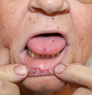
- 👃 Hereditary Haemorrhagic Telangiectasia (HHT):
- Genetic disorder of blood vessels → recurrent nosebleeds, telangiectasias, visceral AVMs.
- Brain AVMs are a recognised complication.

🩺 Clinical Presentation
- Incidental: Often asymptomatic, found on imaging.
- Headache: May be chronic or severe with bleeding.
- Seizures: Common in young adults with cortical AVMs.
- Neurological deficits: Visual changes, weakness, numbness, speech impairment.
- Tinnitus: Audible bruit in large AVMs.
⚠️ Complications
- Intracerebral or subarachnoid haemorrhage.
- Progressive neurological deficits from bleed or mass effect.
- Seizure disorders.
- Hydrocephalus if CSF pathways are obstructed.
📊 Spetzler–Martin Grading (surgical risk)
- Nidus size: Small (<3 cm) = 1; Medium (3–6 cm) = 2; Large (>6 cm) = 3.
- Brain eloquence: Non-eloquent = 0; Eloquent = 1.
- Venous drainage: Superficial = 0; Deep = 1.
- 🔺 Higher total = higher surgical risk.
🔍 Investigations
- 🖼️ CT: First-line in suspected ICH; may show high-density lesion or calcification.
- 🧲 MRI: Best for structural detail; T2 “flow voids” suggest vascular lesion.
- 🩸 Cerebral angiography: Gold standard for diagnosis and planning.
- ⚡ EEG: In seizure presentations.
⚖️ Management
- Conservative: For asymptomatic/low-grade AVMs → monitor with imaging.
- Surgical resection: For accessible AVMs (Grades I–II), especially small, cortical lesions.
- Radiosurgery: SRS or focal beam → causes progressive vessel fibrosis (obliteration in 1–3 yrs).
- Endovascular embolisation: Blocks feeders; often combined with surgery or radiosurgery.
📑 AHA Recommendations
- Unruptured AVMs: Annual bleed risk ~1%. Conservative approach often safer than intervention.
- Ruptured AVMs: Higher rebleed risk (~5%/yr). CTA/MRA/DSA essential; management depends on lesion and patient risk.
📉 Prognosis
- Smaller, surgically accessible AVMs → better outcome after resection.
- Deep venous drainage or associated aneurysm → worse prognosis, higher bleed risk.
📖 References
💡 Exam Pearl: AVMs are direct artery–vein connections → high-pressure venous rupture. Think “young patient, seizures or bleed, MRI flow voids”.
Categories
- A Level
- About
- Acute Medicine
- Anaesthetics and Critical Care
- Anatomy
- Anatomy and Physiology
- Biochemistry
- Book
- Cardiology
- Collections
- CompSci
- Crib Sheets
- Dental
- Dermatology
- Differentials
- Drugs
- ENT
- Education
- Electrocardiogram
- Embryology
- Emergency Medicine
- Endocrinology
- Ethics
- Foundation Doctors
- GCSE
- Gastroenterology
- General Practice
- Genetics
- Geriatric Medicine
- Guidelines
- Gynaecology
- Haematology
- Hepatology
- Immunology
- Infectious Diseases
- Infographic
- Investigations
- Lists
- Mandatory Training
- Medical Students
- Microbiology
- Nephrology
- Neurology
- Neurosurgery
- Nutrition
- OSCE
- OSCEs
- Obstetrics
- Obstetrics Gynaecology
- Oncology
- Ophthalmology
- Oral Medicine and Dentistry
- Orthopaedics
- Paediatrics
- Palliative
- Pathology
- Pharmacology
- Physiology
- Procedures
- Psychiatry
- Public Health
- Radiology
- Renal
- Respiratory
- Resuscitation
- Revision
- Rheumatology
- Statistics and Research
- Stroke
- Surgery
- Toxicology
- Trauma and Orthopaedics
- USMLE
- Urology
- Vascular Surgery
