
| Download the amazing global Makindo app: ✅ Means NICE/National Guidelines 2026 compliant Android | Apple | |
|---|---|
| MEDICAL DISCLAIMER: Educational use only. Not for diagnosis or management. See below for full disclaimer. |
Lung Abscess
Related Subjects: | Assessing Breathlessness | Fever - Pyrexia of unknown origin
⚠️ Poor Prognosis: Advanced age, aspiration risk, immunosuppression, bronchial obstruction, and underlying malignancy are markers of poor outcome.
📖 About
- Lung abscess = localised collection of pus within the pulmonary parenchyma resulting from suppurative necrosis of lung tissue and cavity formation with an air–fluid level.
- Classically follows aspiration pneumonia in patients with altered consciousness (e.g., alcohol intoxication, seizures, stroke, anaesthesia).
- Represents the “end‑point” of severe infection: tissue necrosis → liquefaction → bronchial communication → cavity drainage.
- Commonly affects the posterior segments of upper lobes and superior segments of lower lobes due to gravity during aspiration.
🧬 Pathophysiology
- Aspiration of anaerobic bacteria → local infection and microvascular thrombosis → tissue necrosis.
- Liquefactive necrosis leads to formation of a cavity containing pus, bacteria, and necrotic debris.
- Drainage into a bronchus → expectoration of purulent, foul‑smelling sputum (“fetid abscess”).
- Progression may form a bronchopleural fistula or rupture into the pleural space → secondary empyema.
📂 Classification
- Primary lung abscess: Occurs in previously healthy lungs, usually after aspiration of oropharyngeal contents.
- Secondary lung abscess: Occurs due to pre‑existing lung or systemic disease (e.g., obstructing carcinoma, bronchiectasis, HIV, diabetes, immunosuppression).
- Acute: < 6 weeks; Chronic: > 6 weeks or persistent cavity.
🦠 Microbiology / Causes
- Aspiration (anaerobes ≈ 80 %): Prevotella, Bacteroides, Fusobacterium, Peptostreptococcus.
- Facultative bacteria: Staphylococcus aureus, Streptococcus pyogenes, Klebsiella, Pseudomonas.
- Immunocompromised hosts: Nocardia, Aspergillus, Candida, Mycobacterium tuberculosis.
- Post‑obstructive: Bronchial carcinoma, foreign body, mediastinal mass.
- Haematogenous (septic emboli): Right‑sided endocarditis, infected jugular thrombophlebitis, I.V. drug use (often Staph. aureus).
🩺 Clinical Features
- Fever, chills, weight loss, malaise.
- Persistent productive cough with foul‑smelling purulent sputum or “sputum flooding” when cavity drains.
- Haemoptysis common due to erosion of bronchial arteries.
- Pleuritic chest pain if peripheral lesion abuts pleura.
- Halitosis and metallic taste from anaerobic flora.
- Clubbing and cachexia in chronic infection.
🧾 Differential Diagnosis
- Cavitating squamous carcinoma (thick irregular wall, spiculated margins).
- Tuberculosis with apical cavities; often thinner walls, satellites.
- Wegener’s granulomatosis / vasculitis.
- Pulmonary infarction secondary to embolism with sterile cavitation.
- Cavitating pneumonia (Staph aureus, Klebsiella) vs true abscess = liquefied, thick‑walled cavity + air–fluid level.
🔎 Investigations
- Blood tests: Leukocytosis, ↑ ESR/CRP, ± anaemia of chronic disease.
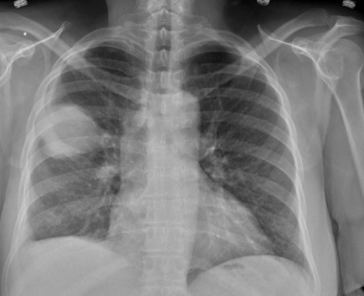
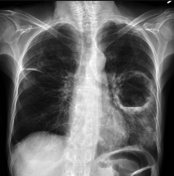
- Chest X‑ray: Cavity with air–fluid level within consolidation, usually posterior segments of upper / superior of lower lobes.
- CT Thorax (contrast HRCT): Defines extent, number of lesions, wall thickness (typically > 2 mm), detects neoplasm or empyema.
- Bronchoscopy: Rule out endobronchial obstruction or neoplasm; may permit lavage or drainage.
- Microbiology:
- Sputum / BAL / aspirate cultures for aerobes and anaerobes.
- Blood cultures before antibiotics.
- Aspiration for Gram stain or AFB if TB suspected.
💊 Management - Multidisciplinary Approach
- General / Supportive:
- Postural drainage - dependent segment uppermost to facilitate drainage gravity‑wise (3–5× daily).
- Physiotherapy and chest percussion to mobilise secretions.
- Hydration and nutritional support to counter catabolic state.
- Meticulous oral care to reduce anaerobic floral load.
- Antibiotic Therapy: (IV → PO 6–8 weeks till radiologic resolution)
- First‑line: Co‑amoxiclav 1.2 g IV TDS or amoxicillin‑clavulanate PO 625 mg TDS if mild.
- Penicillin‑allergic: Clindamycin 600 mg IV/PO QDS.
- Aerobic Gram‑negatives suspected: Add ciprofloxacin 750 mg BD or levofloxacin 500 mg BD.
- MRSA risk / post‑influenza: Add vancomycin or linezolid pending sensitivities.
- Fungal / Nocardia: Voriconazole / TMP‑SMX based on culture.
- Re‑evaluation: Persistent fever > 10–14 days → repeat CT for non‑draining cavity or secondary malignancy.
- Surgical / Interventional Indications:
- Abscess > 6 cm or > 6 weeks non‑responsive to therapy.
- Massive haemoptysis.
- Suspected bronchial obstruction or neoplasm after bronchoscopy.
- Developing empyema / bronchopleural fistula → thoracostomy or lobectomy.
⚠️ Complications
- Empyema (thoracic infection extends to pleural space).
- Bronchopleural fistula → persistent air leak and pneumothorax.
- Massive haemoptysis from erosion of bronchial arteries.
- Sepsis / septic embolisation to brain or other organs.
- Chronic fibrocystic changes or mycetoma formation in residual cavity.
📋 Table – Diagnostic and Therapeutic Pearls
| Feature | Clue / Finding | Clinical Pearl |
|---|---|---|
| Location | Posterior upper / superior lower lobes | Due to supine aspiration. |
| Sputum odour | Foul, putrid | Suggests anaerobic infection. |
| Radiology | Thick‑walled cavity with air–fluid level | Contrast CT to exclude malignancy. |
| Response time | 2–3 weeks to clinical improve, radiologic resolution in 6–8 weeks | Persisting fever → exclude obstruction or empyaema. |
| Drainage need | Large > 6 cm or poorly draining | Aspiration / lobectomy considered. |
📚 References
🧑⚕️ 3 Clinical Cases - Lung Abscess 🫁🕳️
- Case 1 - Aspiration Abscess 🍷: 62‑year‑old man with chronic alcohol use, poor dentition, foul purulent sputum. CXR shows right lower‑lobe air–fluid cavity. Teaching: Aspiration of oropharyngeal flora is the commonest cause; treat with weeks of anaerobic cover and postural drainage.
- Case 2 - Post‑pneumonia (Klebsiella necrosis) 🦠: 47‑year‑old woman after severe Klebsiella pneumonia develops cavity in right upper lobe. Teaching: Gram‑negative organisms can cause necrotising tissue destruction; antibiotics 6–8 weeks, re‑image if non‑resolving.
- Case 3 - Immunocompromised Fungal Abscess 🎗️: 55‑year‑old leukaemia patient on chemotherapy with bilateral cavitating nodules. CT confirms Aspergillus infection. Teaching: Neutropenia predisposes to multi‑focal fungal abscesses; requires voriconazole ± surgical control and immune recovery.
| The content on this website is provided for educational and informational purposes only to support exam preparation (e.g., MLA, MRCP, USMLE) and learning. This is NOT medical advice, diagnosis, treatment, or professional guidance. It does not replace consultation with a qualified healthcare professional, official guidelines (e.g., NICE, GMC, BNF), or supervised clinical practice. Always verify information with current, authoritative sources. Makindo and its contributors accept no liability for any reliance on this content, including errors, omissions, or any resulting harm, loss, or consequences. By using this site, you agree to these terms. |
|
|
Categories
- About
- Acute Medicine
- Anaesthetics and Critical Care
- Anatomy
- Anatomy and Physiology
- Biochemistry
- Book
- Cardiology
- Collections
- CompSci
- Crib Sheets
- Critical care
- Dental
- Dermatology
- Differentials
- Drugs
- ENT
- Electrocardiogram
- Embryology
- Emergency Medicine
- Endocrinology
- Ethics
- Foundation Doctors
- GCSE
- Gastroenterology
- General Practice
- Genetics
- Geriatric Medicine
- Geriatrics
- Guidelines
- Haematology
- Hepatology
- Immunology
- Infectious Diseases
- Infographic
- Investigations
- Lists
- MRCP
- Mandatory Training
- Medical Students
- Microbiology
- Nephrology
- Neurology
- Neurosurgery
- Nutrition
- OSCE
- Obstetrics Gynaecology
- Oncology
- Ophthalmology
- Oral Medicine and Dentistry
- Orthopaedics
- Paediatrics
- Palliative
- Palliative Care
- Pathology
- Pharmacology
- Physiology
- Procedures
- Psychiatry
- Public Health
- Radiology
- Respiratory
- Resuscitation
- Revision Topics
- Rheumatology
- Statistics and Research
- Stroke
- Surgery
- Toxicology
- Trauma and Orthopaedics
- USMLE
- Urology
- Vascular Surgery