Related Subjects:
|Nikolsky's sign
|Koebner phenomenon
|Erythema Multiforme
|Pyoderma gangrenosum
|Erythema Nodosum
|Dermatitis Herpetiformis
|Lichen Planus
|Acanthosis Nigricans
|Acne Rosacea
|Acne Vulgaris
|Alopecia
|Vitiligo
|Urticaria
|Basal Cell Carcinoma
|Malignant Melanoma
|Squamous Cell Carcinoma
|Mycosis Fungoides (Sezary Syndrome)
|Xeroderma pigmentosum
|Bullous Pemphigoid
|Pemphigus Vulgaris
|Seborrheic Dermatitis
|Pityriasis/Tinea versicolor infections
|Pityriasis rosea
|Scabies
|Dermatomyositis
|Toxic Epidermal Necrolysis
|Stevens-Johnson Syndrome
|Atopic Eczema/Atopic Dermatitis
|Psoriasis
Vitiligo 🎨 may be part of the polyglandular autoimmune syndrome, alongside conditions like type 1 diabetes, autoimmune adrenal insufficiency, and autoimmune thyroid disease.
📖 About
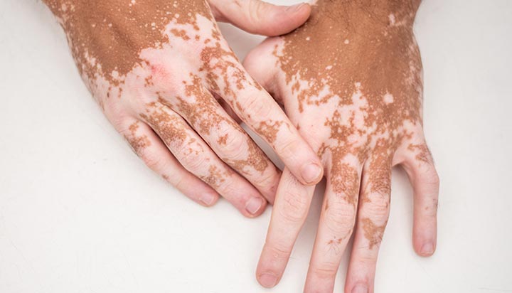
- Characterised by acquired depigmented patches of skin due to melanocyte destruction.
- May be a benign finding in some individuals but has a strong association with autoimmune disease (15× increased risk).
- Onset is often in childhood or young adulthood, with a chronic and relapsing course.
- In rare cases, it can follow an autosomal dominant inheritance pattern.
🔬 Aetiology & Pathophysiology
- Autoimmune destruction of melanocytes by anti-melanocyte antibodies and autoreactive T-cells.
- Skin biopsy → absence of melanocytes in depigmented patches.
- Genetic predisposition + environmental triggers (stress, trauma, sunburn) play a role.
- Koebner phenomenon: new lesions appear at sites of trauma.
🤝 Associations
- Endocrine: Thyroid disease (Hashimoto’s, Graves’), Addison’s disease, diabetes mellitus, hypoparathyroidism.
- Haematological: Pernicious anaemia (B12 deficiency).
- Dermatological: Alopecia areata, psoriasis, lichen sclerosus.
- Ocular: Uveitis, retinal pigment changes.
- Often seen within autoimmune polyglandular syndromes.
👀 Clinical Features
- Well-demarcated, milky-white depigmented patches of skin.
- Symmetrical distribution common; often affects face, hands, elbows, knees, and genitalia.
- Hyperpigmented borders may surround depigmented patches.
- Hair within affected patches may also turn white/grey (leukotrichia).
- Lesions may become itchy or inflamed after sun exposure.
🧪 Investigations
- Primarily a clinical diagnosis.
- Blood tests for associated autoimmune disease:
- FBC & B12 → pernicious anaemia.
- TFTs → autoimmune thyroid disease.
- U&E & calcium → adrenal / parathyroid involvement.
- Autoantibody screen → thyroid, adrenal, parietal cell, IF antibodies.
- Wood’s lamp (UV light) examination → patches fluoresce bright white.
💊 Management
- Education & support: chronic condition, variable progression, no risk of skin cancer from vitiligo itself.
- Cosmetic camouflage (make-up, self-tan products) for psychological support.
- Topical corticosteroids or calcineurin inhibitors (e.g., tacrolimus) → early lesions.
- Phototherapy: Narrowband UVB or PUVA may induce repigmentation in some cases.
- Depigmentation therapy (monobenzone) may be considered for extensive disease (>50% body surface area).
- Counselling: high psychological impact; screen for depression, especially in adolescents.
- Regular monitoring for associated endocrine disorders.
🔮 Prognosis
- Unpredictable course: some patients stabilise, others show progressive spread.
- Repigmentation may occur spontaneously but is often incomplete.
- Chronic condition requiring long-term psychological and dermatological support.
