
| Download the amazing global Makindo app: ✅ Means NICE/National Guidelines 2026 compliant Android | Apple | |
|---|---|
| MEDICAL DISCLAIMER: Educational use only. Not for diagnosis or management. See below for full disclaimer. |
Anterior / Medial Medullary Infarct (Dejerine Syndrome)
Related Subjects: |Anatomy and Physiology of the Brain |Cryptogenic stroke |Anterior / Medial Medullary Infarct (Dejerine Syndrome)
An anterior (medial) medullary infarct is a brainstem stroke of the medial medulla, usually due to occlusion of the anterior spinal artery or paramedian branches of the vertebral artery. It classically produces a triad of contralateral hemiparesis, contralateral loss of vibration and joint position sense, and ipsilateral hypoglossal palsy. Recognising this pattern helps distinguish it from the more common lateral medullary (Wallenberg) syndrome.
🧬 Vascular Anatomy & Pathophysiology
- The medial medulla is supplied by the anterior spinal artery and small paramedian branches of the vertebral arteries.
- Occlusion causes infarction of:
- Corticospinal tract → contralateral limb weakness.
- Medial lemniscus → contralateral loss of vibration and proprioception.
- Hypoglossal nucleus or exiting XII nerve fibres → ipsilateral tongue weakness.
- Atherosclerosis of the vertebral artery, cardioembolism, or vertebral dissection may underlie the event, especially in younger patients.
📌 Clinical Features (Classic Triad)
- Contralateral hemiparesis (arm and leg) – often pyramidal distribution; face may be relatively spared.
- Contralateral loss of vibration and joint position sense – due to medial lemniscus involvement.
- Ipsilateral hypoglossal palsy – tongue deviates towards the lesion on protrusion, with dysarthria and swallowing difficulty.
Additional/variant features:
- Ataxia from involvement of descending cerebellar pathways.
- Respiratory or cardiovascular instability in extensive lesions.
- Usually no Horner’s syndrome or spinothalamic sensory loss – those point more to lateral medullary infarction.
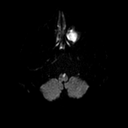
🩻 Imaging
- MRI with DWI is the modality of choice, often showing a “V-shaped” or paramedian lesion in the medial medulla on axial images.
- CTA/MRA should assess vertebral arteries and the origin of the anterior spinal artery for stenosis, occlusion, or dissection.
- CT head is frequently normal early or may show only subtle changes; MRI is often needed for confirmation.
💊 Management
- Acute management follows usual hyperacute stroke protocols – assess for IV thrombolysis and/or thrombectomy according to timing and imaging.
- Secondary prevention as per ischaemic stroke: antiplatelet or anticoagulation (if cardioembolic), statin, BP and risk-factor control, smoking cessation.
- Rehabilitation focuses on bulbar function (swallow, speech), limb weakness, and gait/balance training.
- Monitor for respiratory compromise and aspiration risk; early SALT and nutrition input are often needed.
🧑⚕️ Teaching Pearls
- Think “medial medulla = XII + pyramids + medial lemniscus” → tongue, power, and dorsal-column sensation.
- Crossed signs (ipsilateral cranial nerve, contralateral body) are a big clue to a brainstem lesion.
- Differential includes lateral medullary syndrome, high cervical cord lesions, and internal capsule strokes – the tongue sign helps localise.
| The content on this website is provided for educational and informational purposes only to support exam preparation (e.g., MLA, MRCP, USMLE) and learning. This is NOT medical advice, diagnosis, treatment, or professional guidance. It does not replace consultation with a qualified healthcare professional, official guidelines (e.g., NICE, GMC, BNF), or supervised clinical practice. Always verify information with current, authoritative sources. Makindo and its contributors accept no liability for any reliance on this content, including errors, omissions, or any resulting harm, loss, or consequences. By using this site, you agree to these terms. |
|
|
Categories
- About
- Acute Medicine
- Anaesthetics and Critical Care
- Anatomy
- Anatomy and Physiology
- Biochemistry
- Book
- Cardiology
- Collections
- CompSci
- Crib Sheets
- Critical care
- Dental
- Dermatology
- Differentials
- Drugs
- ENT
- Electrocardiogram
- Embryology
- Emergency Medicine
- Endocrinology
- Ethics
- Foundation Doctors
- GCSE
- Gastroenterology
- General Practice
- Genetics
- Geriatric Medicine
- Geriatrics
- Guidelines
- Haematology
- Hepatology
- Immunology
- Infectious Diseases
- Infographic
- Investigations
- Lists
- MRCP
- Mandatory Training
- Medical Students
- Microbiology
- Nephrology
- Neurology
- Neurosurgery
- Nutrition
- OSCE
- Obstetrics Gynaecology
- Oncology
- Ophthalmology
- Oral Medicine and Dentistry
- Orthopaedics
- Paediatrics
- Palliative
- Palliative Care
- Pathology
- Pharmacology
- Physiology
- Procedures
- Psychiatry
- Public Health
- Radiology
- Respiratory
- Resuscitation
- Revision Topics
- Rheumatology
- Statistics and Research
- Stroke
- Surgery
- Toxicology
- Trauma and Orthopaedics
- USMLE
- Urology
- Vascular Surgery