Makindo Medical Notes"One small step for man, one large step for Makindo" |
|
|---|---|
| Download all this content in the Apps now Android App and Apple iPhone/Pad App | |
| MEDICAL DISCLAIMER: The contents are under continuing development and improvements and despite all efforts may contain errors of omission or fact. This is not to be used for the assessment, diagnosis, or management of patients. It should not be regarded as medical advice by healthcare workers or laypeople. It is for educational purposes only. Please adhere to your local protocols. Use the BNF for drug information. If you are unwell please seek urgent healthcare advice. If you do not accept this then please do not use the website. Makindo Ltd. |
Syringomyelia
-
| About | Anaesthetics and Critical Care | Anatomy | Biochemistry | Cardiology | Clinical Cases | CompSci | Crib | Dermatology | Differentials | Drugs | ENT | Electrocardiogram | Embryology | Emergency Medicine | Endocrinology | Ethics | Foundation Doctors | Gastroenterology | General Information | General Practice | Genetics | Geriatric Medicine | Guidelines | Haematology | Hepatology | Immunology | Infectious Diseases | Infographic | Investigations | Lists | Microbiology | Miscellaneous | Nephrology | Neuroanatomy | Neurology | Nutrition | OSCE | Obstetrics Gynaecology | Oncology | Ophthalmology | Oral Medicine and Dentistry | Paediatrics | Palliative | Pathology | Pharmacology | Physiology | Procedures | Psychiatry | Radiology | Respiratory | Resuscitation | Rheumatology | Statistics and Research | Stroke | Surgery | Toxicology | Trauma and Orthopaedics | Twitter | Urology
Related Subjects:
|Syringomyelia
|Syringobulbia
|Dandy Walker syndrome
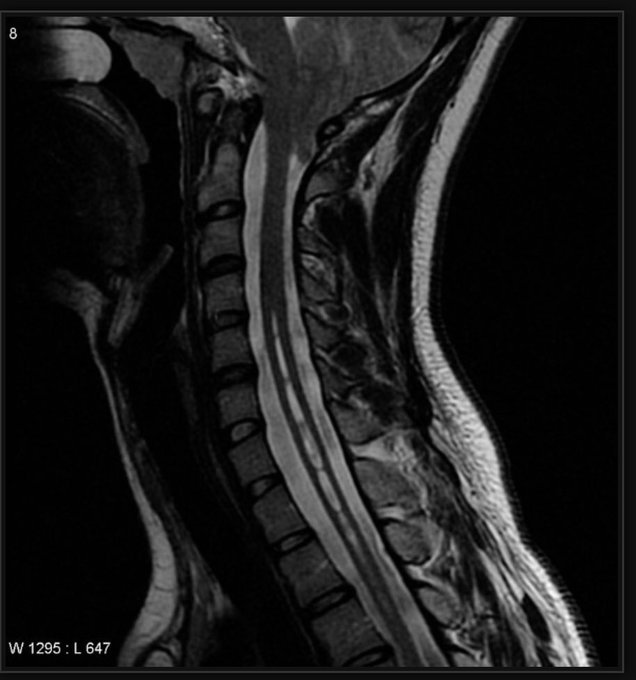
🧠 Syringomyelia is a chronic, progressive neurological condition characterized by the formation of a fluid-filled cavity (syrinx) within the spinal cord.
Over time, the syrinx may expand, damaging the spinal cord and disrupting cerebrospinal fluid (CSF) flow, leading to progressive neurological symptoms.
📖 About
🧬 Aetiology
⚠️ Causes

🩺 Clinical Features
📊 Comparison: Syringomyelia vs Syringobulbia
Feature
Syringomyelia
Syringobulbia
Definition
Fluid-filled cavity (syrinx) within the spinal cord
Fluid-filled cavity within the brainstem (medulla)
Common Association
Chiari I malformation, trauma, tumours
Chiari I malformation, extension of syringomyelia, brainstem tumours
Main Structures Affected
Spinothalamic tracts → pain & temperature loss
Cranial nerve nuclei & brainstem tracts → bulbar & cranial nerve palsies
Classic Clinical Signs
🌀 Cape-like loss of pain & temperature over shoulders/arms
Weakness & wasting of hand muscles🎭 Onion-skin facial sensory loss
🗣 Bulbar palsy (dysphagia, dysarthria)
😮 Horner’s syndrome
Cranial Nerves
Usually spared
Involvement of CN VII, IX, X, XI, XII → facial weakness, hoarseness, tongue wasting
Cerebellar Involvement
Occasional (if syrinx is large)
More likely due to brainstem–cerebellar connections → ataxia
Investigations
MRI spine
MRI brain & spine (to check medulla and extension)
Management
Posterior fossa decompression, syrinx shunting, treat cause
Posterior fossa decompression (Chiari), syrinx shunting, tumour resection if present
🧪 Investigations
💊 Management
📉 Prognosis
📚 References