
| Download the amazing global Makindo app: ✅ Means NICE/National Guidelines 2026 compliant Android | Apple | |
|---|---|
| MEDICAL DISCLAIMER: Educational use only. Not for diagnosis or management. See below for full disclaimer. |
Keloids
Related Subjects: |Seborrheic Keratosis |Keratoacanthoma |Keloids
Some studies suggest that increased expression of Transforming Growth Factor-beta (TGF-β), a cytokine involved in wound healing, plays a pivotal role in keloid formation. This dysregulated fibroblast activity leads to excessive collagen deposition and abnormal scar growth.
🔬 Pathophysiology
- 🧬 Keloids result from excess fibroblast activity and overproduction of collagen following skin injury or inflammation.
- ❌ Unlike hypertrophic scars, keloids extend beyond wound boundaries and may continue to enlarge over months to years.
- 📉 Collagen fibers are disorganized and thickened, creating a raised, firm lesion.
- 👨👩👧 Genetic predisposition plays a role-more common in darker skin tones, suggesting an inherited abnormality of wound healing.
📊 Epidemiology
- ☀️ Higher prevalence in African, Hispanic, and Asian populations.
- 👥 Commonest in ages 10–30 years.
- 📉 Rare after age 30 as collagen synthesis declines.
- 👩 Slightly more common in women (possibly due to ear piercings).
- 👪 Family history increases risk.
🩺 Differential Diagnosis
Several skin lesions can mimic keloids:
- Hypertrophic scars: Confined to wound margins; usually regress after 12–18 months.
- Squamous Cell Carcinoma (SCC): Consider if lesion is ulcerating, irregular, or rapidly enlarging.
- Dermatofibroma: Firm nodules, but non-progressive.
- Melanocytic nevi: Raised pigmented lesions mistaken for keloids.
- Basal Cell Carcinoma (BCC): Can mimic scar-like raised lesions.
- Others: Warts, epidermal cysts, or inflamed nodules.
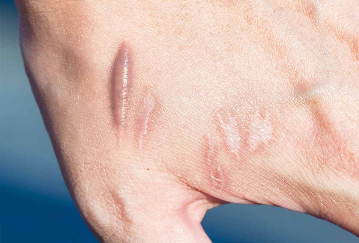
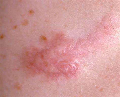
🧾 Clinical Presentation
- Firm, shiny papules or nodules (skin-colored, pink, red, or bluish).
- Often arise from trauma: 💉 piercings, ✂️ surgery, 🤕 burns, or acne scars; but can appear spontaneously.
- Grow beyond original wound edges, sometimes forming claw-like extensions.
- Symptoms: pruritus, tenderness, pain, or stinging.
- Sites: earlobes, shoulders, sternum, scapular region, and upper arms. ❌ Never on palms/soles.
- Functional impact: over joints → restrict mobility.
🧪 Investigations
- Primarily a clinical diagnosis.
- 🔎 Biopsy only if features are atypical or suspicion of malignancy (e.g., ulceration, rapid growth).
- Microbiology if superinfection suspected.
- Ultrasound: occasionally used to assess depth before surgery.
💊 Management
- Conservative (watchful waiting): If asymptomatic and not cosmetically concerning.
- Intralesional corticosteroid injections: 1st line-reduce collagen synthesis, flatten lesions, relieve itch/pain. Multiple sessions needed.
- Cryotherapy: Effective for small/superficial lesions; often combined with steroid injections.
- Silicone gel/sheets & compression therapy: Create pressure to reduce recurrence after surgery.
- Surgical excision: Used for large lesions but ⚠️ high recurrence unless combined with steroids, cryotherapy, or radiotherapy.
- Laser therapy: Improves appearance (flattening, reducing redness), often adjunctive.
- Radiotherapy: Considered post-excision for resistant/recurrent lesions; use sparingly (risk–benefit).
- Emerging therapies: Interferon injections, 5-fluorouracil, bleomycin, and botulinum toxin show promise in resistant cases.
📌 Clinical Pearls
- 🚨 Always differentiate from hypertrophic scars-location and growth pattern are key.
- 🔄 Recurrence after excision is high (up to 45–100%) without adjunctive therapy.
- 🧬 Genetic and ethnic predisposition → important for counselling patients about risk.
- 👂 Ear keloids are particularly common post-piercing.
✅ Conclusion
Keloids are fibroproliferative disorders of wound healing characterized by uncontrolled collagen deposition, extending beyond the wound edges. They are common in darker skin tones, occur most often in young adults, and may be itchy, painful, or cosmetically distressing. First-line treatment is intralesional corticosteroid injection, with adjunctive therapies (silicone, cryotherapy, laser, radiotherapy) often required. Awareness of high recurrence rates is essential for patient counselling and long-term management.