Related Subjects:
|Subarachnoid Haemorrhage
|Perimesencephalic Subarachnoid haemorrhage
|Haemorrhagic stroke
|Cerebellar Haemorrhage
|Putaminal Haemorrhage
|Thalamic Haemorrhage
|ICH Classification and Severity Scores
|Brain Herniation syndromes
|Epidural Haematoma
⏱️ Timely recognition and neurosurgical evacuation can be lifesaving in cerebellar haemorrhage, offering patients the potential for excellent recovery.
📖 About
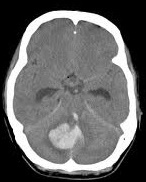
- Cerebellar haemorrhage accounts for ~10% of all intracerebral haemorrhages.
- Causes acute, often persistent vertigo, nausea, vomiting, and imbalance, sometimes mimicking labyrinthitis or intoxication.
- Deterioration can be rapid due to brainstem compression or acute hydrocephalus.
📍 Location & Prognosis
- Midline (vermis) lesions: Higher risk - deep cerebellar nuclei and 4th ventricle involvement, often severe.
- Hemispheric lesions: Lateral bleeds usually have a better prognosis if detected early.
⚡ Causes
- Hypertensive rupture of penetrating arteries (most common).
- Cerebral amyloid angiopathy (elderly).
- Substance misuse (cocaine, amphetamines).
- Arteriovenous malformations, cavernomas.
- Haemorrhagic tumours or metastases.
- Anticoagulant use, thrombocytopenia, or clotting disorders.
- Post-spinal/neurosurgical complications, spontaneous intracranial hypotension.
🧑⚕️ Clinical Presentation
- Sudden severe headache, nausea, vertigo, vomiting.
- Ipsilateral cerebellar signs: limb ataxia, intention tremor, nystagmus.
- Cranial nerve VI & VII involvement possible → diplopia, facial weakness.
- Truncal ataxia: inability to walk unaided, frequent falls.
- Severe cases: brainstem compression → coma, pinpoint pupils, abnormal breathing (Cheyne–Stokes).
🚩 Poor Prognostic Indicators
- Haematoma diameter >3 cm 📏
- Acute hydrocephalus (4th ventricle obstruction)
- Brainstem compression, herniation
- Midline location or intraventricular extension
- Low GCS at presentation
🔍 Differential Diagnosis
- Brainstem infarction (posterior circulation stroke)
- Peripheral vestibular disorders (labyrinthitis, vestibular neuritis)
- Alcohol intoxication or drug toxicity (esp. anticonvulsants)
- Multiple sclerosis relapse (rare mimic)
🧪 Investigations
- Bloods: FBC, coagulation screen (INR if warfarin), U&E, LFTs, glucose.
- ECG, chest X-ray (perioperative/medical optimisation).
- Urgent non-contrast CT: gold standard for diagnosis; defines bleed size, location, hydrocephalus, mass effect.
- MRI: in stable patients, helps detect underlying lesions (AVM, tumour, cavernoma).
🛠️ Management
- Stabilisation (ABC): Airway protection, oxygenation, BP control, and ICU/ITU consideration.
- Surgical evacuation:
- Indicated if clot >3 cm, GCS <14, or acute hydrocephalus.
- Suboccipital craniectomy + clot evacuation can be lifesaving.
- External ventricular drain (EVD) may relieve hydrocephalus, often alongside clot evacuation.
- Medical therapy:
- Osmotic therapy (Mannitol 1 g/kg) as a bridge in raised ICP.
- Reversal of anticoagulation (e.g., Vitamin K + PCC for warfarin).
- Cautious BP lowering to reduce rebleed risk.
- Conservative management: Consider if clot <3 cm, GCS preserved, and no hydrocephalus, but under close neurosurgical observation.
- Rehabilitation: Multidisciplinary input (physio, OT, speech therapy). Recovery can be remarkable compared to supratentorial bleeds.
- Palliative care: In cases with profound brainstem involvement and coma, discussions around ceiling of care may be necessary.
