Cardiac Echocardiography: Basics and Uses
🫀 Cardiac echocardiography ("echo") is a non-invasive ultrasound test that provides real-time images of the heart. It is the first-line imaging tool for assessing cardiac structure and function, offering critical insights into valves, chambers, and haemodynamics.
📖 Introduction
- 🎵 Ultrasound waves generate moving heart images in real time.
- 🩺 Non-invasive (transthoracic) and risk-free for most patients.
- 🎬 Provides dynamic images → useful for assessing function and flow.
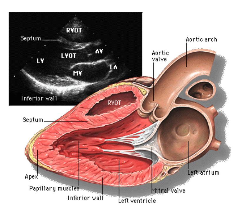
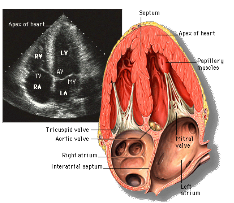
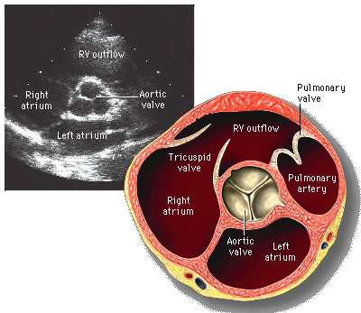
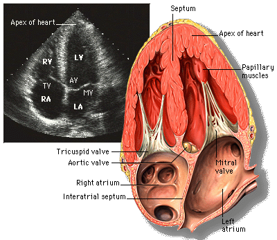
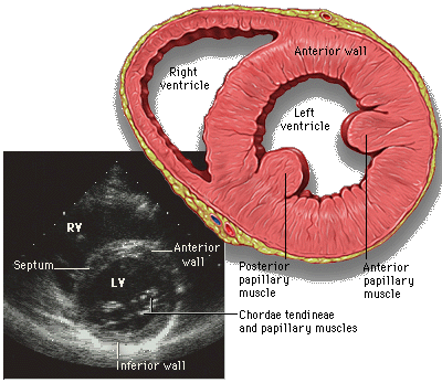
👁️ Different Echo Views
🧩 Types of Cardiac Echocardiography
- 🟦 TTE (Transthoracic Echo) → standard, quick, painless, bedside.
- 🟨 TEE (Transoesophageal Echo) → clearer images, especially for prosthetic valves, endocarditis, left atrial thrombus.
- 🏃 Stress Echo → evaluates inducible ischaemia during exercise/pharmacological stress.
- 🌊 Doppler Echo → quantifies flow across valves & chambers (stenosis, regurgitation).
- 🌀 3D Echo → high-resolution anatomical detail (surgical planning, congenital heart disease).
🔑 Key Uses
- 📊 Measure ejection fraction & wall motion → heart failure diagnosis/prognosis.
- 🫀 Evaluate valves: stenosis, regurgitation, prolapse.
- 💧 Detect pericardial effusion or tamponade.
- 🧬 Assess congenital heart disease.
- 🩸 Identify sources of emboli (e.g., left atrial thrombus, PFO).
- 🔍 Monitor cardiac surgery outcomes (valve replacement, repair, shunts).
🌟 Advantages
- 🙌 Non-invasive and safe.
- 🎥 Real-time functional imaging.
- 🌍 Widely available and cheaper than MRI/CT.
- ☢️ No radiation exposure.
⚠️ Limitations
- 📉 Variable quality in obesity, COPD, or chest wall deformity.
- 🔲 Poor acoustic windows can limit image clarity.
- 🧪 TEE is semi-invasive → rare complications (e.g., oesophageal injury).
🧾 Indications for Echocardiogram
- 🔎 Diagnosis: Valvular disease, cardiomyopathies, congenital heart disease, pericardial effusion, cardiac tumours.
- 📈 Function: LV/RV size, EF, wall motion, cardiac output.
- ❗ Symptoms: Dyspnoea, chest pain, palpitations, syncope, new murmurs.
- 🛠️ Treatment Monitoring: Post-valve surgery, heart failure therapy response.
- 🛡️ Screening: Familial cardiomyopathies, suspected endocarditis, risk stratification.
💡 Clinical Pearls
- 💥 Aortic stenosis: Echo = gold standard for valve area, gradient, LV function.
- 🌊 Mitral regurgitation: Doppler echo quantifies severity & regurgitant volume.
- 🫀 Hypertrophic cardiomyopathy: Echo shows asymmetric septal hypertrophy & SAM (systolic anterior motion) of the mitral valve.
- 🩸 Endocarditis: TEE is far more sensitive than TTE for detecting vegetations.
