
| Download the amazing global Makindo app: ✅ Means NICE/National Guidelines 2026 compliant Android | Apple | |
|---|---|
| MEDICAL DISCLAIMER: Educational use only. Not for diagnosis or management. See below for full disclaimer. |
Pyoderma gangrenosum ✅
Related Subjects: |Cellulitis |Impetigo |Pyoderma gangrenosum |Pemphigus Vulgaris |Toxic Epidermal Necrolysis |Stevens-Johnson Syndrome |Necrotising fasciitis |Gas Gangrene (Clostridium perfringens) |Anatomy of Skin |Skin Pathology and lesions |Skin and soft tissue and bone infections |Lymphangitis
💥 Always consider Pyoderma Gangrenosum (PG) when assessing ulcerated skin lesions. PG is a rare, non-infectious, inflammatory neutrophilic dermatosis characterized by painful ulcers. It is often associated with systemic diseases like IBD, rheumatoid arthritis, and haematologic disorders.
ℹ️ About Pyoderma Gangrenosum
- ⚠️ Often mistaken for common ulcers - always think beyond “just venous ulcers.”
- 👩⚕️ Consider differential diagnoses including skin cancers, atypical infections, and vasculitis.
- 🧪 Urgent referral to dermatology if suspected; consider biopsy to exclude other causes.
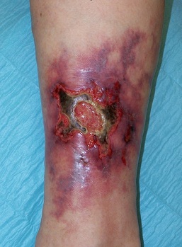
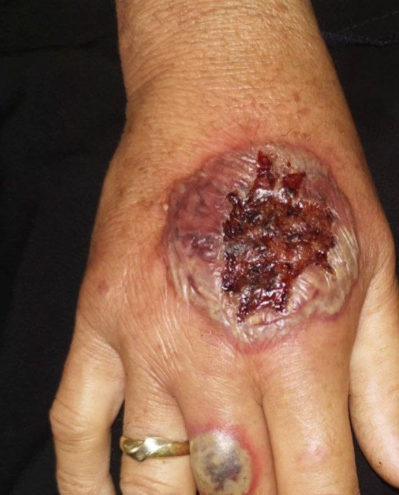
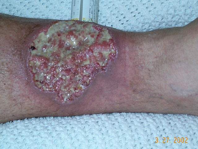
🩺 Clinical Features
- 🔥 Pain: Severe pain, often disproportionate to appearance.
- 🟣 Edges: Undermined, violaceous/blue borders.
- ⚫ Base: Necrotic with purulent/granulomatous tissue.
- 📏 Size: May start small → rapidly expanding ulcers.
- 📍 Sites: Lower legs, surgical wounds, stomas (pathergy phenomenon).
- 🔄 Onset: Often begins as papules, pustules, or vesicles → ulcerates.
- 💥 Trigger: Minor trauma (pathergy) can precipitate.
- 🔗 Associated Diseases: RA, UC, Crohn’s, myeloma, haematologic malignancy.
- 💊 Drugs: Rarely hydroxyurea, GM-CSF.
🧬 Causes and Associations
- ❓ Idiopathic (~20–50%).
- 🧻 IBD: Crohn’s, ulcerative colitis.
- 🩸 Haematologic: Lymphoma, leukaemia, myeloma, myeloproliferative disease.
- 🛡️ Autoimmune: RA, primary biliary cirrhosis.
- 🦠 Other: Hepatitis, HIV, monoclonal gammopathies.
🔍 Investigations
- 📊 FBC, ESR, CRP (raised inflammatory markers).
- 🔬 Skin biopsy (from ulcer edge) → neutrophilic dermatosis (helps exclude other causes but not pathognomonic).
- 🧫 Exclude infection (cultures, swabs).
- 📋 Screen for underlying systemic disease (IBD, RA, haematology referral as needed).
🩹 Management
- 🚫 Avoid trauma: Surgery/skin grafting/debridement can worsen due to pathergy.
- 💊 Topical therapy: Potent/super-potent corticosteroids (e.g., clobetasol), tacrolimus ointment.
- 💊 Pain relief: Analgesia is essential (often very painful ulcers).
- 🩼 Wound care: Non-adhesive dressings, prevent secondary infection.
💉 Systemic Treatments
- 🌟 Corticosteroids: First-line for moderate–severe disease (high-dose oral prednisolone initially).
- 💊 Immunosuppressants: Ciclosporin, methotrexate, dapsone, tetracyclines (e.g., minocycline; steroid-sparing).
- 🧬 Biologics: TNF-α inhibitors (infliximab, adalimumab) - effective esp. in IBD-related/refractory PG.
- 🦠 Antibiotics: Only if secondary infection present (not primary therapy).
🚨 Key Teaching Point: PG is a neutrophilic dermatosis, not an infection. Surgical debridement often makes it worse (pathergy). Always treat underlying systemic disease. Urgent dermatology referral is recommended for suspected cases.
🩺 Case 1 - Associated with Inflammatory Bowel Disease
A 32-year-old woman with ulcerative colitis develops a rapidly enlarging, extremely painful ulcer on her left shin. The lesion has a violaceous undermined edge and a purulent base. She has no signs of cellulitis. Management: 💊 High-dose corticosteroids or ciclosporin; optimise underlying IBD therapy. Wound care and pain relief are essential. Avoid: ❌ Surgical debridement (pathergy phenomenon can worsen lesions); avoid unnecessary antibiotics unless secondary infection is proven.
🩺 Case 2 - Pyoderma Gangrenosum with Rheumatoid Arthritis
A 60-year-old man with long-standing rheumatoid arthritis presents with an ulcer on his right calf, which started as a pustule and progressed to a painful necrotic ulcer with bluish borders. Blood cultures are negative. Management: 🩺 Systemic immunosuppression (steroids, ciclosporin, biologics such as TNF-α inhibitors in refractory cases). Multidisciplinary approach with dermatology and rheumatology input. Avoid: ❌ Mistaking for infective cellulitis and performing repeated surgical interventions; avoid stopping essential RA medications without rheumatology advice.
📚 Key References (as of March 2026)
- British Association of Dermatologists (BAD) Patient Information Leaflet: Pyoderma gangrenosum (updated October 2025).
- Primary Care Dermatology Society (PCDS) Clinical Guidance: Pyoderma gangrenosum (updated February 2026).
- George C et al. Pyoderma gangrenosum – a guide to diagnosis and management. Clin Med (Lond). 2019 (widely referenced in UK practice).
- Recent surveys/meta-analyses confirm steroids/ciclosporin first-line; biologics in refractory/IBD-linked (e.g., UKDCTN prescribing survey 2024; systematic reviews 2025).
| The content on this website is provided for educational and informational purposes only to support exam preparation (e.g., MLA, MRCP, USMLE) and learning. This is NOT medical advice, diagnosis, treatment, or professional guidance. It does not replace consultation with a qualified healthcare professional, official guidelines (e.g., NICE, GMC, BNF), or supervised clinical practice. Always verify information with current, authoritative sources. Makindo and its contributors accept no liability for any reliance on this content, including errors, omissions, or any resulting harm, loss, or consequences. By using this site, you agree to these terms. |
|
|
Categories
- About
- Acute Medicine
- Anaesthetics and Critical Care
- Anatomy
- Anatomy and Physiology
- Biochemistry
- Book
- Cardiology
- Collections
- CompSci
- Crib Sheets
- Critical care
- Dental
- Dermatology
- Differentials
- Drugs
- ENT
- Electrocardiogram
- Embryology
- Emergency Medicine
- Endocrinology
- Ethics
- Foundation Doctors
- GCSE
- Gastroenterology
- General Practice
- Genetics
- Geriatric Medicine
- Geriatrics
- Guidelines
- Haematology
- Hepatology
- Immunology
- Infectious Diseases
- Infographic
- Investigations
- Lists
- MRCP
- Mandatory Training
- Medical Students
- Microbiology
- Nephrology
- Neurology
- Neurosurgery
- Nutrition
- OSCE
- Obstetrics Gynaecology
- Oncology
- Ophthalmology
- Oral Medicine and Dentistry
- Orthopaedics
- Paediatrics
- Palliative
- Palliative Care
- Pathology
- Pharmacology
- Physiology
- Procedures
- Psychiatry
- Public Health
- Radiology
- Respiratory
- Resuscitation
- Revision Topics
- Rheumatology
- Statistics and Research
- Stroke
- Surgery
- Toxicology
- Trauma and Orthopaedics
- USMLE
- Urology
- Vascular Surgery