
| Download the amazing global Makindo app: ✅ Means NICE/National Guidelines 2026 compliant Android | Apple | |
|---|---|
| MEDICAL DISCLAIMER: Educational use only. Not for diagnosis or management. See below for full disclaimer. |
Hyphaema (US Hyphema)
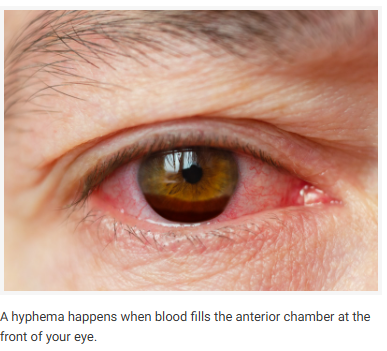
Hyphaema is the presence of blood within the anterior chamber of the eye, the space between the cornea and the iris. This condition is usually the result of trauma but can also occur spontaneously in certain medical conditions. Here is an overview of hyphaema, including its causes, symptoms, diagnosis, treatment options, and potential complications.
Causes of Hyphaema
- Trauma :
- Blunt trauma to the eye is the most common cause, leading to blood vessel rupture.
- Penetrating eye injuries.
- Spontaneous Hyphaema :
- Blood disorders such as haemophilia or sickle cell disease.
- Vascular abnormalities in the eye.
- Neovascularization due to diabetic retinopathy.
- Surgical Procedures :
- Complication of intraocular surgeries.
- Medications :
- Anticoagulants and blood thinners increasing bleeding risk.
It may present as a fine suspension of red blood cells in aqueous (micro-hyphaema) or as a blood-aqueous fluid level (macro-hyphaema)
Symptoms of Hyphaema
- Visible blood in the anterior chamber of the eye.
- Decreased vision or blurry vision.
- Pain in the affected eye.
- Increased sensitivity to light (photophobia).
- Redness of the eye.
Diagnosis of Hyphaema
- Clinical Examination :
- Detailed eye examination by an ophthalmologist.
- Assessment of visual acuity and intraocular pressure.
- Slit-Lamp Examination :
- Allows detailed inspection of the anterior chamber and detection of blood.
- Imaging Studies :
- Ultrasound B-scan to assess the posterior segment if the view is obscured.
- Laboratory Tests :
- Blood tests to check for underlying disorders (e.g., clotting disorders).
Treatment Options for Hyphaema
- Conservative Management :
- Bed rest with head elevation to facilitate blood settling.
- Protective eye shield to prevent further injury.
- Avoiding activities that could increase intraocular pressure (e.g., bending, lifting).
- Medications :
- Topical corticosteroids to reduce inflammation.
- Topical cycloplegics to reduce pain and photophobia.
- Antifibrinolytic agents to prevent re-bleeding in some cases.
- Surgical Intervention :
- Paracentesis to remove blood from the anterior chamber if intraocular pressure is high or if there is corneal blood staining.
- Anterior chamber washout for severe or non-resolving hyphaemas.
Complications of Hyphaema
- Increased intraocular pressure (secondary glaucoma).
- Staining of the cornea with blood (corneal blood staining).
- Permanent vision loss if not treated promptly and appropriately.
- Re-bleeding, which is more likely within the first few days after the initial injury.
Prevention and Management
- Wearing protective eyewear during activities that could result in eye injury.
- Managing underlying medical conditions that could contribute to spontaneous hyphaema.
- Regular follow-up with an ophthalmologist for individuals at risk.
Summary
Hyphaema is the accumulation of blood in the anterior chamber of the eye, usually due to trauma but sometimes resulting from other medical conditions. Early diagnosis and appropriate treatment are crucial to prevent complications such as increased intraocular pressure and permanent vision loss. Treatment ranges from conservative management to surgical intervention, depending on the severity of the condition. Preventive measures, such as wearing protective eyewear and managing underlying conditions, can help reduce the risk of hyphaema.