Related Subjects:
|Neurological History taking
|Causes of Stroke
|Ischaemic Stroke
|Subarachnoid Haemorrhage
|Small Vessel Disease
|Vascular Dementia
|Capsular and Pontine Warning Syndromes
|Dementias
|CADASIL
|CARASIL
|Cerebral Arterial Perfusion and Clinical Correlates
|Anterior circulation Brain
|Posterior circulation Brain
|Acute Stroke Assessment (ROSIER&NIHSS)
|Carotid Artery dissection
|Vertebral artery dissection
|Acute Stroke Assessment (ROSIER&NIHSS)
|Atrial Fibrillation
|Atrial Myxoma
|Causes of Stroke
|Ischaemic Stroke
|Cancer and Stroke
|Cerebral Venous thrombosis
|Cardioembolic stroke
|CT Basics for Stroke
|Endocarditis and Stroke
|Haemorrhagic Stroke
|Stroke Thrombolysis
|Hyperacute Stroke Care
|AP of the Brain
|Cryptogenic stroke
|Carotid Web
|Anterior / Medial Medullary Infarct (Dejerine Syndrome)
🫀 Internal Carotid Artery (ICA)
- Cervical Segment: In the neck, runs without branches. Closely associated with the sympathetic plexus → disruption can cause Horner’s syndrome.
- Petrous Segment: Enters skull via the foramen lacerum, travels anteriorly.
- Cavernous Segment: Runs within the cavernous sinus alongside CN VI (abducens). Gives off superior hypophyseal arteries to the posterior pituitary.
- Supraclinoid Segment: Pierces dura, then bifurcates into the Anterior Cerebral Artery (ACA) and Middle Cerebral Artery (MCA).
👁️ Ophthalmic Artery
- Gives rise to the central retinal artery, essential for vision (occlusion → amaurosis fugax).
- Also supplies scalp around the eye, frontal sinus, and ethmoidal sinuses.
- Important collateral with branches of the maxillary artery in ICA occlusion.
🔗 Posterior Communicating Artery
- Links anterior and posterior circulations (ICA ↔ PCA).
- Runs close to CN III → aneurysms here can cause a pupil-involving third nerve palsy.
- Also supplies thalamus, hypothalamus, and caudate tail.
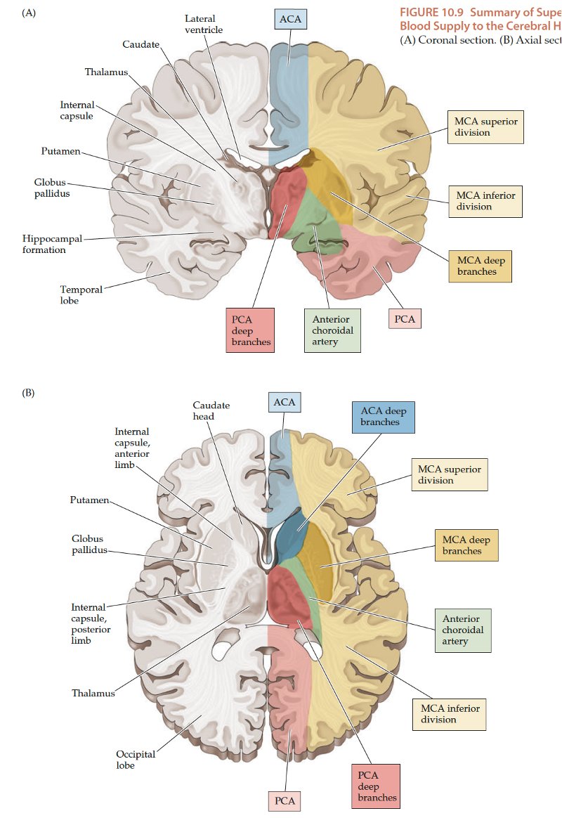
🟡 Anterior Choroidal Artery
- Supplies key deep structures:
- Posterior limb of internal capsule (motor & sensory tracts).
- Choroid plexus of lateral ventricle.
- Optic tract & lateral geniculate body → occlusion may cause contralateral homonymous hemianopia.
- Globus pallidus, substantia nigra, and midbrain regions.
🟥 Anterior Cerebral Artery (ACA)
- Arches around the corpus callosum to supply the medial surfaces of the frontal and parietal lobes → leg weakness is a hallmark of ACA stroke.
ACA – A1 Segment
- From ICA bifurcation to the Anterior Communicating Artery (AComA).
- Medial Lenticulostriates: Supply anterior limb of internal capsule & basal ganglia.
- Anterior Communicating Artery: Joins left & right ACA, common site of berry aneurysms.
- Recurrent Artery of Heubner: Supplies head of caudate & anterior limb of internal capsule.
- Pericallosal Branch: Continuation of ACA, supplies medial cortex & corpus callosum (branches include orbitofrontal, polar frontal, callosomarginal).
🟦 Middle Cerebral Artery (MCA)
- M1 Segment: Horizontal portion, from ICA bifurcation to the insula.
- Occlusion → large hemispheric infarcts.
- Hyperdense MCA sign may be seen on CT.
- Lateral Lenticulostriates: Small penetrating vessels branching at right angles, supplying basal ganglia, posterior limb of internal capsule, and optic radiations (Meyer’s loop).
- Occlusion → lacunar strokes, prone to lipohyalinosis in hypertension.
- M2 Segment: Within Sylvian fissure.
- A thrombus here may give the “dot sign” on CT.
- M3 Segments: Cortical branches divided into:
- Upper Division: Prefrontal, precentral, central, postcentral, parietal (motor & sensory cortex).
- Lower Division: Temporal & occipital gyri, angular gyrus (language functions in dominant hemisphere).
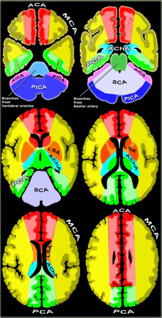
⭕ Circle of Willis
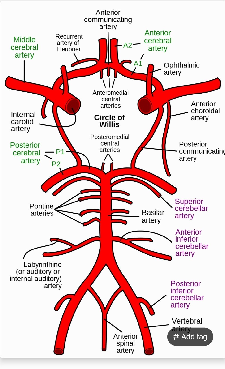
The Circle of Willis provides critical collateral circulation. Aneurysms here account for up to 85% of subarachnoid haemorrhages, most commonly at the junction of the anterior communicating artery, PComA–ICA junction, or MCA bifurcation.
