
| Download the amazing global Makindo app: ✅ Means NICE/National Guidelines 2026 compliant Android | Apple | |
|---|---|
| MEDICAL DISCLAIMER: Educational use only. Not for diagnosis or management. See below for full disclaimer. |
Foreign Body in Eye
Related Subjects: |Episcleritis |Scleritis |Assessing a Red eye |Acute Angle Closure Glaucoma |Allergic and Infective Conjunctivitis |Anterior and Posterior Uveitis |Herpes simplex keratitis (HSK) |Acute Blepharitis |Corneal Abrasion |Foreign Body in Eye
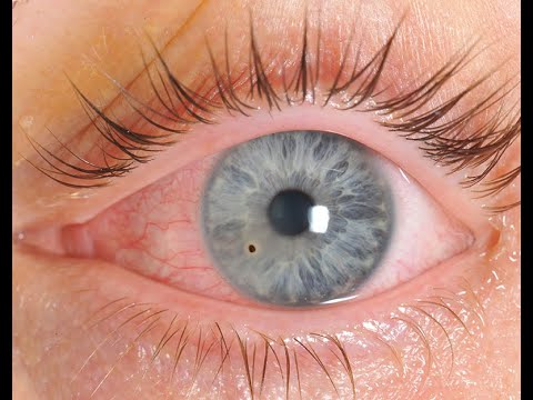
Corneal and conjunctival foreign bodies (FBs) are common ophthalmic presentations, especially in metal workers, DIY enthusiasts, and children. ⚠️ They can cause pain, photophobia, and watering, and if neglected may lead to infection, scarring, and vision loss. Early, careful removal and aftercare are essential.
🛠️ Instruments
- Gloves 🧤, sterile cotton bud, and a 21–25G needle (mainstay).
- Topical anaesthetic (e.g. Amethocaine 1%).
- Slit lamp 🔬 (essential for safe removal).
- Optional: motorised dental burr for metallic FBs (⚠️ never within central 5 mm of cornea).
- ⚙️ Technique: Bend needle tip to 45°; bevel away from eye to reduce trauma.
- Ensure patient’s forehead is firmly against the slit lamp for stability.
📋 Important Notes
- 📉 Visual acuity may initially be reduced - always test with pinhole + topical anaesthetic if discomfort limits accuracy.
- 🌱 Organic FBs (wood, plant matter) → high infection risk.
- 🧲 Metallic FBs → rust rings ⚠️ → scarring if untreated.
- Rust rings in the visual axis must be removed by ophthalmology only.
- Always consider the possibility of an open globe injury - if suspected → shield, nil by mouth, urgent ophthalmology referral.
🔧 Procedure
- 1️⃣ Instil topical anaesthetic (e.g., Amethocaine 1%), repeat every 30s until painless.
- 2️⃣ Seat patient at slit lamp; stabilise head; ask them to fixate on target 🎯.
- 3️⃣ Focus slit lamp beam - use narrow oblique beam at ~45° to assess FB depth.
- 4️⃣ Approach tangentially with cotton bud or bent needle; lift FB away.
- 5️⃣ For superficial metallic FB → gently “flick” with bevel; burr for rust ring (if outside visual axis).
🩺 Post-Procedure Care
- 💊 Topical antibiotic drops (e.g., chloramphenicol qid for 5–7 days).
- 🌙 Cycloplegic drops (e.g., cyclopentolate 1% bd) for pain relief.
- 💊 Oral analgesia (paracetamol, NSAIDs).
- ❌ Avoid eye padding - binocular vision prevents accidents and padding may ↑ infection risk.
- 🚫 No repeat use of anaesthetic drops → toxic to cornea.
- 📆 Follow-up daily with slit lamp until epithelial defect heals (document size + healing progress).
🚩 Red Flags
- ⚠️ Suspected penetrating eye injury (positive Seidel’s test, irregular pupil, iris prolapse).
- FB in central visual axis or deep stromal cornea.
- Persistent rust ring or residual opacity.
- Infective keratitis → pain ↑, photophobia, hypopyon.
- Reduced vision not improving after removal.
👶 Children
- Often difficult to examine; consider sedation or referral if uncooperative.
- Risk of recurrent rubbing and re-injury → protective shield + parental advice.
- Organic FBs (wood, playground material) more common.
🧓 Elderly
- Assess anticoagulation & bleeding risk 💊.
- May present late due to reduced corneal sensation or confusion.
- Healing may be slower; ensure close ophthalmology follow-up.
📌 Teaching Pearls
- Always document visual acuity before and after any intervention.
- Slit lamp exam is gold standard for safe removal.
- Never attempt deep or central FB removal outside specialist care.
- Discharge advice: return immediately if ↑ pain, redness, discharge, or ↓ vision.
🧾 Clinical Case Examples – Foreign Body in the Eye
A 34-year-old construction worker presents with sudden onset pain, watering, and photophobia in the right eye after drilling metal. On exam, visual acuity is intact, and a small metallic speck is seen on the cornea. 👉 Likely diagnosis: Superficial corneal foreign body (metallic). 👉 Management: Topical anaesthetic, fluorescein staining, removal with cotton bud/needle, tetanus check, topical antibiotic drops.Case 2 – Organic Material 🌾 A 19-year-old farmer reports foreign body sensation and tearing after cutting hay. On slit-lamp exam, a small plant fragment is lodged under the upper eyelid. 👉 Concern: Vegetative matter foreign body (↑ risk fungal keratitis). 👉 Management: Lid eversion and removal, topical antibiotic cover, warn about red flags (increasing pain, vision change), urgent ophthalmology review if keratitis suspected.
Case 3 – Contact Lens Wearer 👁️ A 26-year-old woman wearing contact lenses complains of painful red eye and foreign body sensation after a night out. No visible foreign body but corneal staining with fluorescein shows an epithelial defect. 👉 Likely diagnosis: Contact lens–related corneal abrasion ± retained foreign body. 👉 Management: Urgent removal of lens, ciprofloxacin drops (anti-pseudomonal), ophthalmology referral if worsening.
Case 4 – Penetrating Injury 🚨 A 40-year-old man using a hammer develops sudden severe eye pain and blurred vision. He cannot open his eye. Exam: subconjunctival haemorrhage, peaked pupil, and reduced visual acuity. 👉 Concern: Penetrating globe injury with intraocular foreign body. 👉 Management: Do NOT manipulate. Rigid eye shield, nil by mouth, urgent ophthalmology referral for surgical exploration.
Case 5 – Child with Grit 🧒 A 6-year-old boy is brought by his mother after playing in a sandpit. He is rubbing his left eye, which is red and watering. VA normal for age, small particle found under lower lid. 👉 Likely diagnosis: Superficial eyelid foreign body. 👉 Management: Lid eversion, irrigation with saline, discharge with advice on when to return (worsening pain, blurred vision).
| The content on this website is provided for educational and informational purposes only to support exam preparation (e.g., MLA, MRCP, USMLE) and learning. This is NOT medical advice, diagnosis, treatment, or professional guidance. It does not replace consultation with a qualified healthcare professional, official guidelines (e.g., NICE, GMC, BNF), or supervised clinical practice. Always verify information with current, authoritative sources. Makindo and its contributors accept no liability for any reliance on this content, including errors, omissions, or any resulting harm, loss, or consequences. By using this site, you agree to these terms. |
|
|
Categories
- About
- Acute Medicine
- Anaesthetics and Critical Care
- Anatomy
- Anatomy and Physiology
- Biochemistry
- Book
- Cardiology
- Collections
- CompSci
- Crib Sheets
- Critical care
- Dental
- Dermatology
- Differentials
- Drugs
- ENT
- Electrocardiogram
- Embryology
- Emergency Medicine
- Endocrinology
- Ethics
- Foundation Doctors
- GCSE
- Gastroenterology
- General Practice
- Genetics
- Geriatric Medicine
- Geriatrics
- Guidelines
- Haematology
- Hepatology
- Immunology
- Infectious Diseases
- Infographic
- Investigations
- Lists
- MRCP
- Mandatory Training
- Medical Students
- Microbiology
- Nephrology
- Neurology
- Neurosurgery
- Nutrition
- OSCE
- Obstetrics Gynaecology
- Oncology
- Ophthalmology
- Oral Medicine and Dentistry
- Orthopaedics
- Paediatrics
- Palliative
- Palliative Care
- Pathology
- Pharmacology
- Physiology
- Procedures
- Psychiatry
- Public Health
- Radiology
- Respiratory
- Resuscitation
- Revision Topics
- Rheumatology
- Statistics and Research
- Stroke
- Surgery
- Toxicology
- Trauma and Orthopaedics
- USMLE
- Urology
- Vascular Surgery