
| Download the amazing global Makindo app: ✅ Means NICE/National Guidelines 2026 compliant Android | Apple | |
|---|---|
| MEDICAL DISCLAIMER: Educational use only. Not for diagnosis or management. See below for full disclaimer. |
Cavernous angiomas (Cavernomas)
Related Subjects: |Subarachnoid Haemorrhage |Haemorrhagic stroke
🧠 Introduction
- Cerebral cavernous malformations (CCMs) are the most common vascular malformations of the CNS.
- They are clusters of abnormal, thin-walled blood vessels that may bleed, sometimes extending beyond their fragile walls.
- Prevalence: ~0.1–0.5% of the population; some have a genetic basis.
- More common in Hispanic communities, especially Mexican-Americans.
🔬 Pathophysiology
- CCMs lack large feeding arteries or draining veins (“angiographically occult”).
- Can occur anywhere in the brain or spinal cord; deeper lesions (e.g. brainstem) bleed more often than superficial ones.
- Genetic variants:
- CCM1 → chromosome 7q.
- CCM2 → chromosome 7p.
- CCM3 → chromosome 3p (often more severe, early onset).
🩺 Clinical Presentation
- Many are asymptomatic, found incidentally on MRI.
- When symptomatic → intracerebral haemorrhage, seizures, focal deficits, raised ICP, headaches, or hydrocephalus.
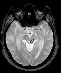
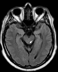
📍 Cavernomas in the Midbrain
Brainstem cavernomas are particularly high risk due to eloquent location and frequent haemorrhage-related morbidity.
🧾 Investigations
- CT/MRI: Characteristic “raspberry” or “popcorn-like” appearance, with or without haemorrhage.
- MRI SWI/GRE: Best for detecting blood products (haemosiderin). Highly sensitive.
- Angiography: Typically negative (lesions are angiographically occult).
📊 MRI Types (Zabramski classification)
- 🟥 Type I: Subacute haemorrhage → hyperintense core on T1/T2.
- 🟧 Type II: “Popcorn” lesion → mixed signals on T1/T2 with hypointense rim.
- 🟨 Type III: Chronic/resolved haemorrhage → isointense core, often familial.
- 🟩 Type IV: Tiny foci, only visible on GRE/SWI, may mimic capillary telangiectasias.
⚖️ Management
- Options: Conservative, microsurgical resection, or stereotactic radiosurgery.
- Surgery indicated: Recurrent haemorrhage, intractable seizures, or progressive neurological deficit (esp. non-eloquent regions).
- Brainstem cavernomas → controversial but some evidence supports resection due to high morbidity if left untreated.
- Conservative management: Annual MRI follow-up if asymptomatic or low-risk.
📖 References
💡 Exam Pearl: Cavernomas are angiographically occult. Think “popcorn lesion” on MRI → consider cavernoma.
| The content on this website is provided for educational and informational purposes only to support exam preparation (e.g., MLA, MRCP, USMLE) and learning. This is NOT medical advice, diagnosis, treatment, or professional guidance. It does not replace consultation with a qualified healthcare professional, official guidelines (e.g., NICE, GMC, BNF), or supervised clinical practice. Always verify information with current, authoritative sources. Makindo and its contributors accept no liability for any reliance on this content, including errors, omissions, or any resulting harm, loss, or consequences. By using this site, you agree to these terms. |
|
|
Categories
- About
- Acute Medicine
- Anaesthetics and Critical Care
- Anatomy
- Anatomy and Physiology
- Biochemistry
- Book
- Cardiology
- Collections
- CompSci
- Crib Sheets
- Critical care
- Dental
- Dermatology
- Differentials
- Drugs
- ENT
- Electrocardiogram
- Embryology
- Emergency Medicine
- Endocrinology
- Ethics
- Foundation Doctors
- GCSE
- Gastroenterology
- General Practice
- Genetics
- Geriatric Medicine
- Geriatrics
- Guidelines
- Haematology
- Hepatology
- Immunology
- Infectious Diseases
- Infographic
- Investigations
- Lists
- MRCP
- Mandatory Training
- Medical Students
- Microbiology
- Nephrology
- Neurology
- Neurosurgery
- Nutrition
- OSCE
- Obstetrics Gynaecology
- Oncology
- Ophthalmology
- Oral Medicine and Dentistry
- Orthopaedics
- Paediatrics
- Palliative
- Palliative Care
- Pathology
- Pharmacology
- Physiology
- Procedures
- Psychiatry
- Public Health
- Radiology
- Respiratory
- Resuscitation
- Revision Topics
- Rheumatology
- Statistics and Research
- Stroke
- Surgery
- Toxicology
- Trauma and Orthopaedics
- USMLE
- Urology
- Vascular Surgery