Related Subjects:
|ECG Basics
|ECG Axis
|ECG Analysis
|ECG LAD
|ECG RAD
|ECG Low voltage
|ECG Pathological Q waves
|ECG ST/T wave changes
|ECG LBBB
|ECG RBBB
|ECG short PR
|ECG Heart Block
|ECG Asystole and P wave asystole
|ECG QRS complex
|ECG ST segment
|ECG: QT interval
|ECG: LVH
|ECG RVH
|ECG: Bundle branch blocks
|ECG Dominant R wave in V1
|ECG Acute Coronary Syndrome
|ECG Narrow complex tachycardia
|ECG Ventricular fibrillation
|ECG Regular Broad complex tachycardia
|ECG Crib sheets
⚡ Key Point: Although a QT interval ≥500 ms is strongly associated with a higher risk of torsades de pointes, there is no absolute "safe" lower threshold. Even modest QT prolongation can be proarrhythmic, especially when combined with bradycardia, electrolyte imbalance, or QT-prolonging drugs.
📖 About
🩺 ECG – QT Interval
- Prolonged QT reflects delayed ventricular repolarisation.
- Abnormal QTc: >450 ms in men and >460 ms in women.
- Excessive prolongation predisposes to Torsades de pointes (TdP) - a polymorphic ventricular tachycardia that may degenerate into ventricular fibrillation.
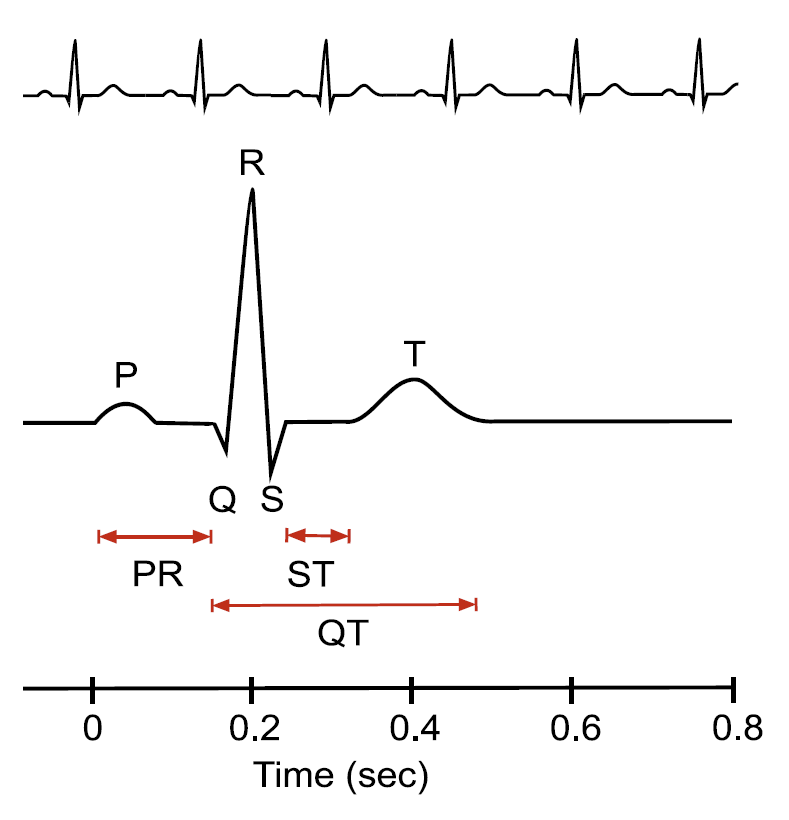
📏 Measuring the QT
- Measure manually in the limb lead showing the clearest T-wave termination.
- From the start of the QRS to the end of the T wave; average across 3–5 beats.
- U waves should be included only if they merge with the T wave.
- In atrial fibrillation, measure several beats with similar RR intervals.
- Preferably assess during peak drug plasma levels when evaluating medication effect.
- Always adjust for heart rate before interpreting QT values.
⚠️ Causes of Long QT
| Category | Examples |
|---|
| Genetic |
- Jervell–Lange Nielsen syndrome – autosomal recessive, associated with congenital deafness 🎧
- Romano–Ward syndrome – autosomal dominant form
|
| Electrolyte Disturbance |
- Hypokalaemia 🧂
- Hypomagnesaemia
- Hypocalcaemia
|
| Drugs |
- Macrolides (e.g. IV erythromycin, clarithromycin)
- Antihistamines (terfenadine, astemizole)
- Antifungals (ketoconazole)
- Antipsychotics (haloperidol, phenothiazines)
- TCAs, lithium, cisapride, halofantrine, amiodarone
- Grapefruit juice (CYP3A4 inhibition 🍊)
|
| Miscellaneous |
- Acute myocardial infarction
- Bradycardia, starvation, myocarditis
- Hypertrophic cardiomyopathy
- Right ventricular dysplasia
- Hypothermia 🧊
|
⬇️ Causes of Short QT
- Hypercalcaemia or hyperkalaemia ⚡
- Digoxin effect (“scooped” ST depression)
- β-blockers, phenytoin, or congenital short-QT syndromes
🧠 Teaching Commentary
QT prolongation represents disordered repolarisation across the ventricular wall. The mid-myocardial “M cells” are especially prone to delayed repolarisation, creating dispersion of recovery times and enabling early after-depolarisations - the substrate for torsades.
Always review drugs, electrolytes, and heart rate before attributing it to a congenital cause. Even mild prolongation in a polypharmacy elderly patient can be dangerous, particularly if combined with bradycardia or hypokalaemia.
🔗 Reference
