
| Download the amazing global Makindo app: ✅ Means NICE/National Guidelines 2026 compliant Android | Apple | |
|---|---|
| MEDICAL DISCLAIMER: Educational use only. Not for diagnosis or management. See below for full disclaimer. |
Acute Renal / Ureteric Colic and Urinary Tract Stones ✅
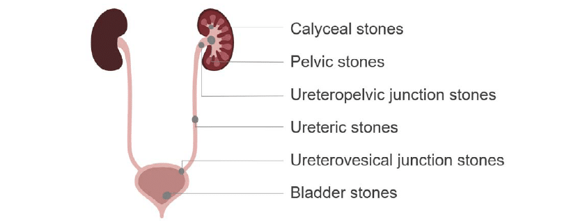
Related Subjects: |Renal Transplantation |Urinary Tract Obstruction |Bladder Stones
Urinary tract stones (urolithiasis) are crystalline deposits that form in the kidneys and may migrate to the ureters, causing obstruction and renal colic. The major complication not to miss is an infected obstructed kidney (stone + infection). If there is fever/systemic illness with obstruction suspicion get early imaging by CT/USS and treat as urgent and escalate for drainage.
- Stones <5 mm (especially distal ureteric): High likelihood (>80–90%) of spontaneous passage within 4–6 weeks with supportive care.
- Stones 5–10 mm (especially distal): Consider medical expulsive therapy (MET) with an alpha-blocker (e.g., tamsulosin 400 micrograms once daily - off-label use) to facilitate passage.
- Stones >10 mm or proximal/mid-ureteric: Lower spontaneous passage rate; often require urology referral for intervention (e.g., within 48 hours if pain uncontrolled or obstruction persists).
- Urgent intervention needed for complicated cases (sepsis, AKI, solitary kidney).
📖 About
- Renal/ureteric colic: Sudden-onset, severe, intermittent colicky pain from ureteric obstruction, typically radiating from loin/flank to groin, scrotum/labia, or inner thigh.
- Pain often causes extreme restlessness (“cannot keep still”), unlike peritonism (where patients lie still).
- Red flags - always exclude: ruptured AAA, appendicitis, ectopic pregnancy, ovarian torsion, obstructed solitary/transplant kidney, urosepsis.

Typical radiation pattern of renal/ureteric colic pain (loin to groin).
📊 Epidemiology
- Annual incidence: ~1–2 per 1000 population; lifetime risk 10–15% (higher in men and certain regions/climates).
- Peak age: 20–60 years; male:female ratio ~2–3:1 (though gap narrowing).
- Recurrence rate: 30–50% within 5–10 years without prevention.
🧬 Pathophysiology of Pain
- Ureteric obstruction → increased intraluminal pressure → distension of renal capsule and ureteric smooth muscle.
- Triggers prostaglandin release → smooth muscle spasm and intense colicky pain.
- Associated nausea/vomiting from vagal/GI stimulation; ileus possible.
- Prolonged obstruction → hydronephrosis → potential renal impairment.
⚠️ Differential Diagnosis / Causes of Obstruction
- Most common: urinary tract calculi.
- Other: sloughed papilla (e.g., papillary necrosis in diabetes, sickle cell, analgesics), blood clot (trauma/tumour), stricture/PUJ obstruction, external compression (e.g., tumours), rare foreign bodies.
🧪 Stone Composition (Approximate %)
- 🧂 Calcium-based (oxalate/phosphate): 70–80% - linked to hypercalciuria, hyperoxaluria, hyperparathyroidism, hypocitraturia.
- 🦠 Struvite (magnesium ammonium phosphate): 10–15% - infection-related (urease-producing bacteria e.g., Proteus, Klebsiella), alkaline urine, often staghorn.
- 🧬 Cystine: 1–2% - genetic cystinuria; recurrent, often bilateral/large.
- 💡 Uric acid: 5–10% - hyperuricosuria (gout, high purine diet, tumour lysis), acidic urine.
- Rare: xanthine, drug-induced (e.g., indinavir, atazanavir).
Not all, but recurrent stones, young age, or atypical features often justify metabolic evaluation and prevention counselling per NG118 pathways.
🎯 Risk Factors
- Low fluid intake / dehydration / hot climates.
- Family history / genetic predisposition.
- Metabolic: primary hyperparathyroidism, gout, obesity, diabetes.
- GI: inflammatory bowel disease (Crohn’s), ileal resection, chronic diarrhoea (enteric hyperoxaluria).
- Drugs: topiramate, acetazolamide, loop diuretics, protease inhibitors, vitamin C/D excess.
- Dietary: high salt, high animal protein, low citrate (low fruit), high oxalate (spinach, nuts), sugary drinks.
🩺 Clinical Features
- Severe colicky flank pain radiating to groin/genitalia; episodic every few minutes.
- Restlessness, diaphoresis; nausea/vomiting common (50–80%).
- Haematuria (micro/macro) in ~80–90%.
- LUTS: frequency, urgency, dysuria if distal stone.
- Red flags: fever/rigors, hypotension, AKI, anuria/oliguria → urgent admission.
🔎 Investigations (NICE NG118 Aligned)
- Bloods: FBC, U&E (check AKI), CRP, calcium, phosphate, urate; consider PTH if hypercalcaemia.
- Urinalysis: dipstick + MC&S; haematuria common; infection markers if febrile.
- Imaging:
- Low-dose non-contrast CT KUB: gold standard (>95% sensitivity/specificity); urgent (within 24 hours) for suspected renal colic in adults.
- Ultrasound: first-line in pregnancy, children/young people (assess hydronephrosis); consider CT if diagnostic uncertainty.
- Plain KUB X-ray: limited (misses uric acid/cystine); useful for radio-opaque stone follow-up.
- Stone analysis: mandatory for all passed/removed stones.
- Metabolic evaluation (recurrent stones): 24-hour urine (volume, pH, Ca, oxalate, citrate, urate, etc.); consider in first-time stone formers with risk factors.
Example low-dose CT KUB: calcified stone in ureter with proximal hydronephrosis (dilated collecting system).
🚑 Indications for Admission / Urgent Urology Review (NICE-aligned)
- Suspected/confirmed urosepsis (fever, rigors, hypotension, elevated CRP/WBC).
- Uncontrolled pain despite analgesia or intractable vomiting.
- Solitary kidney, bilateral obstruction, or transplanted kidney.
- Acute kidney injury / rising creatinine.
- Diagnostic uncertainty (e.g., possible AAA, appendicitis).
- Stone >10 mm with ongoing symptoms or proximal location.
💊 Acute Management (NICE NG118 / CKS)
- Pain relief: NSAID first-line (e.g., diclofenac 75 mg IM/IV/PR/PO); any route effective.
- If NSAIDs contraindicated/not enough: IV paracetamol.
- Opioids (e.g., morphine) only if above fail; avoid routinely.
- Do not use antispasmodics.
- IV fluids if dehydrated/vomiting (monitor for overload).
- Antiemetics: e.g., ondansetron 4–8 mg IV, cyclizine, metoclopramide.
- MET: Consider alpha-blocker (e.g., tamsulosin 400 μg OD) for distal ureteric stones <10 mm (off-label; discuss risks/benefits).
- Watchful waiting: Appropriate for small stones likely to pass; monitor symptoms, renal function, repeat imaging if needed.
- Urgent intervention: Sepsis/obstruction → percutaneous nephrostomy or ureteric stenting; then definitive treatment.
🔨 Definitive Stone Management (NICE NG118)
- Extracorporeal shockwave lithotripsy (SWL): Non-invasive; good for <20 mm renal/upper ureteric stones.
- Ureteroscopy & laser lithotripsy (URS): Preferred for ureteric stones; high success.
- Percutaneous nephrolithotomy (PCNL): For large renal stones (>20 mm) or staghorn calculi.
- Open/laparoscopic: Very rare.
- Timing: Offer intervention within 48 hours if pain ongoing/not tolerated or stone unlikely to pass.
🛡️ Prevention & Long-term Management (NICE NG118)
- General advice (all stone types): Fluid intake to achieve urine output >2–2.5 L/day; limit salt (<6 g/day) and animal protein; normal dietary calcium; increase citrus fruits (↑ citrate).
- Avoid excess vitamin C, sugary drinks; maintain healthy weight.
- Stone-specific:
- Calcium oxalate: potassium citrate if hypocitraturia; thiazides if hypercalciuria + low-salt diet.
- Uric acid: allopurinol if hyperuricosuria; urinary alkalinisation (potassium citrate/sodium bicarbonate).
- Struvite: treat/prevent UTIs; acetohydroxamic acid rare.
- Cystine: high fluid + chelating agents (e.g., tiopronin) if needed.
- Manage underlying conditions (e.g., parathyroidectomy for hyperparathyroidism).
- Follow-up: imaging/biochemistry for recurrent stone formers.
📚 Key References
- NICE NG118: Renal and ureteric stones: assessment and management (Published 8 January 2019; last reviewed 26 February 2021 – current as of 2026)
- NICE CKS: Renal or ureteric colic - acute (Last revised October 2024)
- European Association of Urology (EAU) Urolithiasis Guidelines (updated annually)
- Campbell-Walsh Urology (latest edition)