
🔬 The Gram stain, developed by Hans Christian Gram (1882), is one of the most widely used microbiological techniques. It rapidly differentiates bacteria into Gram-positive 🟣 or Gram-negative 🔴 groups and guides early antibiotic therapy.
📖 About
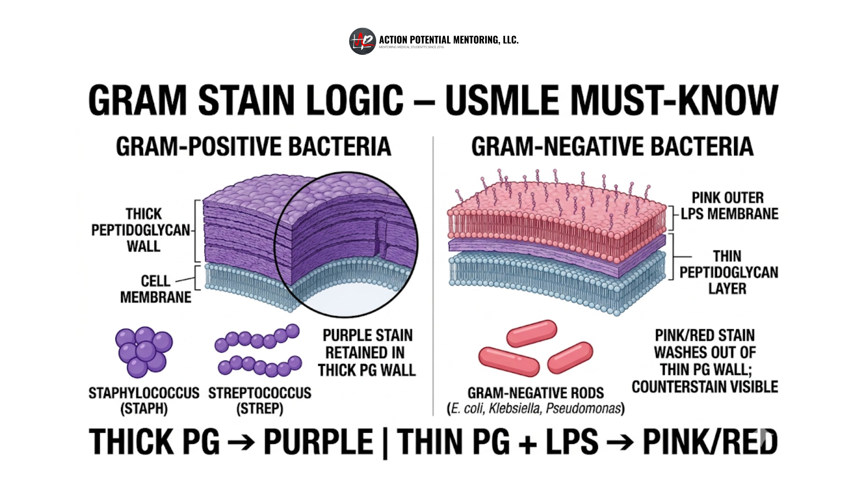
- Classifies bacteria based on their cell wall structure and ability to retain stains.
- Gram-positive: Thick peptidoglycan wall retains crystal violet → purple/violet under the microscope.
- Gram-negative: Thin peptidoglycan wall + outer membrane → loses crystal violet, takes up safranine → pink/red.
- Rapid, cheap, and used worldwide in microbiology labs and clinical settings.
⚙️ Mechanism of Differentiation
- Gram-positive 🟣: Thick peptidoglycan traps the crystal violet–iodine complex even after alcohol wash (e.g., Staphylococcus aureus, Streptococcus pneumoniae).
- Gram-negative 🔴: Alcohol dissolves outer membrane; thin peptidoglycan cannot retain the primary stain, so they take up the counterstain (e.g., E. coli, Neisseria gonorrhoeae).
🧪 Steps in the Gram Stain
- Crystal Violet: Primary stain colours all bacteria violet.
- Iodine: Acts as a mordant, forming a complex with crystal violet inside cells.
- Alcohol/Acetone Wash: Decolourises Gram-negative but not Gram-positive bacteria.
- Safranine: Counterstain gives Gram-negatives a pink/red appearance.
📊 Gram-Positive vs Gram-Negative Bacteria
| Feature | Gram-Positive 🟣 | Gram-Negative 🔴 |
|---|---|---|
| Cell wall | Thick peptidoglycan | Thin peptidoglycan |
| Outer membrane | ❌ Absent | ✅ Present (with lipopolysaccharide endotoxin) |
| Teichoic acids | ✅ Present | ❌ Absent |
| Gram stain colour | Purple/Violet | Pink/Red |
| Examples | Staphylococcus aureus, Streptococcus pneumoniae | E. coli, Pseudomonas aeruginosa, Neisseria gonorrhoeae |
| Antibiotic relevance | Often more susceptible to β-lactams & glycopeptides (e.g., vancomycin) | Outer membrane reduces permeability → may need broader-spectrum agents (e.g., piperacillin-tazobactam) |
💡 Clinical Applications
- Respiratory Infections: Gram-positive S. pneumoniae common in pneumonia.
- Urinary Tract Infections: Gram-negative E. coli most frequent cause.
- Sepsis: Gram-negative organisms like Pseudomonas often drive severe infections.
⚠️ Limitations
- Some bacteria (e.g., Mycobacterium tuberculosis) don’t stain well due to waxy cell walls → require special stains (acid-fast, Ziehl-Neelsen).
- A Gram stain gives a rapid “first impression” but must be confirmed with culture & sensitivity.