
| Download the amazing global Makindo app: ✅ Means NICE/National Guidelines 2026 compliant Android | Apple | |
|---|---|
| MEDICAL DISCLAIMER: Educational use only. Not for diagnosis or management. See below for full disclaimer. |
Abdominal X-Ray (AXR)
📸 Introduction
- An Abdominal X-ray (AXR) is a first-line imaging test used in the evaluation of suspected bowel and urological disease.
- It is quick, inexpensive, and widely available, but sensitivity and specificity are lower than CT. AXR is often a screening or triage tool in the acute setting.
📋 Indications (Suspected Pathology)
- 👉 Large and small bowel obstruction (including volvulus).
- 👉 Perforation of an abdominal viscus (though erect CXR is more sensitive for free air).
- 👉 Constipation – faecal loading assessment.
- 👉 Renal/ureteric calculi (though CT KUB is now gold standard).
- 👉 Foreign bodies (swallowed, inserted, or penetrating).
🛠️ Different Types of AXR Views
- Supine AP: Standard view, patient on their back – best for obstruction, stones, faecal loading.
- Erect AXR: Patient upright; shows air-fluid levels in obstruction and can help detect perforation.
- Decubitus: Patient lies on side; gas-fluid levels rise and can help confirm free air if erect film not possible.
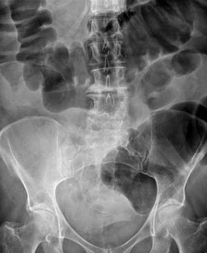
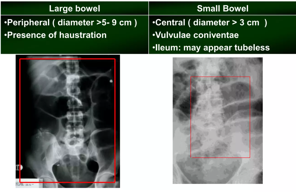
✅ Normal Abdominal X-ray
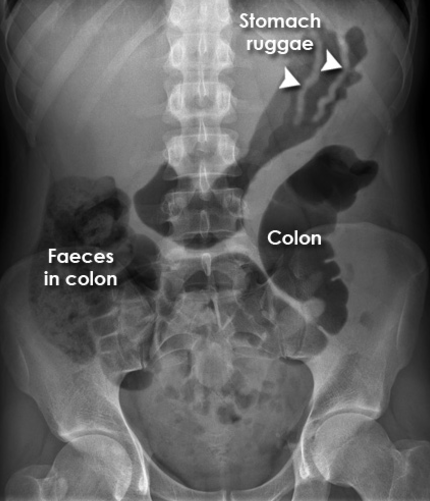
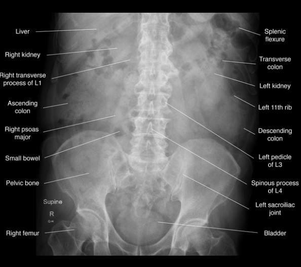
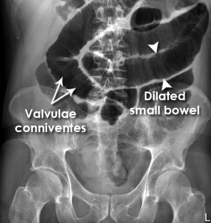
- Small bowel: Central, valvulae conniventes (lines crossing entire width), diameter < 3 cm.
- Large bowel: Peripheral, haustra (do not cross whole lumen), diameter < 6 cm (caecum < 9 cm).
- Stomach: Left upper quadrant gas bubble is normal.
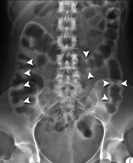
🚨 Large Bowel Obstruction (LBO)
- Grossly dilated colon (>6 cm).
- Haustra visible but stretched and thickened.
- Common causes: colorectal cancer, volvulus, strictures (e.g., diverticular disease).
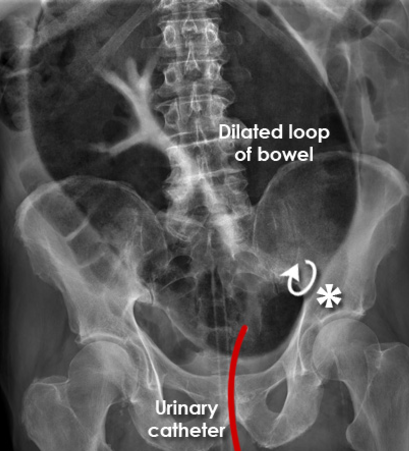
🚨 Small Bowel Obstruction (SBO)
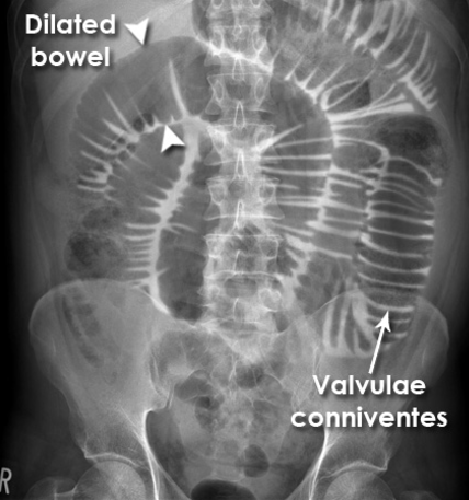
- Central dilated loops >3 cm.
- “Stack of coins” / “step-ladder” appearance with multiple air-fluid levels on erect film.
- Causes: adhesions (most common), hernia, Crohn’s disease.
💥 Perforation
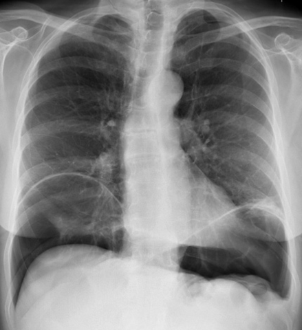
- Free intraperitoneal gas, best seen under diaphragm (erect CXR more sensitive).
- Look for Rigler’s sign (gas outlining both sides of bowel wall).
- Urgent surgical referral needed.
🧩 Ulcerative Colitis – “Thumb Printing”
- Mucosal oedema causes thickened haustral folds → “thumb-print” appearance.
- Seen in severe colitis (IBD or infective).
- Beware of toxic megacolon if gross dilatation present.
🌀 Sigmoid Volvulus
- Massively dilated loop, often extending from pelvis to diaphragm.
- Classic “coffee bean” or “omega loop” sign.
- Common in elderly, institutionalised, or neuropsychiatric patients.
🌀 Caecal Volvulus
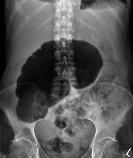
- Dilated caecum, often displaced into left upper quadrant.
- More acute and surgical than sigmoid volvulus.
🔧 Post-Operative Ileus
- Diffuse gaseous distension of small and large bowel.
- No clear transition point (contrast with SBO).
- Common post-op, due to inflammation, analgesia (opioids), or electrolyte imbalance.
📚 Clinical Pearls
- Always assess: gas pattern, bowel size, air-fluid levels, free air, calcifications, soft tissue masses.
- Compare with clinical context – AXR alone rarely definitive.
- In suspected obstruction/perforation, CT abdomen is gold standard for diagnosis and surgical planning.
| The content on this website is provided for educational and informational purposes only to support exam preparation (e.g., MLA, MRCP, USMLE) and learning. This is NOT medical advice, diagnosis, treatment, or professional guidance. It does not replace consultation with a qualified healthcare professional, official guidelines (e.g., NICE, GMC, BNF), or supervised clinical practice. Always verify information with current, authoritative sources. Makindo and its contributors accept no liability for any reliance on this content, including errors, omissions, or any resulting harm, loss, or consequences. By using this site, you agree to these terms. |
|
|
Categories
- About
- Acute Medicine
- Anaesthetics and Critical Care
- Anatomy
- Anatomy and Physiology
- Biochemistry
- Book
- Cardiology
- Collections
- CompSci
- Crib Sheets
- Critical care
- Dental
- Dermatology
- Differentials
- Drugs
- ENT
- Electrocardiogram
- Embryology
- Emergency Medicine
- Endocrinology
- Ethics
- Foundation Doctors
- GCSE
- Gastroenterology
- General Practice
- Genetics
- Geriatric Medicine
- Geriatrics
- Guidelines
- Haematology
- Hepatology
- Immunology
- Infectious Diseases
- Infographic
- Investigations
- Lists
- MRCP
- Mandatory Training
- Medical Students
- Microbiology
- Nephrology
- Neurology
- Neurosurgery
- Nutrition
- OSCE
- Obstetrics Gynaecology
- Oncology
- Ophthalmology
- Oral Medicine and Dentistry
- Orthopaedics
- Paediatrics
- Palliative
- Palliative Care
- Pathology
- Pharmacology
- Physiology
- Procedures
- Psychiatry
- Public Health
- Radiology
- Respiratory
- Resuscitation
- Revision Topics
- Rheumatology
- Statistics and Research
- Stroke
- Surgery
- Toxicology
- Trauma and Orthopaedics
- USMLE
- Urology
- Vascular Surgery