
| Download the amazing global Makindo app: ✅ Means NICE/National Guidelines 2026 compliant Android | Apple | |
|---|---|
| MEDICAL DISCLAIMER: Educational use only. Not for diagnosis or management. See below for full disclaimer. |
Toxic Epidermal Necrolysis (TEN) and Stevens-Johnson syndrome
Related Subjects: |Cellulitis |Pyoderma gangrenosum |Pemphigus Vulgaris |Toxic Epidermal Necrolysis |Stevens-Johnson Syndrome |Necrotising fasciitis |Gas Gangrene (Clostridium perfringens) |Purpura Fulminans |Severe burns |Anatomy of Skin |Skin Pathology and Lesions |Skin, Soft Tissue & Bone Infections
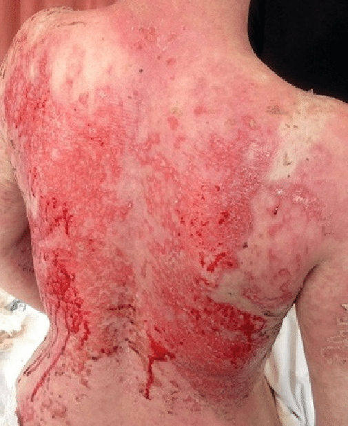
⚠️ Toxic epidermal necrolysis (TEN) is a rare, life-threatening dermatological emergency at the most severe end of the Stevens–Johnson syndrome–toxic epidermal necrolysis spectrum. It is usually a severe drug-induced immune reaction after exposure in which cytotoxic lymphocytes cause widespread keratinocyte death. This produces full-thickness epidermal necrosis, skin detachment and severe mucosal inflammation.
📌 About
- SJS: less than 10% body surface area affected by detached or detachable epidermis.
- SJS–TEN overlap: 10–30% epidermal detachment.
- TEN: more than 30% epidermal detachment.
The body surface area classification refers to detached or detachable epidermis, not the total area of erythematous rash. Extensive epidermal loss produces acute skin failure resembling a major burn. Complications include fluid loss, hypothermia, infection, sepsis, respiratory involvement and multi-organ failure.
🧬 Pathophysiology
SJS and TEN are severe type IV cytotoxic hypersensitivity reactions. A drug or drug metabolite interacts with the immune system in a genetically susceptible patient, activating drug-specific CD8-positive T lymphocytes and natural-killer cells.
- Cytotoxic cells release granulysin, perforin and granzyme B.
- Fas–Fas ligand and tumour necrosis factor pathways may also contribute.
- These mediators cause widespread apoptosis and necrosis of epidermal keratinocytes.
- Full-thickness epidermal necrosis causes separation of the epidermis from the dermis.
- This differs from pemphigus, in which antibodies directly disrupt adhesion between keratinocytes.
🧾 Aetiology
- Most adult cases are caused by medication.
- The reaction usually begins within 4–28 days of starting the causative drug, although it may occur sooner following re-exposure.
- Infections are more important triggers in children than in adults.
- HIV infection, malignancy and immunosuppression are associated with an increased risk.
- Genetic susceptibility is drug- and ancestry-specific, including HLA-B*58:01 with allopurinol and HLA-B*15:02 with carbamazepine in susceptible populations.
💊 Important Culprit Drugs
- Allopurinol
- Co-trimoxazole and other sulphonamide antibiotics
- Carbamazepine
- Lamotrigine
- Phenytoin
- Phenobarbital
- Nevirapine
- Oxicam NSAIDs, such as piroxicam and meloxicam
- Other antibiotics, anticonvulsants, antiretroviral drugs and anticancer treatments have also been implicated.
A detailed drug history should include all prescribed medicines, over-the-counter preparations, herbal treatments and medications stopped during the preceding eight weeks.
🤒 Clinical Features
- Prodrome of fever, malaise, headache, sore throat, cough or myalgia.
- Skin pain and tenderness are prominent and may precede obvious blistering.
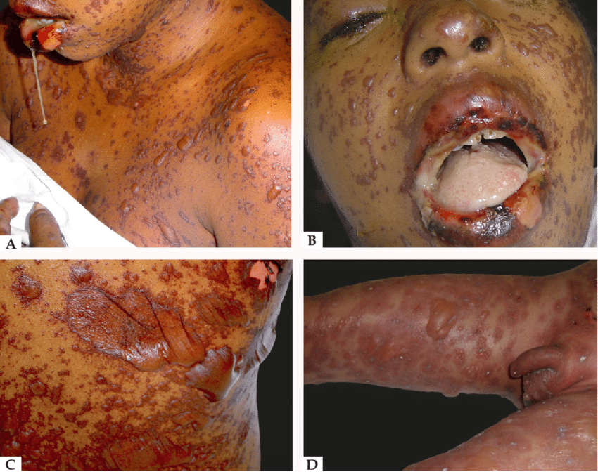
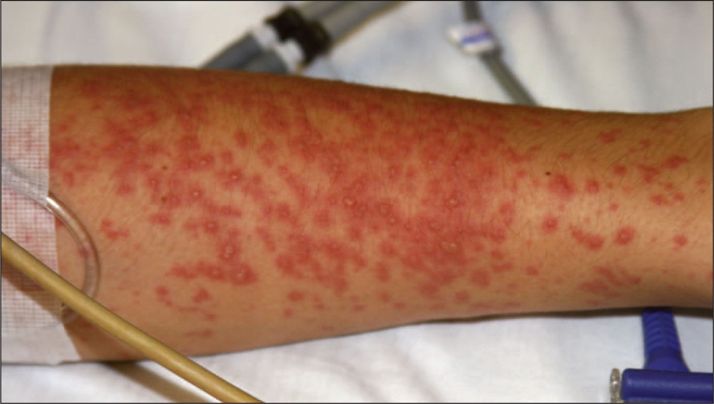
- Dusky erythematous or purpuric macules appear, usually initially on the face and trunk.
- Lesions may resemble atypical target lesions with poorly defined borders.
- Rapid progression to confluent erythema, flaccid blisters and sheets of epidermal detachment.
- Mucosal disease commonly affects the mouth, eyes and genital or urinary tract.
- Oral involvement causes painful erosions, haemorrhagic crusting and difficulty swallowing.
- Ocular disease may cause conjunctivitis, epithelial defects, ulceration and later scarring.
- Genital and urinary involvement may cause painful erosions, dysuria and urinary retention.
- Respiratory epithelial involvement may cause cough, hypoxaemia, bronchial sloughing and respiratory failure.
👉 Nikolsky Sign
Nikolsky sign is usually positive in active SJS or TEN. Gentle lateral pressure applied to erythematous or apparently intact skin causes the superficial epidermis to shear away, leaving a moist erosion. Pressure at the edge of a blister may also cause the blister to extend. A positive sign indicates marked epidermal fragility. It supports the diagnosis but is not pathognomonic for SJS or TEN. It may also be positive in pemphigus vulgaris, staphylococcal scalded skin syndrome and some other blistering disorders. The sign should be tested gently and should not be repeatedly elicited because this may increase epidermal damage.
🔍 Differential Diagnosis
- Staphylococcal scalded skin syndrome: usually affects infants or young children; mucous membranes are spared.
- Generalised bullous fixed drug eruption: sharply demarcated lesions, often recurring at previously affected sites, with less systemic illness.
- DRESS: facial oedema, widespread eruption, eosinophilia, lymphadenopathy and internal-organ inflammation.
- Acute generalised exanthematous pustulosis: numerous superficial pustules, neutrophilia and a shorter drug latency.
- Pemphigus vulgaris: flaccid blisters and prominent mucosal erosions caused by autoantibodies against desmosomal proteins.
- Linear IgA bullous dermatosis.
- Bullous lupus erythematosus.
- Severe phototoxicity or thermal burns.
- Toxic shock syndrome.
🧪 Investigations
- Blood tests: FBC, U&E, glucose, bicarbonate, magnesium, phosphate, LFTs, CRP and coagulation profile.
- Blood gas and lactate if the patient is physiologically unstable.
- Skin biopsy from a fresh lesion for histology.
- Consider a perilesional biopsy for direct immunofluorescence when an immunobullous disorder is possible.
- Histology typically shows full-thickness epidermal necrosis, keratinocyte apoptosis and subepidermal separation.
- Blood, urine and respiratory cultures where infection is suspected.
- Regular microbiological surveillance of sloughy or crusted skin in extensive disease.
- Chest radiograph and respiratory assessment if cough, breathlessness or hypoxaemia is present.
📊 SCORTEN Severity Score
Calculate SCORTEN within the first 24 hours. Each of the following scores one point:
- Age >40 years
- Heart rate >120 beats/minute
- Underlying malignancy
- Detached or detachable epidermis involving >10% body surface area
- Urea >10 mmol/L
- Serum bicarbonate <20 mmol/L
- Glucose >14 mmol/L
SCORTEN estimates mortality risk but should not delay treatment or be used as the sole criterion for deciding the appropriate place of care.
💀 Predicted Mortality by SCORTEN
- Score 0–1: approximately 3%
- Score 2: approximately 12%
- Score 3: approximately 35%
- Score 4: approximately 58%
- Score 5 or more: greater than 90%
🏥 Immediate Management
- Immediately stop the suspected causative drug. Stop all non-essential medication until drug causality has been reviewed. Do not deliberately re-expose the patient to a suspected causative medicine
- Urgent assessment by dermatology with early burns, intensive-care and plastic-surgery involvement. Document the percentage of detached and detachable epidermis using a body surface area chart. Record temperature, heart rate, blood pressure, respiratory rate, oxygen saturation, urine output and body weight.
- Seek urgent advice from a regional SJS–TEN service, burns centre or specialist dermatology service.
- Convene a multidisciplinary team including dermatology, burns or plastic surgery, intensive care, ophthalmology, specialist nursing, pharmacy and microbiology.
- Patients with more than 10% epidermal loss should be admitted without delay to a burns centre or ICU experienced in managing SJS and TEN.
- Calculate SCORTEN but do not allow scoring to delay transfer or supportive treatment.
🩹 Skin and Supportive Care
- Barrier-nurse the patient in a side room using a pressure-relieving mattress.
- Maintain the room temperature at approximately 25–28°C to reduce hypothermia.
- Handle the patient gently and avoid adhesive tapes or unnecessary trauma.
- Clean the skin gently with warmed sterile water or saline.
- Leave non-infected detached epidermis in place where appropriate because it may act as a biological dressing.
- Use non-adherent dressings over denuded dermis.
- Apply greasy emollients according to the specialist skin-care plan.
- Paraffin-containing emollients are flammable when absorbed into clothing or bedding; keep away from cigarettes, naked flames and other ignition sources.
- Provide adequate opioid analgesia, including additional analgesia for movement and dressing changes.
💧 Fluid, Temperature and Circulation
- Establish venous access through unaffected skin where possible.
- Monitor fluid balance, urine output, electrolytes, renal function and acid–base status closely.
- Give warmed intravenous fluids according to clinical losses and physiological endpoints.
- Fluid requirements are often lower than for a thermal burn of equivalent surface area and should be individually titrated.
- Monitor for shock, acute kidney injury and cardiac complications.
- Give venous thromboembolism prophylaxis unless contraindicated.
🥗 Nutrition
- Assess nutritional requirements early.
- Use early enteral nutrition when oral intake is inadequate.
- A nasogastric tube may be required if oral mucosal disease prevents adequate intake.
- Monitor phosphate, magnesium and potassium and consider the risk of refeeding syndrome.
🦠 Infection
- Use strict barrier nursing and appropriate infection-control measures.
- Monitor wounds, temperature, inflammatory markers and cultures.
- Do not give prophylactic systemic antibiotics routinely.
- Give systemic antibiotics when there are clinical or microbiological features of infection.
- Common pathogens include Staphylococcus aureus initially and Gram-negative organisms later in the illness.
👁️ Eye Care
- Arrange ophthalmology assessment as early as possible. May need daily ophthalmological review is required during the acute illness. Use frequent preservative-free ocular lubricant. Perform regular ocular hygiene under ophthalmology guidance. Topical corticosteroids or antibiotics may be required when prescribed by ophthalmology. Corneal epithelial defects, ulceration, pseudomembranes and symblepharon require urgent specialist treatment.
👄 Oral and Genital Care
- Review the mouth and lips daily.
- Use frequent white soft paraffin on the lips.
- Provide saline mouth care and appropriate topical analgesia.
- Assess swallowing and aspiration risk.
- Inspect genital and urinary mucosa daily.
- Use suitable emollients, topical treatment and non-adherent dressings under specialist guidance.
- Consider early gynaecology or urology involvement to reduce the risk of adhesions and scarring.
🫁 Respiratory Involvement
- Monitor oxygen saturation and respiratory symptoms closely.
- Cough, dyspnoea, hypoxaemia or haemoptysis may indicate respiratory epithelial involvement.
- Discuss early with intensive care when respiratory involvement is suspected.
- Fibre-optic bronchoscopy may be required in an ICU or burns centre.
- Use lung-protective ventilation if mechanical ventilation becomes necessary.
💉 Disease-Modifying Treatment
Supportive care and prompt withdrawal of the causative drug remain the foundations of treatment. There is no single universally accepted immunomodulatory treatment for all patients.
- Systemic corticosteroids
- Ciclosporin
- Etanercept or another tumour necrosis factor inhibitor
- Intravenous immunoglobulin
These treatments should be considered only by an experienced specialist SJS–TEN multidisciplinary team. Treatment selection depends on disease stage, comorbidity, infection risk and local specialist protocols and must not delay supportive care.
📋 Discharge and Prevention
- Clearly document the suspected causative drug and related medicines to avoid.
- Provide the patient with written drug-allergy information.
- Update the GP and all relevant hospital records.
- Submit an MHRA Yellow Card report.
- Encourage the patient to carry a drug-allergy card or medical-alert identification.
- Arrange dermatology and ophthalmology follow-up.
- Assess for long-term ocular, oral, genital, respiratory, psychological and functional complications.
- Routine drug provocation or rechallenge is contraindicated.
🖼️ Images
🩺 Example Case 1: Allopurinol-Associated TEN
A 72-year-old man with chronic kidney disease develops fever, painful eyes and oral ulceration three weeks after starting allopurinol. Over 24 hours he develops tender dusky macules, flaccid blisters and epidermal detachment affecting 35% of his body surface area. Gentle lateral pressure causes epidermal shearing.
- The diagnosis is toxic epidermal necrolysis.
- The Nikolsky sign is positive.
- Allopurinol must be stopped immediately and permanently avoided.
- SCORTEN should be calculated within 24 hours.
- He requires immediate specialist burns or ICU care with dermatology and ophthalmology involvement.
🩺 Example Case 2: Lamotrigine-Associated SJS
A 29-year-old woman develops fever, conjunctivitis, painful oral erosions and a dusky target-like rash two weeks after starting lamotrigine. There are scattered blisters with epidermal detachment affecting approximately 6% of body surface area.
- The diagnosis is Stevens–Johnson syndrome because epidermal detachment is below 10%.
- Lamotrigine must be stopped immediately.
- The limited area of detachment does not make the condition minor; mucosal and systemic complications may still be severe.
- She requires urgent specialist assessment and daily ophthalmological review.
🩺 Example Case 3: Positive Nikolsky Sign but Not TEN
An 18-month-old child develops fever, irritability and widespread tender erythema followed by superficial blistering. There is crusting around the mouth, but the oral mucosa itself is normal. Nikolsky sign is positive.
- The likely diagnosis is staphylococcal scalded skin syndrome.
- Sparing of the mucous membranes favours SSSS rather than SJS or TEN.
- The positive Nikolsky sign is not specific for TEN.
- Treatment requires intravenous anti-staphylococcal antibiotics and supportive care.
💡 Clinical Clues
- Think of SJS or TEN when a patient taking a recently introduced high-risk medicine develops skin pain, fever and mucosal erosions.
- Skin pain is often a more important warning symptom than itch.
- A positive Nikolsky sign supports epidermal necrolysis but does not establish the diagnosis.
- The first and most important treatment is immediate withdrawal of the causative drug.
- Never allow the apparently limited early rash to delay specialist referral because progression can be rapid.
📚 References
| The content on this website is provided for educational and informational purposes only to support exam preparation (e.g., MLA, MRCP, USMLE) and learning. This is NOT medical advice, diagnosis, treatment, or professional guidance. It does not replace consultation with a qualified healthcare professional, official guidelines (e.g., NICE, GMC, BNF), or supervised clinical practice. Always verify information with current, authoritative sources. Makindo and its contributors accept no liability for any reliance on this content, including errors, omissions, or any resulting harm, loss, or consequences. By using this site, you agree to these terms. |
|
|
Categories
- About
- Acute Medicine
- Anaesthetics and Critical Care
- Anatomy
- Anatomy and Physiology
- Biochemistry
- Book
- Cardiology
- Collections
- CompSci
- Crib Sheets
- Critical care
- Dental
- Dermatology
- Differentials
- Drugs
- ENT
- Electrocardiogram
- Embryology
- Emergency Medicine
- Endocrinology
- Ethics
- Foundation Doctors
- GCSE
- Gastroenterology
- General Practice
- Genetics
- Geriatric Medicine
- Geriatrics
- Guidelines
- Haematology
- Hepatology
- Immunology
- Infectious Diseases
- Infographic
- Investigations
- Lists
- MRCP
- Mandatory Training
- Medical Students
- Microbiology
- Nephrology
- Neurology
- Neurosurgery
- Nutrition
- OSCE
- Obstetrics Gynaecology
- Oncology
- Ophthalmology
- Oral Medicine and Dentistry
- Orthopaedics
- Paediatrics
- Palliative
- Palliative Care
- Pathology
- Pharmacology
- Physiology
- Procedures
- Psychiatry
- Public Health
- Radiology
- Respiratory
- Resuscitation
- Revision Topics
- Rheumatology
- Statistics and Research
- Stroke
- Surgery
- Toxicology
- Trauma and Orthopaedics
- USMLE
- Urology
- Vascular Surgery