
| Download the amazing global Makindo app: ✅ Means NICE/National Guidelines 2026 compliant Android | Apple | |
|---|---|
| MEDICAL DISCLAIMER: Educational use only. Not for diagnosis or management. See below for full disclaimer. |
Sturge Weber syndrome
Related Subjects: |Subarachnoid Haemorrhage |Dural Arteriovenous Malformations |Pulmonary Arteriovenous malformation |Sturge Weber syndrome
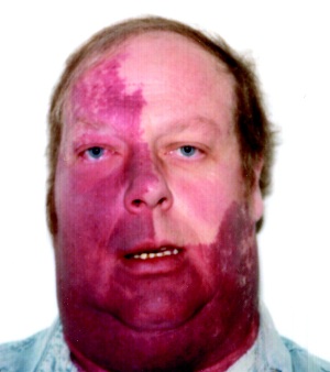
🍷🧠👁️ Sturge–Weber syndrome (SWS) is a sporadic neurocutaneous vascular disorder caused by a post-zygotic (mosaic) somatic mutation. It classically features a facial port-wine stain (capillary malformation), leptomeningeal angiomatosis (pial vascular malformation), and ocular disease (especially glaucoma). Neurological morbidity is mainly driven by chronic venous congestion → cortical ischaemia → seizures, stroke-like episodes and progressive atrophy.
📖 About
- Nature: Congenital, non-inherited (sporadic), due to somatic mosaicism.
- Core triad: 🍷 facial capillary malformation + 🧠 leptomeningeal angioma + 👁️ glaucoma (variable combination).
- Typical distribution: Port-wine stain often involves the ophthalmic (V1) region; risk of brain/eye involvement is higher with V1 (especially upper eyelid) involvement.
🧬 Genetics & Pathophysiology
- Cause: Somatic activating mutation in GNAQ (mosaic) → abnormal vascular development.
- Brain mechanism: Leptomeningeal venous malformation → impaired cortical venous drainage → chronic hypoperfusion and “vascular steal” → gliosis, calcification, and hemispheric atrophy.
- Why seizures? Irritable, hypoperfused cortex + gliosis → lower seizure threshold; early-onset seizures predict worse cognitive outcome.
- Why calcification? Chronic cortical injury → dystrophic gyriform (“tram-track”) calcifications, classically in parieto-occipital cortex.
🧾 Roach Classification (Types)
Most teaching uses the Roach classification (Types I–III), based on whether SWS involves the face (port-wine stain) 🍷, the leptomeninges (brain) 🧠, and/or the eye (glaucoma) 👁️.
- Type I (classic / most common) 🍷🧠 (±👁️)
- Facial port-wine stain + leptomeningeal angioma.
- Glaucoma may occur (often ipsilateral; can be early or later).
- Type II 🍷 (±👁️)
- Facial port-wine stain only (no leptomeningeal involvement).
- Glaucoma may occur.
- Type III 🧠
- Leptomeningeal angioma only (typically no facial port-wine stain).
- Often presents later with seizures/headache/neurological events; glaucoma is uncommon but assess eyes anyway 👁️.
🩺 Clinical Presentation
- Skin 🍷: facial port-wine stain present at birth; commonly V1/V2 distribution.
- Neurology 🧠:
- Seizures (often in infancy/early childhood; focal ± secondary generalisation).
- Stroke-like episodes or transient neurological deficits (weakness, visual symptoms) due to hypoperfusion/venous congestion.
- Hemiparesis (often contralateral to brain involvement), developmental delay, learning difficulties.
- Headache/migraine can be prominent.
- Visual field deficit (occipital involvement) e.g., homonymous hemianopia.
- Eye 👁️:
- Glaucoma (can be congenital/early-onset or develop later).
- Choroidal haemangioma → visual impairment, retinal complications.
🖼️ Imaging & hallmark findings
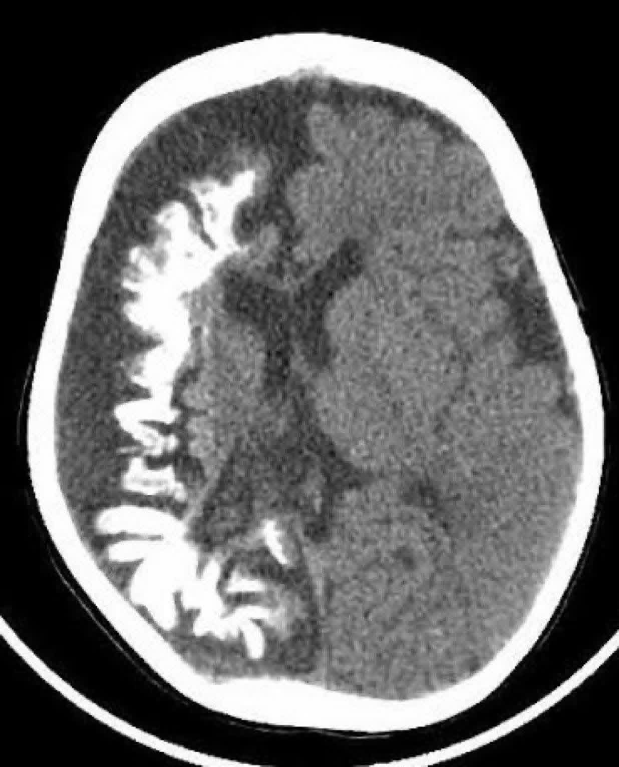
🧠 “Tram-track” calcifications are the classic imaging hallmark of SWS. They represent gyriform cortical/subcortical calcification, often parieto-occipital, with associated hemispheric atrophy and sometimes calvarial thickening.
🧪 Investigations
- 🩻 CT brain: can show gyriform (“tram-track”) calcification and hemispheric volume loss (often later in disease).
- 🧲 MRI brain with contrast (key test): leptomeningeal enhancement, pial angioma, cortical atrophy; consider SWI for venous abnormalities.
- 🩸 MRV/MRA (where available): evaluates venous drainage anomalies and associated vascular features.
- ⚡ EEG: for seizure classification, lateralisation, and presurgical work-up if refractory epilepsy.
- 👁️ Ophthalmology assessment: urgent baseline + ongoing monitoring (IOP, optic nerve, choroidal haemangioma).
⚠️ Complications
- ⚡ Epilepsy (including refractory focal epilepsy; status epilepticus risk).
- 🧠 Progressive neurological impairment: hemiparesis, cognitive decline, developmental delay.
- 🧩 Stroke-like episodes / transient deficits (hypoperfusion/venous congestion).
- 👁️ Glaucoma → optic nerve damage and vision loss if untreated.
- 🩸 Intracranial haemorrhage is less central than in AVMs, but vascular fragility/venous hypertension can contribute to complications.
⚖️ Management (practical, multidisciplinary)
- Seizure control ⚡:
- Early recognition and treatment of focal seizures; optimise anti-seizure medication and adherence.
- Refractory epilepsy → refer to tertiary epilepsy service for presurgical evaluation (EEG + MRI ± PET/SPECT).
- Stroke-like episodes / neurological decline 🧠:
- Supportive management, avoid dehydration and hypotension; treat triggers (infection, poor sleep, missed meds).
- Consider specialist advice regarding antiplatelet use in recurrent stroke-like episodes (practice varies; individualised risk/benefit).
- Ophthalmology 👁️:
- Urgent assessment for glaucoma at diagnosis, then ongoing monitoring.
- Manage glaucoma with drops and/or surgery per ophthalmology; treat choroidal haemangioma complications if present.
- Dermatology / laser therapy 🍷:
- Pulsed dye laser can improve port-wine stain appearance (cosmetic + psychosocial benefit).
- Neurodevelopment & rehab 🧑🏫:
- Early developmental assessment; physiotherapy/OT/speech and language support as needed.
- Education planning and family support are central to long-term outcomes.
📉 Prognosis
- Earlier seizure onset, frequent seizures, and extensive unilateral leptomeningeal involvement are associated with worse neurodevelopmental outcomes.
- Good seizure control and early multidisciplinary care improve function and quality of life.
- Ocular prognosis depends on early detection and control of glaucoma.
💡 Exam Pearls
- 🍷 Port-wine stain in V1 distribution → think SWS and check brain + eyes.
- 🧠 Tram-track calcifications + seizures + hemiparesis → classic triad.
- 👁️ Glaucoma can be early or late → needs ongoing surveillance.
📖 References
| The content on this website is provided for educational and informational purposes only to support exam preparation (e.g., MLA, MRCP, USMLE) and learning. This is NOT medical advice, diagnosis, treatment, or professional guidance. It does not replace consultation with a qualified healthcare professional, official guidelines (e.g., NICE, GMC, BNF), or supervised clinical practice. Always verify information with current, authoritative sources. Makindo and its contributors accept no liability for any reliance on this content, including errors, omissions, or any resulting harm, loss, or consequences. By using this site, you agree to these terms. |
|
|
Categories
- About
- Acute Medicine
- Anaesthetics and Critical Care
- Anatomy
- Anatomy and Physiology
- Biochemistry
- Book
- Cardiology
- Collections
- CompSci
- Crib Sheets
- Critical care
- Dental
- Dermatology
- Differentials
- Drugs
- ENT
- Electrocardiogram
- Embryology
- Emergency Medicine
- Endocrinology
- Ethics
- Foundation Doctors
- GCSE
- Gastroenterology
- General Practice
- Genetics
- Geriatric Medicine
- Geriatrics
- Guidelines
- Haematology
- Hepatology
- Immunology
- Infectious Diseases
- Infographic
- Investigations
- Lists
- MRCP
- Mandatory Training
- Medical Students
- Microbiology
- Nephrology
- Neurology
- Neurosurgery
- Nutrition
- OSCE
- Obstetrics Gynaecology
- Oncology
- Ophthalmology
- Oral Medicine and Dentistry
- Orthopaedics
- Paediatrics
- Palliative
- Palliative Care
- Pathology
- Pharmacology
- Physiology
- Procedures
- Psychiatry
- Public Health
- Radiology
- Respiratory
- Resuscitation
- Revision Topics
- Rheumatology
- Statistics and Research
- Stroke
- Surgery
- Toxicology
- Trauma and Orthopaedics
- USMLE
- Urology
- Vascular Surgery