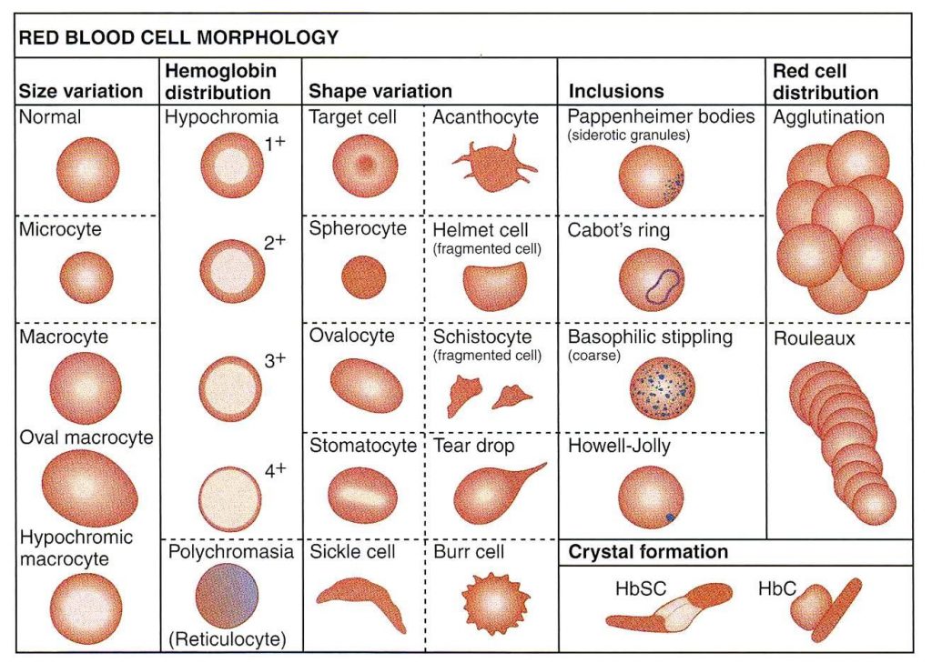
| Microcytic RBCs |
Fe deficiency, thalassaemia trait and syndromes, congenital sideroblastic anaemia, anaemia of chronic disorders |
| Macrocytic RBCs |
Alcohol/liver disease (round macrocytes), MDS, pregnancy and newborn, haemolysis, B12 or folate deficiency, hydroxyurea and antimetabolites (oval macrocytes), acquired sideroblastic anaemia, hypothyroidism, chronic respiratory failure, aplastic anaemia |
| Dimorphic RBCs |
Fe deficiency responding to Fe, mixed Fe and B12/folate deficiency, sideroblastic anaemia, post-transfusion |
| Polychromatic RBCs |
Response to bleeding or haematinic Rx, haemolysis, BM infiltration |
| Spherocytes |
HS, haemolysis, e.g. warm AIHA, delayed transfusion reaction, ABO HDN, DIC, and MAHA, post-splenectomy |
| Pencil/rod cells |
Fe deficiency anaemia, thalassaemia trait and syndromes, PK deficiency |
| Elliptocytes |
Hereditary elliptocytosis, MPD, and MDS |
| Fragmented RBCs |
MAHA, DIC, renal failure, HUS, TTP |
| Teardrop RBCs |
Myelofibrosis, metastatic marrow infiltration, MDS |
| Sickle cells |
Sickle cell anaemia, other sickle syndromes (not sickle trait) |
| Target cells |
Liver disease, Fe deficiency, thalassaemia, HbC syndromes |
| Crenated red cells |
Usually storage or EDTA artifact. Genuine RBC crenation may be seen post-splenectomy and in renal failure |
| Burr cells |
Renal failure |
| Acanthocytes |
Hereditary acanthocytosis, a-β-lipoproteinaemia, McLeod red cell phenotype, PK deficiency, chronic liver disease (esp. Zieve’s) |
| Bite cells |
G6PD deficiency, oxidative haemolysis |
| Basophilic stippling |
Megaloblastic anaemia, lead poisoning, MDS, haemoglobinopathies |
| Rouleaux |
Chronic inflammation, paraproteinaemia, myeloma |
| Reticulocytes |
Bleeding, haemolysis, marrow infiltration, severe hypoxia, response to haematinic therapy |
| Heinz bodies |
Not seen in normals (removed by spleen), small numbers seen post-splenectomy, oxidant drugs, G6PD deficiency, sulfonamides, unstable Hb (Hb Zurich, Köln) |
| Howell–Jolly bodies |
Made of DNA, generally removed by the spleen, dyserythropoietic states, e.g. B12 deficiency, MDS, post-splenectomy, hyposplenism |
| H bodies |
HbH inclusions, denatured HbH (β4 tetramer), stain with methylene blue, seen in HbH disease (− −/− α), less prominent in α thalassaemia trait, not present in normals |
| Hyposplenic blood film |
Howell–Jolly bodies, target cells, occasional nucleated RBCs, lymphocytosis, macrocytosis, acanthocytes |