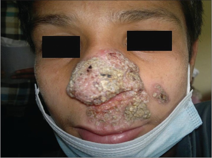
Lupus Vulgaris
Multiple red-brown nodules with a soft, jelly-like consistency are classically described as “apple-jelly nodules” 🍎, a hallmark of Lupus Vulgaris. Severe, chronic cutaneous tuberculosis with scarring, atrophy, and disfigurement if untreated.
📖 About
- Represents cutaneous invasion by Mycobacterium tuberculosis.
- Chronic, progressive, and destructive skin disease.
- Occurs in patients with a moderate to high degree of immunity (reactivation TB).
- More common in females than males 👩.
🧬 Aetiology
- Direct extension from underlying TB focus (lymph node, bone, joint, mucosa).
- Haematogenous or lymphatic spread from a primary TB site.
- Exogenous inoculation (rare).
🩺 Clinical Features
- Reddish-brown plaques with peripheral extension and central healing → atrophy and scarring.
- Apple-jelly nodules 🍎 visible on diascopy (pressing glass slide on lesion).
- Most common sites:
- Head and neck (≈80%).
- Followed by arms, legs, then trunk.
- Lesions may ulcerate or form disfiguring scars.
- Longstanding disease → risk of malignant transformation (SCC).
🔍 Differentials
- Sarcoidosis (also produces “apple-jelly” appearance).
- Leprosy (especially tuberculoid form).
- Lupus erythematosus (DLE).
- Deep fungal infections (sporotrichosis, chromoblastomycosis).
- Cutaneous leishmaniasis.
🧪 Investigations
- Baseline bloods: FBC, U&E, LFTs.
- Screen for systemic TB: CXR, sputum AFB, HIV test.
- Skin biopsy: granulomatous inflammation with caseating necrosis, Ziehl-Neelsen staining (may show few bacilli).
- Culture for Mycobacterium tuberculosis (gold standard, but slow growth).
- Molecular tests: PCR for TB DNA (faster diagnosis).
💊 Management
- Treat as for tuberculosis using standard anti-TB therapy (RIPE regimen: Rifampicin, Isoniazid, Pyrazinamide, Ethambutol).
- Duration: usually 6 months; longer if extensive or resistant disease.
- Reconstructive surgery may be needed for disfigurement or functional impairment.
- Monitor for secondary bacterial infection and skin malignancy in chronic scars.
📚 References
🧾 Clinical Case – Lupus Vulgaris
A 42-year-old woman presents with a slowly enlarging, reddish-brown plaque with an “apple jelly” appearance on her cheek, present for 2 years.
She reports mild itching but no systemic symptoms.
Skin biopsy shows granulomatous inflammation with caseating necrosis, and cultures confirm Mycobacterium tuberculosis.
Diagnosis: Lupus vulgaris, a cutaneous form of TB.
She was started on standard anti-tuberculous therapy with good clinical response.
