
| Download the amazing global Makindo app: ✅ Means NICE/National Guidelines 2026 compliant Android | Apple | |
|---|---|
| MEDICAL DISCLAIMER: Educational use only. Not for diagnosis or management. See below for full disclaimer. |
Anatomy and Physiology of Nerves
Related Subjects: |Anatomy and Physiology of the Brain |Clinically Isolated Syndrome (CIS) |Focal Cortical Dysplasia (FCD) |Infantile Spasms (West Syndrome) |Neurological History taking |Motor Neuron Disease (MND-ALS) |Miller-Fisher syndrome |Guillain Barre Syndrome |Multifocal Motor Neuropathy with Conduction block |Multiple Sclerosis (MS) Demyelination |Transverse myelitis |Acute Disseminated Encephalomyelitis |Progressive Multifocal Leukoencephalopathy (PML) |Inclusion Body Myositis |Cervical spondylosis |Anterior Spinal Cord syndrome |Central Spinal Cord syndrome |Brown-Sequard Spinal Cord syndrome |Spinal Cord Compression |Spinal Cord Haematoma |Spinal Cord Infarction
🧠 Nerve Cells (Neurons): Anatomy & Physiology
Neurons are highly specialised excitable cells designed for rapid communication within the nervous system. They receive, integrate, and transmit electrical signals over long distances and convert these signals into chemical messages at synapses. This capability underpins sensation, voluntary movement, autonomic regulation, cognition, and higher cortical function.
Unlike most cells, neurons are post-mitotic and must function for a lifetime. Structural damage, metabolic failure, or disruption of ion channel function therefore has immediate and often irreversible clinical consequences.
🔬 Basic Anatomy of a Neuron
The anatomy of a neuron is tightly coupled to its function. Each component contributes to signal reception, integration, propagation, or transmission.
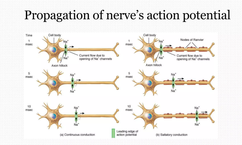
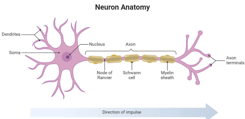
Figure: Typical neuron showing dendrites, soma, axon, myelin sheath, nodes of Ranvier, and axon terminals. This structural layout explains both rapid conduction and directional signalling.
- Dendrites: Branched processes receiving synaptic input; allow spatial and temporal summation of thousands of signals.
- Cell body (soma): Contains nucleus and metabolic machinery; integrates incoming excitatory and inhibitory signals.
- Axon hillock: Trigger zone with high density of voltage-gated sodium channels where action potentials are initiated.
- Axon: Long projection specialised for rapid electrical conduction over distance.
- Myelin sheath: Lipid-rich insulation (oligodendrocytes in CNS, Schwann cells in PNS) that increases conduction speed.
- Nodes of Ranvier: Gaps in myelin rich in ion channels enabling saltatory conduction.
- Axon terminals: Presynaptic endings releasing neurotransmitters into the synaptic cleft.
⚙️ Neuronal Physiology: Resting Membrane Potential
At rest, neurons maintain a stable electrical gradient across their membrane known as the resting membrane potential (approximately −70 mV). This polarity is essential for excitability and action potential generation.
- High intracellular potassium (K⁺) and low intracellular sodium (Na⁺).
- High extracellular sodium (Na⁺) and chloride (Cl⁻).
- Na⁺/K⁺ ATPase: Pumps 3 Na⁺ out and 2 K⁺ in, maintaining ionic gradients.
- Potassium leak channels: Allow passive K⁺ efflux, the main determinant of resting potential.
Small changes in extracellular potassium can significantly alter neuronal excitability, explaining neurological features of hypo- and hyperkalaemia.
⚡ Action Potential: Electrical Signalling
An action potential is a rapid, stereotyped reversal of membrane polarity that propagates along the axon without decrement. It obeys the all-or-nothing principle once threshold is reached.
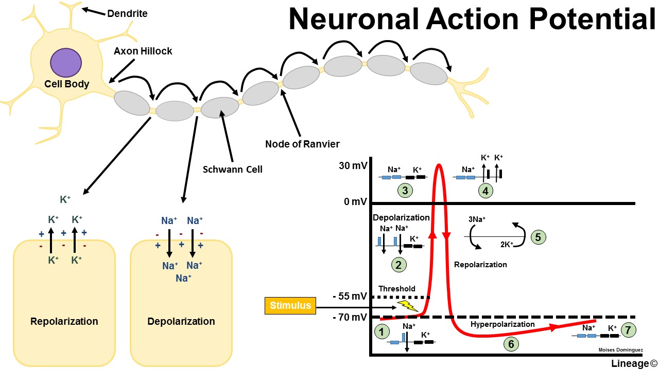
Figure: Phases of the action potential showing depolarisation, repolarisation, and hyperpolarisation driven by voltage-gated sodium and potassium channels.
- Threshold (~−55 mV): Achieved when depolarising inputs exceed inhibitory influences.
- Depolarisation: Rapid opening of voltage-gated Na⁺ channels → Na⁺ influx.
- Peak (~+30 mV): Na⁺ channels inactivate.
- Repolarisation: Voltage-gated K⁺ channels open → K⁺ efflux.
- Hyperpolarisation: Continued K⁺ efflux temporarily overshoots resting potential.
- Return to rest: Gradients restored by pumps and leak channels.
⛔ Refractory Periods
Refractory periods ensure unidirectional propagation and limit the maximum firing frequency of neurons.
- Absolute refractory period: Na⁺ channels are inactivated; no action potential can occur.
- Relative refractory period: Stronger stimulus required due to ongoing hyperpolarisation.
🚀 Conduction of Action Potentials
Action potentials propagate along the axon without loss of amplitude. Conduction velocity depends on axon diameter and myelination.
- Continuous conduction: Seen in unmyelinated fibres; slow and energy-intensive.
- Saltatory conduction: Action potentials jump between nodes of Ranvier in myelinated fibres.
- Myelination increases speed and reduces metabolic cost.
- Larger diameter axons conduct faster due to reduced internal resistance.
Demyelination slows or blocks conduction, explaining weakness, sensory loss, and fatigue in disorders such as multiple sclerosis.
🔄 Synaptic Transmission
At synapses, electrical signals are converted into chemical signals, allowing precise and modifiable communication between cells.
- Action potential reaches presynaptic terminal.
- Voltage-gated Ca²⁺ channels open → Ca²⁺ influx.
- Ca²⁺ triggers neurotransmitter vesicle fusion.
- Neurotransmitters bind postsynaptic receptors.
- Postsynaptic potentials may be excitatory (EPSP) or inhibitory (IPSP).
🧬 Sensory Nerve Fibres
- Ia (A-alpha): Proprioception from muscle spindles; fastest conduction (70–120 m/s).
- Ib (A-alpha): Golgi tendon organs; monitor muscle tension.
- II (A-beta): Touch, pressure, vibration.
- III (A-delta): Fast, sharp pain; cold temperature.
- IV (C fibres): Slow, dull pain; heat; itch; unmyelinated.
🦵 Motor & Autonomic Fibres
- A-alpha: Voluntary skeletal muscle contraction.
- A-gamma: Muscle spindle sensitivity and tone.
- B fibres: Preganglionic autonomic fibres (myelinated).
- C fibres: Postganglionic autonomic fibres (unmyelinated).
🩺 Clinical Integration & Exam Pearls
- Loss of myelination affects fast fibres first → early proprioceptive and motor deficits.
- Sharp pain precedes dull pain due to A-delta then C fibre activation.
- Autonomic neuropathies predominantly involve small C fibres.
- Channelopathies alter excitability rather than structure.
References
| The content on this website is provided for educational and informational purposes only to support exam preparation (e.g., MLA, MRCP, USMLE) and learning. This is NOT medical advice, diagnosis, treatment, or professional guidance. It does not replace consultation with a qualified healthcare professional, official guidelines (e.g., NICE, GMC, BNF), or supervised clinical practice. Always verify information with current, authoritative sources. Makindo and its contributors accept no liability for any reliance on this content, including errors, omissions, or any resulting harm, loss, or consequences. By using this site, you agree to these terms. |
|
|
Categories
- About
- Acute Medicine
- Anaesthetics and Critical Care
- Anatomy
- Anatomy and Physiology
- Biochemistry
- Book
- Cardiology
- Collections
- CompSci
- Crib Sheets
- Critical care
- Dental
- Dermatology
- Differentials
- Drugs
- ENT
- Electrocardiogram
- Embryology
- Emergency Medicine
- Endocrinology
- Ethics
- Foundation Doctors
- GCSE
- Gastroenterology
- General Practice
- Genetics
- Geriatric Medicine
- Geriatrics
- Guidelines
- Haematology
- Hepatology
- Immunology
- Infectious Diseases
- Infographic
- Investigations
- Lists
- MRCP
- Mandatory Training
- Medical Students
- Microbiology
- Nephrology
- Neurology
- Neurosurgery
- Nutrition
- OSCE
- Obstetrics Gynaecology
- Oncology
- Ophthalmology
- Oral Medicine and Dentistry
- Orthopaedics
- Paediatrics
- Palliative
- Palliative Care
- Pathology
- Pharmacology
- Physiology
- Procedures
- Psychiatry
- Public Health
- Radiology
- Respiratory
- Resuscitation
- Revision Topics
- Rheumatology
- Statistics and Research
- Stroke
- Surgery
- Toxicology
- Trauma and Orthopaedics
- USMLE
- Urology
- Vascular Surgery