
| Download the amazing global Makindo app: ✅ Means NICE/National Guidelines 2026 compliant Android | Apple | |
|---|---|
| MEDICAL DISCLAIMER: Educational use only. Not for diagnosis or management. See below for full disclaimer. |
The Face 😀
The face is a highly specialised “mobile skin organ” built for expression, speech, eating, eye protection, breathing through the nose, and social communication. Many facial muscles insert into skin rather than bone, so small motor signals create visible movements. The face is richly supplied by sensory nerves and blood vessels, which is why facial pain is often sharp and well localised, and why facial wounds can bleed heavily but usually heal well.
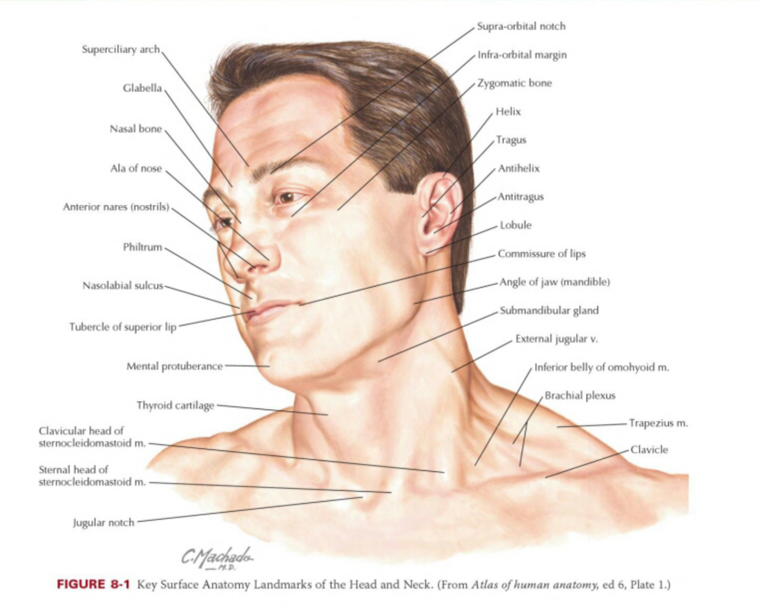
🧴 Skin of the Face
Facial skin has the usual layers (epidermis, dermis, subcutaneous tissue) but varies markedly by region. Eyelid skin is extremely thin with minimal fat, so it swells and bruises easily. The nose and forehead are thicker and more sebaceous, supporting barrier function but predisposing to acne-prone areas. High vascularity and dense innervation explain why inflammation (e.g., cellulitis) can look dramatic and why sensory symptoms are prominent.
- Epidermis: barrier layer; protects from dehydration, pathogens, UV injury.
- Dermis: collagen and elastin for strength and recoil; contains vessels, nerves, glands.
- Subcutaneous tissue: fat compartments shape contour and permit mobility.
- Skin appendages: sebaceous glands, sweat glands, hair follicles (brows/lashes for protection).
🧱 Fascial Planes and the SMAS
The key facial fascial concept is the superficial musculoaponeurotic system (SMAS), a fibromuscular layer that links facial expression muscles to the dermis and transmits movement to the skin. It continues superiorly with the temporoparietal fascia and inferiorly with platysma. These planes influence how swelling, bruising, and infection track and are central to many surgical approaches.
- SMAS: fibromuscular sheet connecting muscles to skin; major “gliding” layer.
- Fat compartments: discrete superficial and deep pads that shape contour and guide spread of oedema/haematoma.
- Parotidomasseteric fascia: tough fascia over masseter/parotid region; important because CN VII branches traverse the parotid gland.
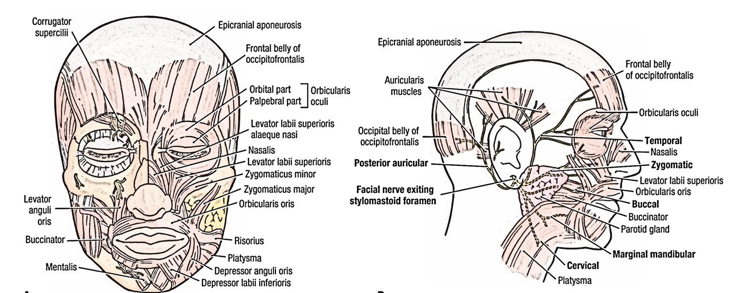
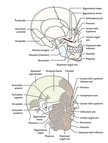
💪 Muscles of the Face
Facial muscles are best grouped into muscles of facial expression (motor supply by facial nerve, CN VII) and muscles of mastication (motor supply by mandibular division of trigeminal nerve, V3). Expression muscles insert into skin to create communication signals; mastication muscles move the mandible to bite and chew.

🎭 Muscles of Facial Expression (Motor = CN VII)
- Scalp and forehead
- Frontalis (occipitofrontalis, frontal belly): elevates eyebrows; wrinkles forehead (surprise).
- Corrugator supercilii: draws eyebrows medially/down (frown).
- Procerus: wrinkles the bridge of the nose.
- Eyelids
- Orbicularis oculi: closes eyelids (blinking and tight closure).
- Clinical pearl: inability to close the eye suggests CN VII; true ptosis suggests CN III (levator palpebrae).
- Nose
- Nasalis: compresses or dilates nostrils (airflow modulation).
- Levator labii superioris alaeque nasi: elevates upper lip; flares nostril.
- Mouth and cheek
- Orbicularis oris: purses lips; seals mouth (speech, feeding).
- Buccinator: presses cheek against teeth; keeps food between occlusal surfaces; blowing/whistling.
- Zygomaticus major/minor: elevates mouth corners (smile).
- Levator labii superioris: elevates upper lip.
- Depressor anguli oris: pulls mouth corners down (sadness).
- Depressor labii inferioris: lowers the lower lip.
- Mentalis: protrudes the lower lip; wrinkles chin (pout).
- Platysma: tenses neck skin; assists lower lip depression and grimace.
🍽️ Muscles of Mastication (Motor = V3)
The main chewing muscles move the mandible at the temporomandibular joint (TMJ). They generate bite force and coordinate elevation, depression, protrusion, retraction, and lateral excursion.
- Masseter: elevates mandible; strong bite.
- Temporalis: elevates mandible; posterior fibres retract.
- Medial pterygoid: elevates; contributes to grinding.
- Lateral pterygoid: protrudes and depresses mandible; key for opening and side-to-side movements.
🧠 Nerve Supply of the Face
Facial innervation divides into motor (expression = CN VII; mastication = V3) and sensation (largely trigeminal nerve, CN V). Autonomic fibres (sympathetic and parasympathetic) hitchhike along nerves and vessels to reach glands and vessels, controlling tears, saliva, sweating, and vascular tone.
⚡ Motor Nerves
- Facial nerve (CN VII): motor to facial expression; emerges from stylomastoid foramen and branches within the parotid gland.
- Main CN VII branches
- Temporal: forehead and upper orbicularis oculi.
- Zygomatic: lower orbicularis oculi and upper cheek.
- Buccal: upper lip, cheek, buccinator.
- Marginal mandibular: lower lip muscles.
- Cervical: platysma.
- Trigeminal (V3): motor to muscles of mastication (plus mylohyoid and anterior belly of digastric).
🖐️ Sensory Nerves (CN V territories)
The trigeminal nerve provides facial sensation in three divisions. This pattern is clinically important for pain syndromes, corneal reflex testing, and local anaesthetic blocks.
- V1 (Ophthalmic): forehead, upper eyelid, dorsum of nose (e.g., supraorbital/supratrochlear).
- V2 (Maxillary): lower eyelid, cheek, lateral nose, upper lip (e.g., infraorbital).
- V3 (Mandibular): lower lip, chin, jawline, parts of temple (e.g., mental nerve).
🌿 Autonomic (Glands and Vessels)
- Parasympathetic
- Lacrimal gland: via CN VII (greater petrosal) → pterygopalatine ganglion.
- Submandibular/sublingual glands: via CN VII (chorda tympani) → submandibular ganglion.
- Parotid gland: via CN IX (lesser petrosal) → otic ganglion → auriculotemporal (V3).
- Sympathetic: vasoconstriction, sweating; fibres from superior cervical ganglion travel along arteries.
🩸 Blood Supply of the Face
The face has a rich blood supply with extensive anastomoses between the external carotid system and the ophthalmic artery (internal carotid). This supports excellent healing but also explains why vascular complications can be significant in certain regions.
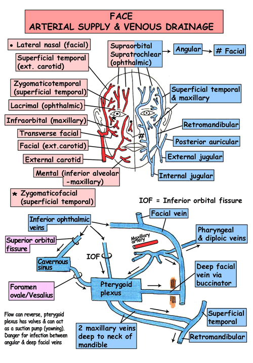
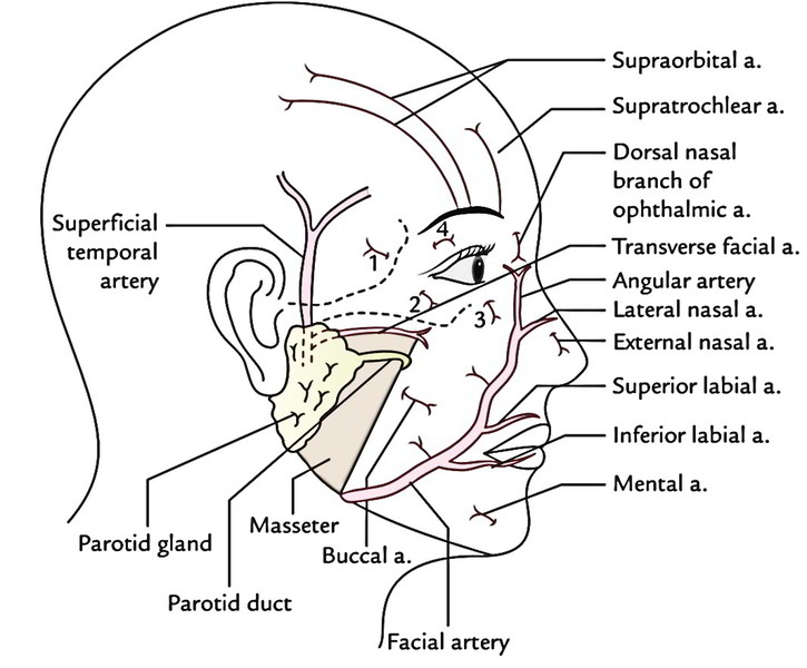
🟥 Arterial Supply
- External carotid artery branches
- Facial artery: major supply to lips and nose; crosses the mandible anterior to masseter; ends as the angular artery near the medial canthus.
- Superficial temporal artery: scalp and temporal region; gives transverse facial artery.
- Maxillary artery: deep face; supplies via infraorbital artery and other branches.
- Internal carotid via ophthalmic artery
- Supraorbital/supratrochlear arteries: forehead and scalp.
- Dorsal nasal artery: bridge of nose.
- Key anastomoses
- Angular (facial) ↔ dorsal nasal (ophthalmic) near medial canthus.
- Facial ↔ infraorbital across the midface.
🟦 Venous Drainage
Facial veins are relatively valveless and connect superficial and deep systems. Clinically, venous connections from the central face can communicate with intracranial venous sinuses via ophthalmic and deep facial pathways, which is why severe infections in certain regions can rarely cause intracranial complications.
- Facial vein: drains much of the face; connects with the angular vein near the medial canthus.
- Angular vein ↔ ophthalmic veins: potential route toward cavernous sinus.
- Retromandibular vein: drains parotid/temporal regions; contributes to external jugular system.
🧫 Lymphatic Drainage
Lymph drainage follows predictable regional patterns and matters for infection spread and malignancy staging.
- Submental nodes: central lower lip and chin.
- Submandibular nodes: cheeks, lateral nose, upper lip, lateral lower lip.
- Preauricular/parotid nodes: lateral eyelids and temporal region.
- Deep cervical nodes: final common pathway for much of facial lymph.
🧩 Integrated Function
Facial expression depends on CN VII motor output, skin elasticity, and the SMAS/fat compartments to transmit force smoothly to the dermis. Sensory input via CN V provides protective reflexes and fine tactile discrimination around the eyes and mouth, supporting feeding and corneal protection. A robust vascular supply supports high metabolic demand and enables rapid healing, while autonomic control adjusts tear/saliva production and cutaneous blood flow (e.g., blushing and pallor).
- Eye protection: orbicularis oculi closes lids; corneal sensation (V1) triggers blink reflex.
- Speech and feeding: orbicularis oris seals lips; buccinator keeps food between teeth; jaw movement via V3.
- Emotional signalling: subtle expression changes via CN VII branches.
- Autonomic effects: sweating and vascular tone changes in stress/emotion.
🩺 Clinical Correlations (High Yield)
- Facial palsy (CN VII): weakness of facial expression; inability to close eye risks exposure keratopathy and corneal injury.
- Trigeminal sensory loss: reduced corneal sensation (V1) increases corneal injury risk; numbness follows V1/V2/V3 territories.
- Parotid surgery: facial nerve branches traverse the parotid gland, so careful anatomy reduces iatrogenic palsy risk.
- Facial lacerations: often bleed heavily but heal well; align aesthetic units and consider nerve/duct injury.
- Central facial venous connections: rare but important intracranial spread risk in severe infections.
Blood supply
Muscles
Expression
| The content on this website is provided for educational and informational purposes only to support exam preparation (e.g., MLA, MRCP, USMLE) and learning. This is NOT medical advice, diagnosis, treatment, or professional guidance. It does not replace consultation with a qualified healthcare professional, official guidelines (e.g., NICE, GMC, BNF), or supervised clinical practice. Always verify information with current, authoritative sources. Makindo and its contributors accept no liability for any reliance on this content, including errors, omissions, or any resulting harm, loss, or consequences. By using this site, you agree to these terms. |
|
|
Categories
- About
- Acute Medicine
- Anaesthetics and Critical Care
- Anatomy
- Anatomy and Physiology
- Biochemistry
- Book
- Cardiology
- Collections
- CompSci
- Crib Sheets
- Critical care
- Dental
- Dermatology
- Differentials
- Drugs
- ENT
- Electrocardiogram
- Embryology
- Emergency Medicine
- Endocrinology
- Ethics
- Foundation Doctors
- GCSE
- Gastroenterology
- General Practice
- Genetics
- Geriatric Medicine
- Geriatrics
- Guidelines
- Haematology
- Hepatology
- Immunology
- Infectious Diseases
- Infographic
- Investigations
- Lists
- MRCP
- Mandatory Training
- Medical Students
- Microbiology
- Nephrology
- Neurology
- Neurosurgery
- Nutrition
- OSCE
- Obstetrics Gynaecology
- Oncology
- Ophthalmology
- Oral Medicine and Dentistry
- Orthopaedics
- Paediatrics
- Palliative
- Palliative Care
- Pathology
- Pharmacology
- Physiology
- Procedures
- Psychiatry
- Public Health
- Radiology
- Respiratory
- Resuscitation
- Revision Topics
- Rheumatology
- Statistics and Research
- Stroke
- Surgery
- Toxicology
- Trauma and Orthopaedics
- USMLE
- Urology
- Vascular Surgery