
| Download the amazing global Makindo app: ✅ Means NICE/National Guidelines 2026 compliant Android | Apple | |
|---|---|
| MEDICAL DISCLAIMER: Educational use only. Not for diagnosis or management. See below for full disclaimer. |
Ventricular Tachycardia VT ✅
Related Subjects: |Electrical Storm (Recurrent VT/VF) |Ventricular Fibrillation |Classical Ventricular Tachycardia |Idiopathic Ventricular Tachycardia |Resuscitation - Adult Tachycardia Algorithm |Resuscitation - Advanced Life Support |Automatic Implantable Cardioverter Defibrillator (AICD) |Brugada Syndrome |Long QT syndrome (LQTS) Acquired |Long QT syndrome (LQTS) Congenital |Torsades de Pointes |Wolff-Parkinson White syndrome (WPW) |Supraventricular Tachycardia (SVT) |Atrial Flutter |Atrial Fibrillation
⚡ Ensure a defibrillator is immediately available, switched on, and ready for use. Follow the Adult Tachycardia Algorithm (Resuscitation Council UK / ALS 2021).
| 📋 Management Summary: IV Access & Defibrillator Preparedness |
|---|
|
ℹ️ Key Points
- 📊 Broad-complex tachycardia (>120 bpm) usually ventricular in origin, commonly post-MI.
- ⚠️ Even seemingly stable patients can deteriorate → always prepare for rapid intervention.
- 🏥 Admit to CCU / high-dependency unit; continuous ECG monitoring and immediate defibrillator access required.
📌 Types of VT
- Sustained VT: ≥30 seconds or requiring intervention due to haemodynamic compromise.
- Non-sustained VT: ≥3 consecutive ventricular beats, lasting <30 seconds, rate >100 bpm.
🩺 Causes of VT
- ❤️ Ischaemic cardiomyopathy (most common)
- 💪 LVH (e.g., hypertension)
- 🫀 Idiopathic dilated cardiomyopathy
- ⚡ Channelopathies: Brugada syndrome, long QT syndromes
- 🧬 ARVC (Arrhythmogenic Right Ventricular Cardiomyopathy)
- 🦠 Myocarditis
- 🏋️ Hypertrophic cardiomyopathy
- 💊 Drugs / toxins: TCAs, digoxin, antiarrhythmics, cocaine
- 🧪 Electrolyte disturbances: hypo-/hyperkalaemia, hypomagnesaemia
- 💉 Phaeochromocytoma
- ➡️ RVOT VT (LBBB morphology + RAD)
👨⚕️ Clinical Presentation
- Palpitations, dizziness, syncope
- 💨 Pulmonary oedema, 🩸 hypotension, chest pain
- JVP: cannon a-waves possible
- Sudden cardiac arrest in severe cases
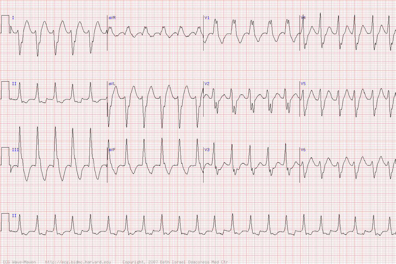
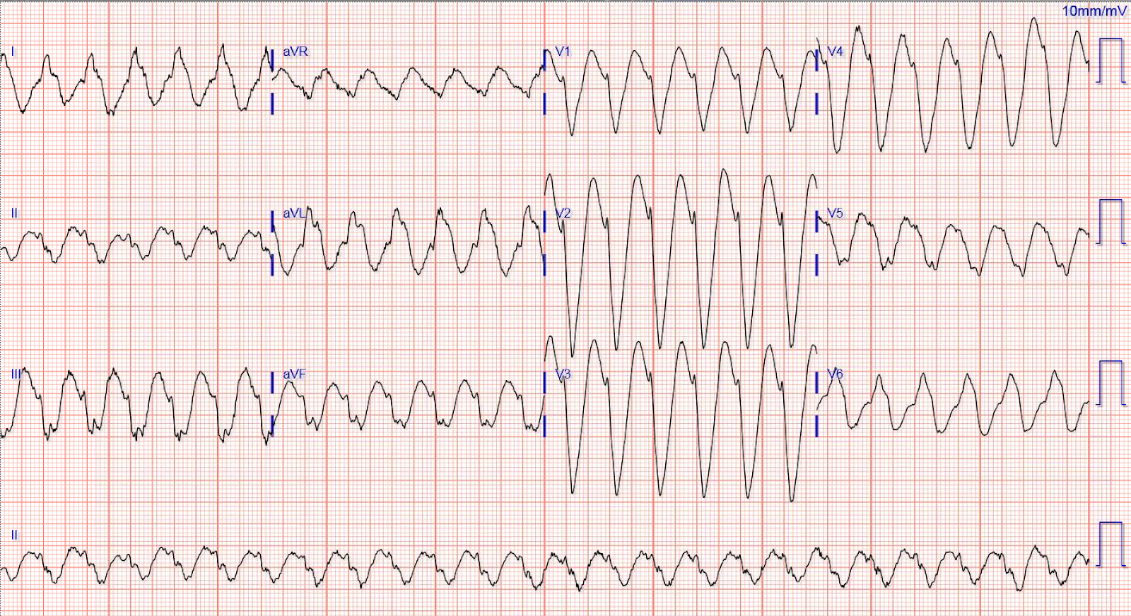
📈 ECG Features
- Wide QRS complex >120 ms
- AV dissociation, fusion/capture beats
- Extreme axis deviation or RS absence in precordial leads
- Monomorphic vs polymorphic (torsades de pointes: polymorphic, twisting QRS around baseline)
12-Lead ECG - Ventricular Tachycardia
🔀 Differential Diagnosis of Regular Wide-Complex Tachycardia
- ⚡ Ventricular Tachycardia (assume VT if unsure)
- ↔️ SVT with bundle branch block
- ↔️ Atrial flutter with BBB
- 🧪 Severe hyperkalaemia
- ⚡ Pre-excited atrial tachycardia (WPW)
🧪 Investigations
- Bloods: FBC, U&E, Mg²⁺, Ca²⁺, lactate, ABG, troponin
- ECG: continuous monitoring, 12-lead for diagnosis
- Echocardiography: assess LV function, structural disease
- Coronary angiography if ACS suspected
🚑 Acute Management (NICE/RCUK ALS Compliant)
- ABC assessment; pulseless → CPR + defibrillation per ALS
- Unstable VT → immediate synchronized cardioversion
- Stable VT → IV antiarrhythmics (amiodarone first-line; lidocaine or procainamide alternatives)
- Torsades de pointes → IV magnesium sulfate 2 g over 15 min; correct K⁺, Mg²⁺, Ca²⁺; temporary pacing if recurrent
- Treat reversible causes: ACS, electrolyte imbalance, hypoxia, drug toxicity
🩺 Long-Term Management
- Beta-blockers for non-sustained VT in structurally normal hearts
- EF <35–30% → consider ICD/AICD for primary prevention
- Antiarrhythmic therapy guided by cardiology/EP team
- Catheter ablation for recurrent, symptomatic VT
- Regular follow-up with ECG, Holter, and echocardiography
⚡ Ventricular Tachycardia Management Flowchart (RCUK/NICE Compliant)
→ CPR + Defibrillation (ALS algorithm)
→ Treat reversible causes (H’s & T’s)
→ Assess haemodynamic stability
- IV access, oxygen, ECG
- Correct electrolytes & reversible causes
- IV Amiodarone 5 mg/kg over 20–60 min (max 300 mg)
- Continuous monitoring, admit to CCU
- Signs: Low BP, shock, syncope, angina, acute HF
- Immediate synchronized DC cardioversion (100–200 J biphasic)
- Conscious → sedation if possible
- Post-shock → IV amiodarone if VT persists
- IV Magnesium sulfate 2 g over 15 min
- Correct K⁺, Mg²⁺, Ca²⁺
- Stop QT-prolonging drugs
- Temporary pacing if recurrent / symptomatic
🩺 Long-Term / Secondary Prevention
- Beta-blockers for non-sustained VT in structurally normal hearts
- EF <35–30% → ICD / AICD for primary prevention
- Antiarrhythmic therapy under cardiology / EP guidance
- Catheter ablation for recurrent VT
- Regular follow-up: ECG, Holter, echocardiography
| The content on this website is provided for educational and informational purposes only to support exam preparation (e.g., MLA, MRCP, USMLE) and learning. This is NOT medical advice, diagnosis, treatment, or professional guidance. It does not replace consultation with a qualified healthcare professional, official guidelines (e.g., NICE, GMC, BNF), or supervised clinical practice. Always verify information with current, authoritative sources. Makindo and its contributors accept no liability for any reliance on this content, including errors, omissions, or any resulting harm, loss, or consequences. By using this site, you agree to these terms. |
|
|
Categories
- About
- Acute Medicine
- Anaesthetics and Critical Care
- Anatomy
- Anatomy and Physiology
- Biochemistry
- Book
- Cardiology
- Collections
- CompSci
- Crib Sheets
- Critical care
- Dental
- Dermatology
- Differentials
- Drugs
- ENT
- Electrocardiogram
- Embryology
- Emergency Medicine
- Endocrinology
- Ethics
- Foundation Doctors
- GCSE
- Gastroenterology
- General Practice
- Genetics
- Geriatric Medicine
- Geriatrics
- Guidelines
- Haematology
- Hepatology
- Immunology
- Infectious Diseases
- Infographic
- Investigations
- Lists
- MRCP
- Mandatory Training
- Medical Students
- Microbiology
- Nephrology
- Neurology
- Neurosurgery
- Nutrition
- OSCE
- Obstetrics Gynaecology
- Oncology
- Ophthalmology
- Oral Medicine and Dentistry
- Orthopaedics
- Paediatrics
- Palliative
- Palliative Care
- Pathology
- Pharmacology
- Physiology
- Procedures
- Psychiatry
- Public Health
- Radiology
- Respiratory
- Resuscitation
- Revision Topics
- Rheumatology
- Statistics and Research
- Stroke
- Surgery
- Toxicology
- Trauma and Orthopaedics
- USMLE
- Urology
- Vascular Surgery