
| Download the amazing global Makindo app: ✅ Means NICE/National Guidelines 2026 compliant Android | Apple | |
|---|---|
| MEDICAL DISCLAIMER: Educational use only. Not for diagnosis or management. See below for full disclaimer. |
Skull Anatomy
The skull provides structural protection for the brain, support for the face, and passage for cranial nerves and major vessels. For clinical practice, skull anatomy is best understood by combining bones, named foramina, transmitted structures, and embryological development. This integrated approach helps explain fracture patterns, cranial nerve deficits, and congenital abnormalities.
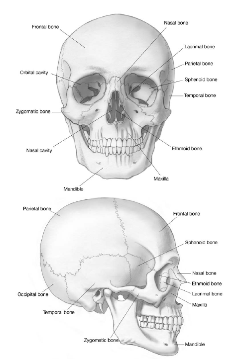
🧩 Bones of the Skull
The skull is divided into the neurocranium (protecting the brain) and the viscerocranium (forming the facial skeleton). The neurocranium consists of eight bones forming the cranial vault and base, while the facial skeleton comprises fourteen bones.
- Neurocranium: Frontal, parietal (×2), temporal (×2), occipital, sphenoid, ethmoid
- Viscerocranium: Maxilla (×2), zygomatic (×2), nasal (×2), lacrimal (×2), palatine (×2), inferior nasal concha (×2), vomer, mandible
🕳️ Skull Base Foramina – High-Yield Anatomy
The skull base contains multiple foramina that transmit cranial nerves and major vessels. Lesions at the skull base often produce characteristic clusters of cranial nerve deficits, making knowledge of these foramina clinically essential.
Anterior Cranial Fossa
- Cribriform plate (ethmoid): Olfactory nerve (CN I) → anosmia, CSF rhinorrhoea if fractured
- Optic canal (sphenoid): Optic nerve (CN II), ophthalmic artery
Middle Cranial Fossa
- Superior orbital fissure: CN III, IV, V1, VI + sympathetic fibres
- Foramen rotundum: Maxillary nerve (V2)
- Foramen ovale: Mandibular nerve (V3)
- Foramen spinosum: Middle meningeal artery (rupture → extradural haematoma)
Posterior Cranial Fossa
- Internal acoustic meatus: CN VII, VIII
- Jugular foramen: CN IX, X, XI + internal jugular vein
- Hypoglossal canal: CN XII
- Foramen magnum: Medulla, vertebral arteries, spinal root of CN XI
🧠 Relations & Clinical Correlations
The skull base is closely related to the brainstem, pituitary gland, cavernous sinus, and major vascular structures. This explains why skull base pathology can produce mixed cranial nerve palsies and autonomic symptoms. For example, cavernous sinus disease may affect eye movements, corneal sensation, and sympathetic fibres simultaneously.
- Cavernous sinus: CN III, IV, V1, V2, VI + internal carotid artery
- Middle meningeal artery runs deep to pterion
- Jugular foramen lesions → dysphagia, hoarseness, shoulder weakness
🧬 Development of the Skull
The skull develops from both neural crest cells and mesoderm, using two distinct ossification processes. The cranial vault forms mainly by intramembranous ossification, allowing postnatal brain growth, while the skull base forms by endochondral ossification, creating a rigid platform for the brain.
- Intramembranous: Flat bones of skull vault (fontanelles allow growth)
- Endochondral: Skull base (sphenoid, occipital)
- Premature
| The content on this website is provided for educational and informational purposes only to support exam preparation (e.g., MLA, MRCP, USMLE) and learning. This is NOT medical advice, diagnosis, treatment, or professional guidance. It does not replace consultation with a qualified healthcare professional, official guidelines (e.g., NICE, GMC, BNF), or supervised clinical practice. Always verify information with current, authoritative sources. Makindo and its contributors accept no liability for any reliance on this content, including errors, omissions, or any resulting harm, loss, or consequences. By using this site, you agree to these terms. |
|
|
Categories
- About
- Acute Medicine
- Anaesthetics and Critical Care
- Anatomy
- Anatomy and Physiology
- Biochemistry
- Book
- Cardiology
- Collections
- CompSci
- Crib Sheets
- Critical care
- Dental
- Dermatology
- Differentials
- Drugs
- ENT
- Electrocardiogram
- Embryology
- Emergency Medicine
- Endocrinology
- Ethics
- Foundation Doctors
- GCSE
- Gastroenterology
- General Practice
- Genetics
- Geriatric Medicine
- Geriatrics
- Guidelines
- Haematology
- Hepatology
- Immunology
- Infectious Diseases
- Infographic
- Investigations
- Lists
- MRCP
- Mandatory Training
- Medical Students
- Microbiology
- Nephrology
- Neurology
- Neurosurgery
- Nutrition
- OSCE
- Obstetrics Gynaecology
- Oncology
- Ophthalmology
- Oral Medicine and Dentistry
- Orthopaedics
- Paediatrics
- Palliative
- Palliative Care
- Pathology
- Pharmacology
- Physiology
- Procedures
- Psychiatry
- Public Health
- Radiology
- Respiratory
- Resuscitation
- Revision Topics
- Rheumatology
- Statistics and Research
- Stroke
- Surgery
- Toxicology
- Trauma and Orthopaedics
- USMLE
- Urology
- Vascular Surgery