Related Subjects:
| Vascular Surgery: Introduction
| Acute Limb Ischaemia
| Ankle-Brachial Pressure Index (ABPI) and Peripheral Vascular Disease
| Peripheral Arterial Disease (PAD)
| Waterlow Score
🦵 Leg Ulcers are chronic wounds of the lower limb caused by impaired blood supply, pressure, or systemic disease.
They are important to recognise, as correct diagnosis guides treatment - compression can heal venous ulcers but is dangerous in severe arterial disease.
📚 In exams, you should always consider venous vs arterial vs neuropathic causes first, but remember rarer systemic and malignant ulcers.
🧬 Pathophysiology
- Venous ulcers: Venous hypertension from valve incompetence or DVT → capillary leak → oedema → tissue hypoxia → ulceration.
- Arterial ulcers: Atherosclerotic obstruction → ischaemia → tissue necrosis, especially at distal pressure points.
- Neuropathic ulcers: Loss of protective sensation (e.g., diabetes) → repeated trauma/pressure at plantar sites.
- Inflammatory/autoimmune ulcers: Vasculitis or neutrophilic dermatoses (e.g., pyoderma gangrenosum).
- Malignant ulcers: Neoplastic transformation of chronic wounds (e.g., Marjolin’s ulcer).
📝 History Taking
- ⏳ Duration, recurrence, progression of ulcer.
- ⚡ Pain: severe rest pain (arterial), minimal pain (venous), painless (neuropathic).
- 🧑⚕️ PMH: diabetes, PVD, DVT, varicose veins, autoimmune disease, IBD.
- 💊 Drugs: anticoagulants, steroids, immunosuppressants.
- 👣 Risk factors: smoking, immobility, trauma, footwear.
👀 Examination
- 📍 Site: medial malleolus (venous), toes/heel (arterial), plantar foot (neuropathic).
- 🔍 Appearance: shallow/granulating (venous), punched-out (arterial), undermined edge (pyoderma), rolled edge (malignant).
- 🩸 Circulation: palpate pulses, capillary refill, measure ABPI.
- 🦶 Neurology: sensation, vibration, proprioception (for neuropathy).
- 🌍 Surrounding skin: eczema, lipodermatosclerosis, atrophy blanche, cellulitis.
🔎 Investigations
- 🩺 ABPI (Ankle–Brachial Pressure Index): >0.8 safe for compression; <0.5 suggests severe arterial disease.
- 🧪 Bloods: FBC, glucose/HbA1c, ESR/CRP (vasculitis, infection).
- 🩸 Doppler ultrasound: venous reflux, arterial stenosis.
- 📷 Imaging: angiography or CTA/MRA if arterial disease suspected.
- 🧫 Biopsy: if non-healing, atypical, or suspected malignant ulcer.
🟢 Venous Ulcers
- Description: Above medial malleolus, may extend circumferentially, sometimes bilateral.
- Features: Granulating base, oedema, varicosities, haemosiderin pigmentation, lipodermatosclerosis.
- Treatment: Compression therapy if ABPI >0.8, elevation, dressings, emollients/steroids for eczema, optimise nutrition/mobility.
🔴 Arterial Ulcers
- Description: Toes, heel, lateral malleolus; “punched-out”, cold, very painful.
- Features: Low ABPI, absent pulses, shiny hairless skin.
- Treatment: Urgent vascular referral → angioplasty, bypass. Analgesia. Amputation if non-salvageable.
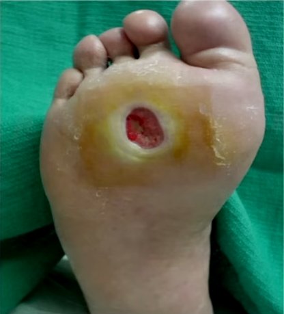
🦶 Neuropathic Ulcers
- Description: Plantar pressure sites (heel, metatarsal heads), painless.
- Features: Loss of sensation, common in diabetes, alcoholism.
- Treatment: Pressure offloading (orthotics), regular podiatry, debridement, optimise glycaemic control.
🟠 Vasculitis Ulcers
- Description: Over tibia, irregular, inflamed.
- Features: Associated systemic signs: rash, arthralgia, renal impairment.
- Treatment: Immunosuppression (steroids, azathioprine, biologics), specialist input.
🟣 Pyoderma Gangrenosum
- Description: Painful, violaceous undermined edge. Pathergy (worsens after trauma/debridement).
- Features: Linked to IBD, RA, haematological disease.
- Treatment: ❌ Avoid surgical debridement. Systemic steroids, ciclosporin, biologics. Specialist referral essential.
🧫 Malignant Ulcers
- Description: Non-healing ulcer, may arise from chronic venous ulcer (Marjolin’s).
- Features: Raised/rolled edge, contact bleeding, resistant to standard care.
- Treatment: Biopsy, wide local excision ± oncological therapy.
⚡ Traumatic Ulcers
- Description: From injury, esp. in diabetics or neuropathy.
- Features: At trauma/pressure sites.
- Treatment: Prevent recurrence, optimise footwear, wound care.
📊 Comparison Table: Venous vs Arterial vs Neuropathic Ulcers
|
|
| Feature |
Venous 🟢 |
Arterial 🔴 |
Neuropathic 🦶 |
| Typical Site |
Medial malleolus, gaiter area |
Toes, heel, lateral malleolus |
Plantar pressure points (metatarsal heads, heel) |
| Pain |
Mild–moderate, relieved by elevation |
Severe, worse at night/rest, relieved by hanging leg down |
Painless (loss of sensation) |
| Ulcer Edge |
Sloping, irregular |
Punched-out, well defined |
Variable, often surrounded by callus |
| Surrounding Skin |
Oedema, haemosiderin pigmentation, varicosities, eczema |
Shiny, hairless, cool, poor cap refill |
Normal or callused, may have Charcot changes |
| Pulses |
Present |
Absent/weak |
Present (unless coexisting PVD) |
| ABPI |
>0.8 (normal) |
<0.8 (low) |
Usually normal |
| Management |
Compression therapy, dressings, elevation |
Revascularisation (angioplasty/bypass), analgesia |
Offloading (orthotics), debridement, glycaemic control |
Neuropathic Ulcer
📚 Summary & Exam Pearls
- 📍 Site + appearance often guides diagnosis.
- 🧮 Always measure ABPI before applying compression.
- 🩺 “Ulcer + varicose veins” → think venous.
“Painful punched-out toe ulcer” → think arterial.
“Painless plantar ulcer in diabetic” → think neuropathic.
- 🔬 Non-healing or atypical ulcers → always biopsy to rule out malignancy.
