
| Download the amazing global Makindo app: ✅ Means NICE/National Guidelines 2026 compliant Android | Apple | |
|---|---|
| MEDICAL DISCLAIMER: Educational use only. Not for diagnosis or management. See below for full disclaimer. |
Ultrasound Physics & Imaging
⚛️ Physics Basics🩻
- 🔊 Ultrasound = sound waves above human hearing (>20 kHz). Medical imaging typically uses 1–20 MHz.
- 📡 When waves pass through tissues, they are transmitted or reflected depending on differences in acoustic impedance (density × speed of sound).
- ➡️ The greater the impedance difference at a tissue boundary, the more reflection occurs (bright echo).
- 🔬 Transducer: contains piezoelectric crystals. About 1% emit ultrasound; 99% detect echoes.
- 🧴 Coupling gel eliminates air between probe and skin, allowing transmission into the body.
- 📈 Frequency trade-off:
- High frequency → high resolution, shallow penetration (e.g., superficial structures, MSK).
- Low frequency → deeper penetration, lower resolution (e.g., abdomen, pelvis).
- 🖥️ Echoes displayed as 2D image with characteristic appearances:
- Bone & calculi: bright white with acoustic shadow beneath.
- Fluid (blood, urine, bile, water): black (anechoic).
- Solid organs: grey (variable echogenicity).
- Interfaces: may appear brighter (acoustic enhancement).
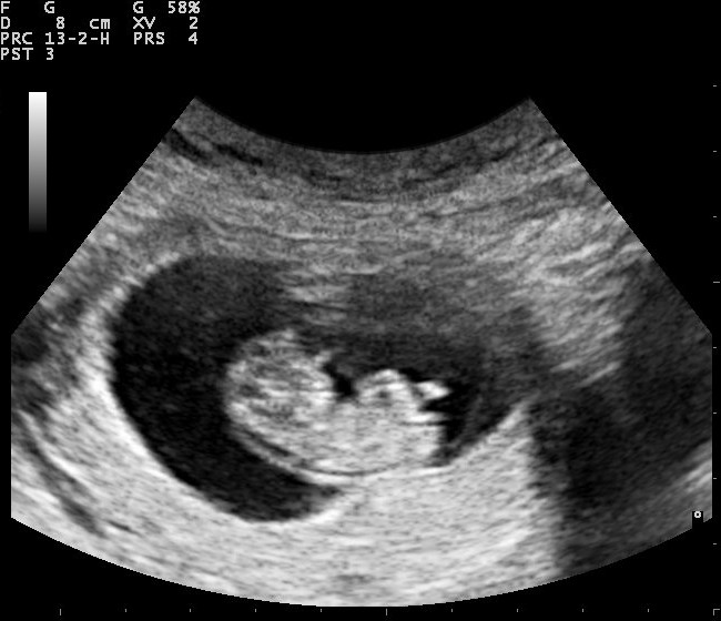
👶 Obstetric Imaging
Fetus seen in utero with ultrasound.
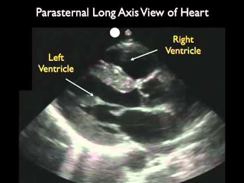
❤️ Cardiac Imaging
Heart seen on echocardiography (dynamic imaging of chambers, valves, flow).
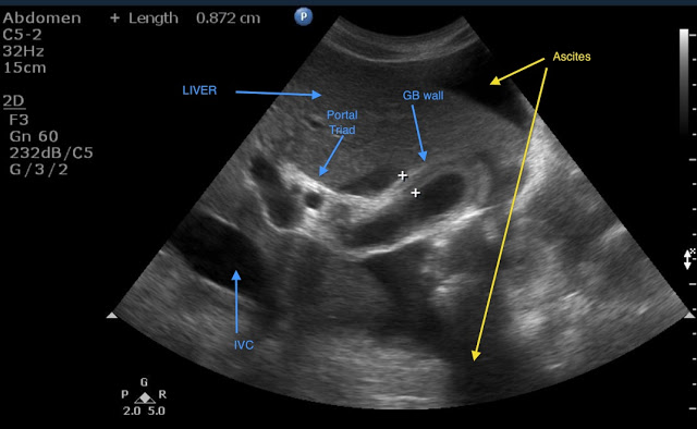
🍃 Hepatobiliary Imaging
Liver, gallbladder, bile ducts and related structures on ultrasound.
📌 Exam Pearls
- Bone & gas = enemies of ultrasound (reflect/scatter waves, blocking deeper view).
- FAST scan in trauma = free intraperitoneal or pericardial fluid.
- Doppler ultrasound = assesses flow direction & velocity.
- Always optimise gain, depth, and frequency for best image.
| The content on this website is provided for educational and informational purposes only to support exam preparation (e.g., MLA, MRCP, USMLE) and learning. This is NOT medical advice, diagnosis, treatment, or professional guidance. It does not replace consultation with a qualified healthcare professional, official guidelines (e.g., NICE, GMC, BNF), or supervised clinical practice. Always verify information with current, authoritative sources. Makindo and its contributors accept no liability for any reliance on this content, including errors, omissions, or any resulting harm, loss, or consequences. By using this site, you agree to these terms. |
|
|
Categories
- About
- Acute Medicine
- Anaesthetics and Critical Care
- Anatomy
- Anatomy and Physiology
- Biochemistry
- Book
- Cardiology
- Collections
- CompSci
- Crib Sheets
- Critical care
- Dental
- Dermatology
- Differentials
- Drugs
- ENT
- Electrocardiogram
- Embryology
- Emergency Medicine
- Endocrinology
- Ethics
- Foundation Doctors
- GCSE
- Gastroenterology
- General Practice
- Genetics
- Geriatric Medicine
- Geriatrics
- Guidelines
- Haematology
- Hepatology
- Immunology
- Infectious Diseases
- Infographic
- Investigations
- Lists
- MRCP
- Mandatory Training
- Medical Students
- Microbiology
- Nephrology
- Neurology
- Neurosurgery
- Nutrition
- OSCE
- Obstetrics Gynaecology
- Oncology
- Ophthalmology
- Oral Medicine and Dentistry
- Orthopaedics
- Paediatrics
- Palliative
- Palliative Care
- Pathology
- Pharmacology
- Physiology
- Procedures
- Psychiatry
- Public Health
- Radiology
- Respiratory
- Resuscitation
- Revision Topics
- Rheumatology
- Statistics and Research
- Stroke
- Surgery
- Toxicology
- Trauma and Orthopaedics
- USMLE
- Urology
- Vascular Surgery