
| Download the amazing global Makindo app: Android | Apple | |
|---|---|
| MEDICAL DISCLAIMER: Educational use only. Not for diagnosis or management. See below for full disclaimer. |
Anatomy of Spinal Column
Related Subjects: |Anatomy of Skin |Anatomy of the Hand |Anatomy of the Thorax |Anatomy of Muscle Groups |Anatomy of Arteries |Anatomy of Spinal Column
🦴 Bony Anatomy of the Spine
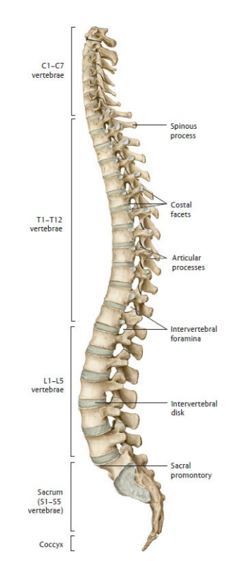
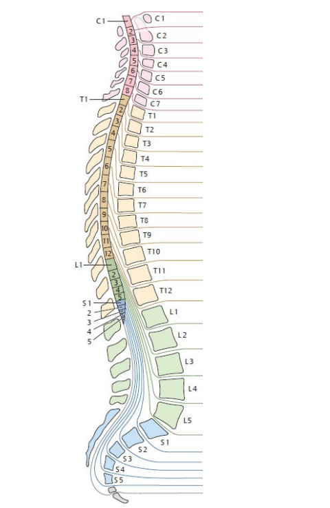
The vertebral column (spine) is the central structural axis of the skeleton. It supports body weight, protects the spinal cord, enables movement, and maintains posture. It is composed of 33 vertebrae, grouped into regions reflecting both form and function.
🧱 Regions of the Spine
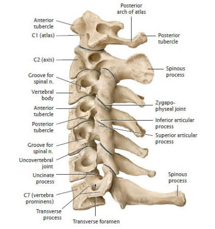
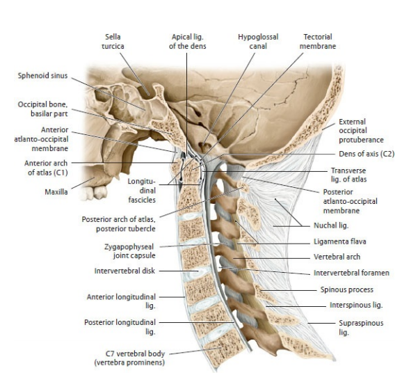
- Cervical (C1–C7):
- Supports the skull and permits a wide range of motion - flexion, extension, rotation, and lateral flexion.
- Atlas (C1): ring-shaped, supports the skull via the occipital condyles.
- Axis (C2): contains the dens (odontoid process) acting as a pivot for rotation.
- C7 - the vertebra prominens - is easily palpable at the neck base.
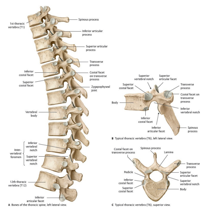
- Thoracic (T1–T12):
- Articulates with ribs to form the thoracic cage.
- Provides stability and protection for thoracic viscera (heart, lungs).
- Limited flexion and extension due to rib attachment and coronal facet orientation.
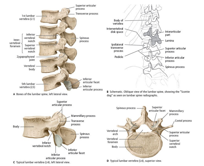
- Lumbar (L1–L5):
- Large, kidney-shaped vertebral bodies bear the greatest load.
- Facet joints oriented sagittally - allowing flexion and extension but limiting rotation.
- Common site for disc herniation and mechanical back pain.
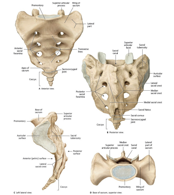
- Sacral (S1–S5):
- Five fused vertebrae forming the sacrum.
- Transmits body weight to the pelvis via sacroiliac joints.
- Contains sacral foramina for exit of sacral nerves.
- Coccygeal (3–5 fused):
- Forms the coccyx or tailbone - vestigial structure providing attachment for pelvic floor muscles (coccygeus, levator ani).
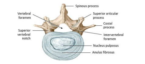
🧩 Structure of a Typical Vertebra
- Vertebral Body: Cylindrical, weight-bearing anterior part. Covered by hyaline cartilage endplates; separated by intervertebral discs.
- Vertebral Arch: Formed by pedicles and laminae; encloses the vertebral foramen (spinal canal).
- Processes:
- Spinous Process: Posterior projection for muscle/ligament attachment.
- Transverse Processes: Lateral projections; in thoracic vertebrae, articulate with ribs.
- Articular Processes: Superior and inferior facets form synovial facet (zygapophyseal) joints, guiding motion.
💠 Intervertebral Discs
- Located between vertebral bodies (C2–S1).
- Nucleus pulposus: Gelatinous centre acting as a shock absorber.
- Annulus fibrosus: Concentric fibrocartilaginous rings that contain and stabilise the nucleus.
- Degeneration or herniation may compress spinal nerves, causing radiculopathy.
🩻 Curvatures of the Spine
- Cervical and lumbar lordosis: Convex anteriorly, develop postnatally (secondary curves).
- Thoracic and sacral kyphosis: Concave anteriorly, primary fetal curves.
- These curvatures distribute axial load and provide flexibility and shock absorption.
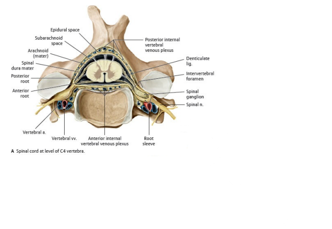
🧠 Neural Anatomy - Spinal Cord and Nerves
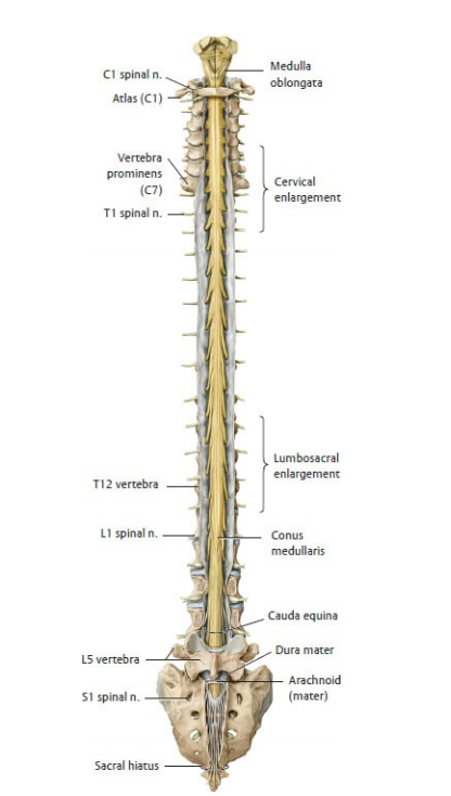
The spinal cord is a continuation of the medulla oblongata, ending at the L1–L2 vertebral level in adults. It lies within the vertebral canal and is protected by meninges and cerebrospinal fluid (CSF).
- Spinal Nerves: 31 pairs - 8 cervical, 12 thoracic, 5 lumbar, 5 sacral, 1 coccygeal.
- Each forms from:
- Anterior (ventral) root: Motor (efferent) fibres from anterior horn.
- Posterior (dorsal) root: Sensory (afferent) fibres with cell bodies in dorsal root ganglia.
- Nerves exit through intervertebral foramina.
- C1–C7 exit above corresponding vertebrae; C8 exits below C7.
- T1–Co1 exit below corresponding vertebrae.
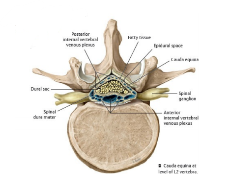
- Below L1, nerve roots descend in the dural sac forming the cauda equina.
- The terminal filum (pia mater extension) anchors the cord to the coccyx.
🧬 Spinal Cord Physiology
- Grey Matter: Butterfly-shaped core; contains neuronal cell bodies.
- Anterior horn: motor neurons.
- Posterior horn: sensory interneurons.
- Lateral horn: sympathetic neurons (T1–L2).
- White Matter: Myelinated ascending and descending tracts.
- Ascending (sensory): dorsal columns, spinothalamic, spinocerebellar tracts.
- Descending (motor): corticospinal (pyramidal), rubrospinal, vestibulospinal tracts.
- Reflex Arcs: Automatic responses mediated by spinal circuits (e.g. patellar reflex at L3–L4).
- Dermatomes: Areas of skin supplied by a single spinal nerve; clinically used to localise lesions.
- Myotomes: Muscle groups innervated by a specific spinal segment.
🩸 Vascular Supply of the Spinal Cord
- Anterior spinal artery: Formed from vertebral branches; supplies anterior two-thirds of cord (motor tracts).
- Posterior spinal arteries (2): Supply posterior one-third (sensory tracts).
- Radicular arteries: Reinforce blood supply; the largest is the artery of Adamkiewicz (T9–L2).
- Venous drainage via internal vertebral venous plexus - valveless, facilitating metastatic spread (Batson’s plexus).
⚙️ Biomechanics and Function of the Spine
- Support: Bears axial load and transmits body weight to the pelvis.
- Protection: Vertebral canal shields the spinal cord and meninges.
- Movement: Facilitates flexion, extension, lateral flexion, and rotation; greatest in cervical and lumbar regions.
- Shock Absorption: Achieved via intervertebral discs and curvature elasticity.
- Posture and Balance: Paraspinal muscles (erector spinae, multifidus) and ligaments maintain upright posture.
🪶 Ligaments of the Spine
- Anterior longitudinal ligament: Prevents hyperextension.
- Posterior longitudinal ligament: Prevents hyperflexion; runs within the vertebral canal.
- Ligamentum flavum: Elastic; connects laminae, aids recoil after flexion.
- Interspinous and supraspinous ligaments: Limit flexion.
- Nuchal ligament: Thickened supraspinous ligament in cervical region; supports head.
🧪 Clinical Correlations
- Disc Herniation: Protrusion of nucleus pulposus compressing spinal nerve root - e.g. L5/S1 → sciatica.
- Spinal Stenosis: Narrowing of vertebral canal causing neurogenic claudication.
- Scoliosis: Lateral curvature with vertebral rotation.
- Kyphosis: Exaggerated thoracic curvature (common in osteoporosis).
- Lordosis: Increased lumbar curvature (seen in obesity, pregnancy).
- Spinal Cord Injury: Lesion level determines neurological deficit (e.g. C4 injury → diaphragmatic paralysis).
🧫 Embryological and Evolutionary Notes
- Derived from the sclerotome (paraxial mesoderm).
- Spinal cord initially extends entire vertebral length - ascends during growth, forming cauda equina.
- Curvatures develop postnatally as a functional adaptation for bipedal stance.
📚 Summary of Key Visuals
Cervical Region
Thoracic Region
Lumbar Region
Sacral Region
Skull and Atlas-Axis Relationship
Spinal Cord Anatomy
Spinal Segments
Intervertebral Disc
Cervical Cord (C4)
Cauda Equina (L4)
🧠 Teaching Tip: The spine is not a rigid pillar but a dynamic organ of support, protection, and motion. Each vertebra contributes to a delicate balance between stability and flexibility, while the spinal cord transforms mechanical input into neural output - the living bridge between brain and body. Understanding this integration of structure and function is the foundation of all clinical neurology and musculoskeletal medicine.
| The content on this website is provided for educational and informational purposes only to support exam preparation (e.g., MLA, MRCP, USMLE) and learning. This is NOT medical advice, diagnosis, treatment, or professional guidance. It does not replace consultation with a qualified healthcare professional, official guidelines (e.g., NICE, GMC, BNF), or supervised clinical practice. Always verify information with current, authoritative sources. Makindo and its contributors accept no liability for any reliance on this content, including errors, omissions, or any resulting harm, loss, or consequences. By using this site, you agree to these terms. |
|
|
Categories
- About
- Acute Medicine
- Anaesthetics and Critical Care
- Anatomy
- Anatomy and Physiology
- Biochemistry
- Book
- Cardiology
- Collections
- CompSci
- Crib Sheets
- Critical care
- Dental
- Dermatology
- Differentials
- Drugs
- ENT
- Electrocardiogram
- Embryology
- Emergency Medicine
- Endocrinology
- Ethics
- Foundation Doctors
- GCSE
- Gastroenterology
- General Practice
- Genetics
- Geriatric Medicine
- Geriatrics
- Guidelines
- Haematology
- Hepatology
- Immunology
- Infectious Diseases
- Infographic
- Investigations
- Lists
- MRCP
- Mandatory Training
- Medical Students
- Microbiology
- Nephrology
- Neurology
- Neurosurgery
- Nutrition
- OSCE
- Obstetrics Gynaecology
- Oncology
- Ophthalmology
- Oral Medicine and Dentistry
- Orthopaedics
- Paediatrics
- Palliative
- Palliative Care
- Pathology
- Pharmacology
- Physiology
- Procedures
- Psychiatry
- Public Health
- Radiology
- Respiratory
- Resuscitation
- Revision Topics
- Rheumatology
- Statistics and Research
- Stroke
- Surgery
- Toxicology
- Trauma and Orthopaedics
- USMLE
- Urology
- Vascular Surgery