
| Download the amazing global Makindo app: ✅ Means NICE/National Guidelines 2026 compliant Android | Apple | |
|---|---|
| MEDICAL DISCLAIMER: Educational use only. Not for diagnosis or management. See below for full disclaimer. |
Extradural haemorrhage
Quick facts | emergency priorities
Clinical summary: This entry requires a disease-specific recognition, red-flag and emergency-management review. Refer to the underlying article and current local pathway; do not use this panel as a treatment protocol.
Browse the Emergency Medicine textbook: Emergency Medicine: Environmental emergencies (specialty landing page only; no disease-level match confirmed)
References: Existing source link (not yet checked)
Editorial draft: the full historic text, doses, and pathways have not been clinically signed off. Cross-check BNF/BNFC and local protocols.
🩺 UK targeted safety update — 4 November 2026
Extradural haemorrhage: imaging and neurosurgical urgency
- Suspected intracranial bleeding after head trauma warrants urgent assessment, GCS/neurological monitoring and CT according to NICE NG232.
- A lucid interval is neither required nor reliably present; a deteriorating patient needs immediate resuscitation and neurosurgical discussion.
- Look for anticoagulant exposure, coagulopathy, seizures and evolving signs of raised intracranial pressure.
UK source: guidance / safety update. Only the specific points above were source checked. All other historical text, doses and procedural instructions remain unvalidated: verify current BNF/BNFC and local guidance before clinical use.
🧠 The classic presentation of an Extradural Haematoma (EDH) is: ➡️ Head injury → brief loss of consciousness → lucid interval → rapid decline in GCS. 🚨 This is a neurosurgical emergency requiring urgent CT imaging and intervention.
💡 About
- EDH = bleeding between the dura mater and skull, confined by the skull’s periosteal suture lines.
- Classically arterial, most often from the middle meningeal artery.
- Rapidly progressive → may cause brain herniation if untreated.
- Accounts for ~2% of head injuries, typically in young adults (20–30 years), often after high-impact trauma.
🔎 Aetiology & Causes
- Rupture of the middle meningeal artery (or vein).
- Associated with temporal/parietal skull fractures.
- Common causes:
- Falls 🪂
- Road traffic collisions 🚗
- Assaults 🥊
- Sports injuries (e.g., skiing) ⛷️
- Rarely: birth trauma in neonates 👶
🩺 Clinical Features
- Initial head trauma ± scalp wound.
- Lucid interval → temporary recovery → rapid neurological deterioration.
- ↓ GCS, confusion, drowsiness, then coma.
- Focal neurology (e.g., hemiparesis, dilated ipsilateral pupil from CN III palsy).
- Raised ICP signs: Cheyne–Stokes respiration, bradycardia, hypertension (Cushing’s triad).
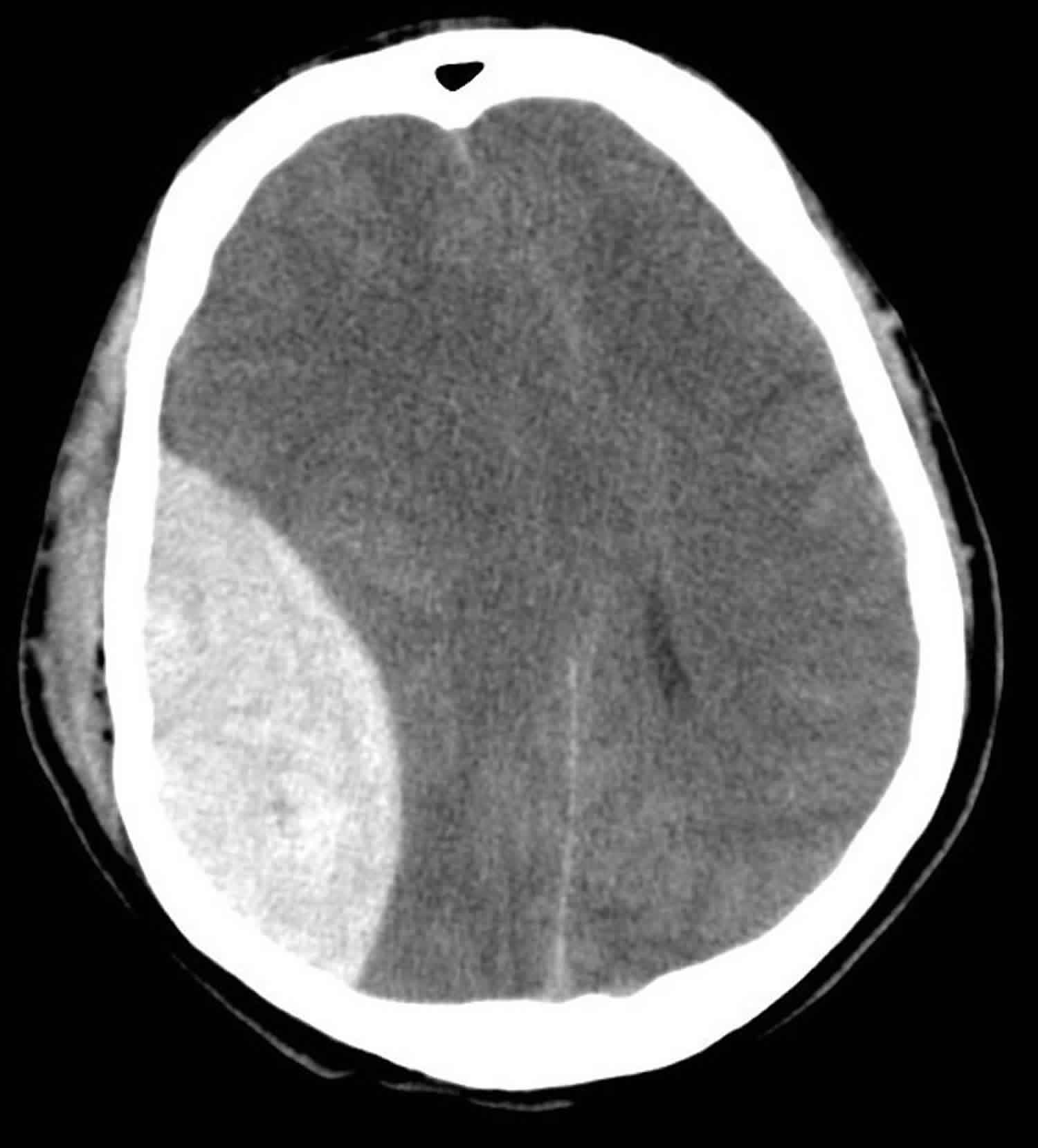
🧪 Investigations
- CT Head (Gold Standard):
- Biconvex (lens-shaped) hyperdense lesion.
- Confined by suture lines (unlike subdural haematoma).
- May show midline shift ± skull fracture.
- Bloods: FBC, U&E, LFTs, clotting screen (relevant if anticoagulated).
⚡ Management
- Initial ABC resuscitation: Secure airway, give O₂, IV fluids. Intubate if GCS < 8.
- Urgent neurosurgical referral in all suspected cases.
- Reverse coagulopathy (vitamin K, PCC if on warfarin; stop antiplatelets if safe).
- Surgery:
- Immediate evacuation indicated if clot >30 cm³, thickness >15 mm, midline shift >5 mm, or GCS ≤8 with focal deficit.
- Craniotomy = definitive treatment.
- Burr hole may be lifesaving if deterioration occurs before theatre access.
- Post-operative: ICU care with ICP monitoring, neuro-rehab as required.
📋 Key Exam Tips
✅ Lucid interval is the buzzword for EDH (but not always present).
✅ CT: biconvex/lens-shaped bleed, confined by sutures.
✅ Differentiate from Subdural Haematoma (crescent-shaped, not limited by sutures).
✅ Always think middle meningeal artery rupture in temporal fracture.
| The content on this website is provided for educational and informational purposes only to support exam preparation (e.g., MLA, MRCP, USMLE) and learning. This is NOT medical advice, diagnosis, treatment, or professional guidance. It does not replace consultation with a qualified healthcare professional, official guidelines (e.g., NICE, GMC, BNF), or supervised clinical practice. Always verify information with current, authoritative sources. Makindo and its contributors accept no liability for any reliance on this content, including errors, omissions, or any resulting harm, loss, or consequences. By using this site, you agree to these terms. |
|
|
Categories
- Acute Medicine
- Anaesthetics and Critical Care
- Anatomy
- Anatomy and Physiology
- Biochemistry
- Cardiology
- Clinical Genetics
- Clinical Immunology
- Collections
- Critical Care
- Critical care
- Dental
- Dermatology
- Differentials
- Electrocardiogram
- Embryology
- Emergency Medicine
- Endocrinology
- ENT
- Gastroenterology
- Gastroenterology & Hepatology
- General Practice
- Genetics
- Geriatric Medicine
- Guidelines
- Gynaecology
- Haematology
- Hepatology
- Immunology
- Infectious Diseases
- Infographic
- Investigations
- Lists
- Microbiology
- Nephrology
- Neurology
- Neurology & Stroke
- Neurosurgery
- Obstetrics
- Obstetrics Gynaecology
- Oncology
- Ophthalmology
- Oral Medicine and Dentistry
- OSCE
- Paediatrics
- Palliative
- Palliative Medicine
- Pathology
- Pharmacology
- Physiology
- Procedures
- Psychiatry
- Public Health
- Radiology
- Renal Medicine
- Respiratory
- Respiratory Medicine
- Resuscitation
- Revision Topics
- Rheumatology
- Stroke
- Surgery
- Toxicology
- Trauma and Orthopaedics
- Urology
- Vascular Surgery