
| Download the amazing global Makindo app: ✅ Means NICE/National Guidelines 2026 compliant Android | Apple | |
|---|---|
| MEDICAL DISCLAIMER: Educational use only. Not for diagnosis or management. See below for full disclaimer. |
Anomalous Coronary Arteries
Quick facts | emergency priorities
Clinical summary: This entry requires a disease-specific recognition, red-flag and emergency-management review. Refer to the underlying article and current local pathway; do not use this panel as a treatment protocol.
Browse the Cardiology textbook: Cardiology: Presentations (specialty landing page only; no disease-level match confirmed)
Condition-specific reference needed. NICE guidance index is a discovery route, not a disease-specific citation.
Editorial draft: the full historic text, doses, and pathways have not been clinically signed off. Cross-check BNF/BNFC and local protocols.
Related Subjects: |Brugada Syndrome |Long QT syndrome (LQTS) Acquired |Long QT syndrome (LQTS) Congenital |Torsades de Pointes |Ventricular Fibrillation |Ventricular Tachycardia |Resuscitation - Adult Tachycardia Algorithm |Automatic Implantable Cardioverter Defibrillator (AICD)
ℹ️ About Anomalous Coronary Arteries
- Rare congenital variants affecting coronary origin or course.
- Most are harmless incidental findings, but certain subtypes carry risk of myocardial ischemia or sudden death.
- High-risk features include interarterial course (between aorta and pulmonary artery), intramural segment, slit-like ostium, acute take-off angle, or proximal narrowing - these can cause compression during exercise.
📊 Incidence & Epidemiology
These anomalies are uncommon but increasingly detected thanks to advanced imaging. Prevalence varies by detection method and anomaly type.
- Overall Prevalence: ~0.3–1.3% in angiography/CT studies; up to 1–5.6% in some reports; higher in autopsy/specialized imaging (many asymptomatic cases undetected).
- Age & Gender: ALCAPA often presents in infancy with severe symptoms; anomalous origin from opposite sinus (e.g., right from left) may appear in adolescence/adulthood. Slight male predominance in some high-risk types linked to sudden events.
- Sudden Death Risk: Major cause of sudden cardiac death in young athletes (<35 years); highest with interarterial course (especially left from right sinus); right from left sinus also risky but lower than left anomalies.
- Detection Trends: Rising due to CT coronary angiography (CCTA), MRI, and awareness; key in screening athletes or symptomatic young people.
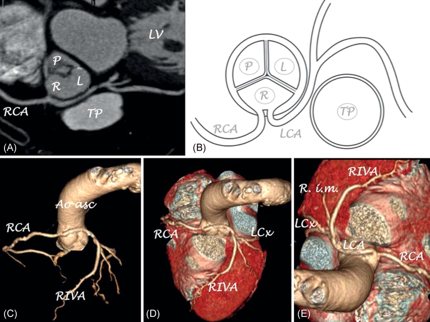
🔍 Main Types of Anomalous Coronary Arteries
| Type | Description | Risk Level | Key Notes |
|---|---|---|---|
| ALCAPA
(Anomalous Left Coronary Artery from Pulmonary Artery) |
Left coronary arises from pulmonary artery instead of aorta. | High (malignant in infancy) | Infants: heart failure, ischemia; surgery essential early. Rare in adults if survived untreated. |
| Anomalous Right Coronary Artery from Left Sinus
(R-AAOCA/ARCA) |
RCA originates from left sinus; often interarterial course. | Moderate–High | Common high-risk type; compression risk during exercise; sudden death link. |
| Anomalous Left Coronary Artery from Right Sinus
(L-AAOCA) |
Left (or left main) from right sinus; interarterial common. | Very High | Strongest SCD association in young; often surgical indication. |
| Single Coronary Artery | One artery supplies entire heart via branches. | Variable | May be benign or cause ischemia depending on course/distribution. |
| Other Variants | e.g., High/low interarterial, retroaortic, prepulmonic, subpulmonic. | Mostly Benign (except interarterial) | Interarterial = high-risk; others usually low-risk. |
High-risk = interarterial + features like intramural/slits/acute angle.
🩺 Symptoms & Presentation
Symptoms vary widely - many people have none 😴.
- Asymptomatic (most common).
- Exertional chest pain/angina, shortness of breath, fatigue.
- Palpitations, syncope, or arrhythmias.
- In severe cases: myocardial infarction or sudden cardiac arrest ⚡.
- Infants (ALCAPA): poor feeding, sweating, irritability, heart failure signs.
Symptoms often exertion-related; high-risk anomalies worse during intense activity.
🛠️ Diagnostic Investigations
Accurate imaging is key to confirm anomaly, assess course/risk features, and guide management. Echocardiography first-line; advanced imaging essential.
- ECG: Initial screen; may show ischemia/arrhythmias (often nonspecific).
- Echocardiography ❤️: First-line, especially kids; views origin/course; stress echo for ischemia.
- CT Coronary Angiography (CCTA) 📸: Gold standard noninvasive; detailed anatomy, high-risk features (interarterial, intramural); low radiation with modern protocols.
- Cardiac MRI/CMR: No radiation; excellent for anatomy, function, perfusion, scarring; great for follow-up.
- Invasive Coronary Angiography: Reserved for complex cases or pre-intervention; real-time but invasive.
- Exercise Stress Testing: Assess inducible ischemia in active/suspected patients; helps risk-stratify.
(Recent guidelines [2025 ACC/AHA] emphasize CCTA/MRI for detailed evaluation in suspected anomalies.)
💉 Treatment Options
Depends on type, symptoms, ischemia evidence, high-risk anatomy, age, and activity level. Conservative for low-risk; surgery for high-risk/symptomatic.
- Conservative/Medical: Asymptomatic low-risk → observation, risk factor control (BP, lipids), beta-blockers for symptoms; restrict intense sports if uncertain.
- Surgical Correction (preferred for high-risk):
- Reimplantation: Move anomalous artery to correct sinus (common for AAOCA).
- Unroofing: Open intramural segment + neo-ostium creation.
- Other: PA translocation, patch augmentation; CABG rarely (competitive flow issues).
- Lifestyle & Monitoring: Activity restriction (avoid competitive sports if high-risk); regular follow-up imaging/tests; shared decision-making.
Surgery safe/effective in expert centers; relieves symptoms in most.
📈 Outcomes & Prognosis
- Benign anomalies: excellent long-term with monitoring; normal life.
- High-risk (esp. untreated interarterial): risk of SCD, but early detection/surgery dramatically improves outlook.
- Post-surgery: low mortality, symptom relief in >95%; return to activity possible after clearance.
- Individualized: based on anomaly, age, symptoms - specialist (adult congenital/pediatric cardiology) input essential.
🔚 Summary
Anomalous coronary arteries range from harmless variants to life-threatening causes of ischemia/SCD. Early detection via advanced imaging (CCTA/MRI key), risk stratification (high-risk features), and tailored management (surgery for symptomatic/high-risk) save lives - especially in young/active people. Always refer to cardiologist for personalized assessment! 🚀
| The content on this website is provided for educational and informational purposes only to support exam preparation (e.g., MLA, MRCP, USMLE) and learning. This is NOT medical advice, diagnosis, treatment, or professional guidance. It does not replace consultation with a qualified healthcare professional, official guidelines (e.g., NICE, GMC, BNF), or supervised clinical practice. Always verify information with current, authoritative sources. Makindo and its contributors accept no liability for any reliance on this content, including errors, omissions, or any resulting harm, loss, or consequences. By using this site, you agree to these terms. |
|
|
Categories
- Acute Medicine
- Anaesthetics and Critical Care
- Anatomy
- Anatomy and Physiology
- Biochemistry
- Cardiology
- Clinical Genetics
- Clinical Immunology
- Collections
- Critical Care
- Critical care
- Dental
- Dermatology
- Differentials
- Electrocardiogram
- Embryology
- Emergency Medicine
- Endocrinology
- ENT
- Gastroenterology
- Gastroenterology & Hepatology
- General Practice
- Genetics
- Geriatric Medicine
- Guidelines
- Gynaecology
- Haematology
- Hepatology
- Immunology
- Infectious Diseases
- Infographic
- Investigations
- Lists
- Microbiology
- Nephrology
- Neurology
- Neurology & Stroke
- Neurosurgery
- Obstetrics
- Obstetrics Gynaecology
- Oncology
- Ophthalmology
- Oral Medicine and Dentistry
- OSCE
- Paediatrics
- Palliative
- Palliative Medicine
- Pathology
- Pharmacology
- Physiology
- Procedures
- Psychiatry
- Public Health
- Radiology
- Renal Medicine
- Respiratory
- Respiratory Medicine
- Resuscitation
- Revision Topics
- Rheumatology
- Stroke
- Surgery
- Toxicology
- Trauma and Orthopaedics
- Urology
- Vascular Surgery