MAKINDO MEDICINEClinical textbook for medical students and doctors
For education and revision only. Not individual medical advice; check current NICE/BNF and local guidance before clinical use. Clinical safety and terms.
Chapter 191 of 268
Images
MAKINDO MEDICINE · Cardiology191 / 268

Integrated cardiology chapter

Alphabetical topic index (A–Z) all 331 topics

Each entry jumps to its section in this chapter.

A

B

C

D

E

F

G

H

I

K

L

M

N

O

P

R

S

T

U

V

W

GMC MLA cardiovascular coverage map 30 listed conditions · direct topic links

The 2026 GMC content map lists these conditions under “Heart and vasculature”; this is a map of section locations, not a claim that all content has been medically validated. The content map also tests practical skills and patient presentations.

Part 1 of 12 · 37 topics

1. Ischaemic Heart Disease and Acute Coronary Syndromes

🫀 Ischaemic heart disease (IHD), also called coronary heart disease, occurs when the heart muscle does not get enough oxygen for what it is being asked to do. The usual cause is atherosclerosis narrowing the coronary arteries, but spasm, microvascular disease, dissection and high demand can do the same.
The spectrum runs from stable angina (predictable pain on exertion) to acute coronary syndromes (a sudden clot), heart failure and sudden death.
⚠️ Clinical priority: Recognise a blocked coronary artery quickly. Every minute it stays closed, more heart muscle dies.


1.01 What Is IHD?

Ischaemia means that a tissue is receiving too little blood flow for its needs. In the heart this produces two broad clinical pictures:

  • Chronic coronary disease: A fixed narrowing limits blood flow. At rest there is enough oxygen, but during exercise demand outstrips supply and the patient gets angina. The muscle recovers when demand falls, so no permanent damage occurs. This is stable angina.
  • Acute coronary syndrome (ACS): A plaque suddenly ruptures and a clot forms on it, abruptly cutting off flow. If the blockage lasts long enough, heart muscle dies: this is a myocardial infarction (MI).

The same disease can also cause:

  • Ischaemic cardiomyopathy: Heart failure from previous infarcts.
  • Arrhythmias: Ischaemic or scarred muscle can trigger dangerous rhythms.
  • Sudden cardiac death: Usually from ventricular fibrillation.
  • Non-obstructive ischaemia: Vasospastic and microvascular angina, where the large arteries are not significantly narrowed.

💡 Clinical Pearl: Think of IHD as a problem of supply versus demand. Stable angina is a demand problem against a fixed supply; ACS is a sudden collapse in supply.

↑ Chapter index

1.02 Coronary Anatomy

The right and left coronary arteries arise from the aortic root just above the aortic valve. Knowing which artery supplies which territory lets you predict both the ECG changes and the likely complications.

  • Left main stem (LMS): Short trunk that divides into the LAD and circumflex. Significant disease here endangers most of the left ventricle.
  • Left anterior descending (LAD): Supplies the anterior wall, the front of the septum and the apex. Blockage causes an anterior MI. A proximal LAD occlusion can destroy a large area of muscle, causing heart failure and shock.
  • Left circumflex (LCx): Supplies the lateral wall and, in some people, the posterior or inferior wall. Blockage causes a lateral or posterior MI.
  • Right coronary artery (RCA): Supplies the right ventricle, the inferior wall and, in most people, the SA and AV nodes. Blockage causes an inferior MI, often with bradycardia or heart block. A proximal occlusion can also cause RV infarction.
  • Dominance: The artery that gives rise to the posterior descending artery is "dominant" — the RCA in about 85% of people.
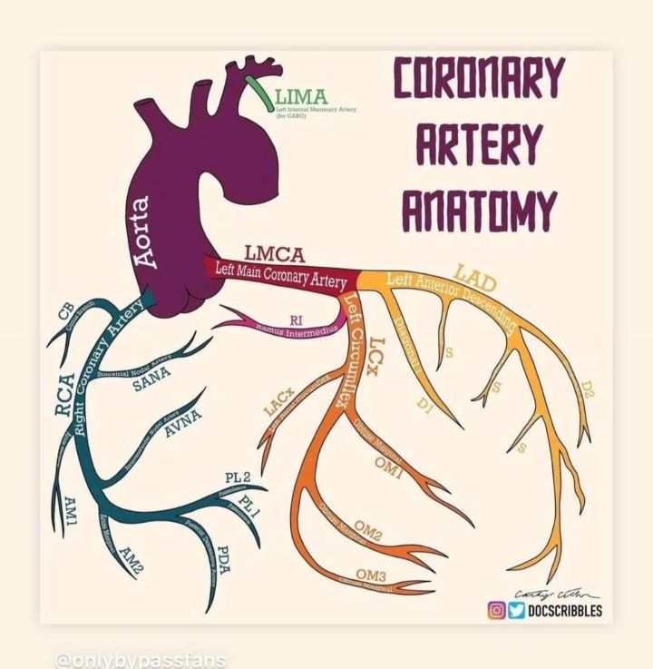
Coronary artery anatomy

💡 Clinical Pearl: Inferior MI → think bradycardia, heart block and RV infarction (RCA supplies the nodes and RV). Large anterior MI → think heart failure, shock and VT (big LV territory).

↑ Chapter index

1.03 Atherosclerosis and Plaques

Mechanism: Atherosclerosis is a slow, inflammatory disease of the artery wall that begins in childhood and progresses over decades. It develops in stages:

  • Endothelial damage: Smoking, high blood pressure, diabetes and high cholesterol damage the inner lining (endothelium), making it "leaky" and sticky.
  • LDL entry: LDL cholesterol particles pass into the vessel wall and become oxidised.
  • Inflammation: Monocytes enter the wall, become macrophages and engulf the oxidised LDL, turning into lipid-laden foam cells.
  • Fatty streak: Collections of foam cells form the earliest visible lesion, already present in many teenagers.
  • Mature plaque: Smooth muscle cells migrate in and lay down collagen, forming a fibrous cap over a soft, lipid-rich necrotic core.
  • Advanced plaque: The plaque may calcify, bleed internally, ulcerate or rupture.

Stable versus vulnerable plaque. Plaques behave very differently depending on their structure:

  • Stable plaque: Thick fibrous cap and small lipid core, often calcified. It narrows the artery gradually and causes exertional angina, but rarely ruptures.
  • Vulnerable plaque: Thin cap, large lipid core and active inflammation. It may cause little narrowing but can rupture, exposing its highly thrombogenic contents to blood. Platelets stick, a clot forms and the artery blocks, causing ACS.
  • Plaque erosion: The endothelium over a plaque is lost without the cap rupturing. This can also trigger a clot and is relatively more common in younger patients and women.

💡 Important Concept: Heart attacks often arise from plaques that were not severely narrowing the artery beforehand. This is why stenting a stable narrowing does not reliably prevent MI, whereas statins (which stabilise all plaques) do.

↑ Chapter index

1.04 Oxygen Supply and Demand

Ischaemia occurs whenever oxygen delivery falls below what the heart muscle needs. Treatments for angina work by either reducing demand or improving supply.

  • Supply depends on: Coronary blood flow and perfusion pressure, haemoglobin, oxygen saturation, and the length of diastole.
  • Demand depends on: Heart rate, contractility, wall tension, afterload (blood pressure) and preload (ventricular filling).
  • Why diastole matters: The left ventricle is mainly perfused during diastole, because in systole the contracting muscle squeezes its own small vessels shut.
  • Why tachycardia is harmful: A fast heart rate both increases demand and shortens diastole, reducing supply. This is why beta blockers and rate control are so effective.
  • Non-coronary causes of ischaemia: Severe aortic stenosis, hypertrophic cardiomyopathy, severe anaemia, hypoxia, fast arrhythmias, shock, coronary spasm, coronary embolism, spontaneous coronary artery dissection and vasculitis (e.g. Kawasaki disease).

💡 Clinical Pearl: The subendocardium (inner layer) is the most vulnerable to ischaemia. It is furthest from the epicardial arteries and under the highest pressure during systole. This is why demand ischaemia typically causes ST depression.

↑ Chapter index

1.05 Risk Factors and Risk Assessment

Non-modifiable risk factors:

  • Increasing age.
  • Male sex (risk in women rises after the menopause).
  • Family history of premature coronary disease (a first-degree male relative under 55 or female under 65).
  • Familial hypercholesterolaemia.
  • South Asian ancestry.

Modifiable risk factors:

  • Smoking (including passive smoking).
  • Raised LDL cholesterol.
  • Hypertension.
  • Diabetes.
  • Obesity, especially central.
  • Physical inactivity.
  • Diet high in saturated fat.
  • Chronic kidney disease.
  • Excess alcohol.
  • Psychosocial stress and deprivation.

Risk-enhancing factors:

  • Raised lipoprotein(a).
  • Chronic inflammatory disease (e.g. rheumatoid arthritis, SLE).
  • Previous pre-eclampsia or gestational diabetes.
  • Persistent albuminuria.
  • Obstructive sleep apnoea.
  • Severe mental illness.

UK risk assessment (NICE NG238):

  • QRISK3: Estimates 10-year cardiovascular risk in people without known cardiovascular disease.
  • Primary prevention: If QRISK3 is 10% or more, offer atorvastatin 20 mg daily after a shared discussion.
  • Secondary prevention: People who already have IHD do not need a QRISK score; they all qualify for high-intensity statin therapy.
  • Lipids: Measure total cholesterol, HDL, non-HDL cholesterol, LDL and triglycerides.
  • Familial hypercholesterolaemia: Suspect it with total cholesterol above 7.5 mmol/L and a personal or family history of premature coronary disease.

💡 Clinical Pearl: There is no "safe" cholesterol threshold — risk rises continuously with lifetime exposure to LDL. Light drinking should not be recommended for heart protection; the apparent benefit in observational studies is due to confounding.

↑ Chapter index

1.06 Stable Angina

Mechanism: A fixed coronary narrowing (usually more than 70% of the lumen) allows enough flow at rest but not during exertion. When demand rises, the muscle beyond the narrowing becomes ischaemic and causes pain. When demand falls, the ischaemia resolves without lasting damage — so troponin does not rise.

Clinical pattern: NICE describes three characteristic features of anginal pain:

  • Constricting discomfort in the front of the chest, neck, jaw, shoulders or arms.
  • Brought on by physical exertion.
  • Relieved by rest or GTN within about 5 minutes.

The number of features present defines the type of pain:

  • Typical angina: All three features.
  • Atypical angina: Two features.
  • Non-anginal chest pain: One or none.

Pain is often predictable, occurring at the same level of activity, and is worse in cold weather, after heavy meals or with emotional stress. Some patients, especially older people and those with diabetes, report breathlessness or fatigue instead of pain ("angina equivalent").

Examination: Often normal. Look for causes and contributors:

  • Hypertension.
  • Aortic stenosis (ejection systolic murmur).
  • Anaemia.
  • Heart failure.
  • Signs of hyperlipidaemia (xanthelasma, tendon xanthomata, corneal arcus in the young).

💡 Teaching Pearl: Angina that is new, increasing in frequency, occurring at rest or lasting more than 15 minutes is no longer stable. Treat it as possible ACS.

↑ Chapter index

1.07 Investigating Stable Angina

Investigations aim to (1) confirm that the symptoms are due to coronary disease, (2) find contributing conditions and (3) identify patients who would benefit from revascularisation.

  • Baseline tests: A 12-lead ECG, which is often normal between episodes, though Q waves suggest a previous MI. Bloods: FBC (anaemia), U&E, HbA1c, lipid profile and thyroid function if indicated.
  • Echocardiogram: If there is a murmur, suspected heart failure or a previous MI.
  • CT coronary angiography (CTCA): NICE's first-line test when typical or atypical angina cannot be excluded clinically. It shows plaques, calcium and narrowings non-invasively, and a normal CTCA effectively rules out obstructive disease. Heavy calcification reduces accuracy.
  • Functional (ischaemia) testing: Used when CTCA shows a narrowing of uncertain significance, or is non-diagnostic. Options include stress echocardiography, stress perfusion cardiac MRI, myocardial perfusion scintigraphy (SPECT) and CT-derived fractional flow reserve (FFR-CT).
  • Invasive coronary angiography: For patients whose symptoms persist despite medical therapy, or whose non-invasive tests suggest high-risk anatomy. It allows PCI at the same sitting. Pressure-wire measurements (FFR or iFR) tell whether an intermediate narrowing actually limits flow.

💡 Clinical Pearl: Anatomy is not the same as physiology. A narrowing that looks moderate may cause significant ischaemia, while one that looks severe may not limit flow. Pressure-wire testing settles the question.

↑ Chapter index

1.08 Drug Treatment of Stable Angina

Treatment has two separate goals, and every patient needs both:

  • Relieve symptoms: Reduce oxygen demand or improve supply.
  • Prevent MI and death: Use antiplatelet, statin and risk-factor treatment.

Step 1 — Sublingual GTN for every patient.

  • How it works: GTN dilates veins, reducing preload and therefore oxygen demand.
  • Dose: 400 micrograms (one spray) sublingually. Repeat after 5 minutes if needed.
  • When to call 999: If pain persists 5 minutes after the second dose.
  • Before exertion: It can be taken before activities known to cause angina.
  • Side effects: Headache, flushing and dizziness. Sit down when taking it.
  • Contraindication: Do not give within 24–48 hours of a PDE-5 inhibitor (e.g. sildenafil) because of the risk of profound hypotension.

Step 2 — First-line: a beta blocker OR a calcium channel blocker, chosen according to comorbidities.

  • Beta blockers slow the heart rate and reduce contractility, lowering demand and lengthening diastole.
    • Bisoprolol: 2.5–5 mg once daily initially, usually 5–10 mg once daily (max 20 mg).
    • Atenolol: 100 mg daily (once daily or 50 mg twice daily).
    • Metoprolol: 50–100 mg two to three times daily.
    • Target: Resting heart rate of 55–60 bpm.
    • Avoid or use caution: Asthma, heart block, severe bradycardia and decompensated heart failure.
  • Calcium channel blockers dilate arteries, lowering afterload and improving coronary flow; the rate-limiting types also slow the heart.
    • Amlodipine (dihydropyridine): 5 mg once daily, increased to 10 mg if needed. Safe to combine with a beta blocker.
    • Diltiazem (rate-limiting): standard-release 60 mg three times daily (60 mg twice daily in older people), max 360 mg/day. Modified-release preparations differ, so prescribe by brand.
    • Verapamil (rate-limiting): 80–120 mg three times daily.
    • Avoid diltiazem and verapamil in LV systolic dysfunction or conduction disease.

Step 3 — If symptoms are not controlled: Switch to the other class, or combine a beta blocker with a dihydropyridine CCB (e.g. amlodipine).

⚠️ Dangerous combination: Never combine a beta blocker with verapamil (risk of complete heart block and asystole). Diltiazem with a beta blocker should only be used with caution and monitoring.

Step 4 — Add-on drugs if two drugs fail or are not tolerated. NICE advises adding a third anti-anginal only while awaiting revascularisation, or when revascularisation is not appropriate.

  • Long-acting nitrates: Reduce preload. Tolerance develops with continuous exposure, so leave a nitrate-free period each day.
    • Isosorbide mononitrate 10 mg twice daily initially, usually 20–40 mg twice daily, given asymmetrically (e.g. 8 am and 2 pm).
    • Or modified-release 25–60 mg once daily in the morning (max 120 mg daily).
    • GTN patch 5–15 mg/24 h with an 8–12-hour patch-free period.
    • Contraindicated with PDE-5 inhibitors.
  • Ivabradine: Slows the sinus node only, without lowering blood pressure. Only works in sinus rhythm.
    • 5 mg twice daily (2.5 mg twice daily if aged 75 or over), increased after 3–4 weeks to a max of 7.5 mg twice daily.
    • Start only if resting heart rate is 70 bpm or more.
    • Reduce the dose if the heart rate stays below 50 bpm; stop if there is no improvement after 3 months.
    • Contraindicated in AF. Do not combine with diltiazem or verapamil.
  • Nicorandil: Potassium-channel opener with nitrate-like effects.
    • 10 mg twice daily (5 mg twice daily if prone to headache), usually 10–20 mg twice daily, max 30 mg twice daily.
    • Can cause ulceration of the mouth, gut, skin or eyes — stop if this develops (MHRA warning).
    • Contraindicated with PDE-5 inhibitors.
  • Ranolazine: Blocks the late sodium current, improving symptoms with little effect on heart rate or blood pressure.
    • 375 mg twice daily for 2–4 weeks, then 500 mg twice daily, max 750 mg twice daily.
    • Avoid with strong CYP3A4 inhibitors or a creatinine clearance below 30 mL/min.
    • Prolongs the QT interval.

Preventing MI and death (all patients):

  • Aspirin: 75 mg daily (consider, balancing bleeding risk).
  • High-intensity statin: Atorvastatin 80 mg daily.
  • ACE inhibitor: If there is diabetes, hypertension, CKD or LV dysfunction.
  • Risk-factor control: Treat blood pressure and diabetes; stop smoking; exercise regularly; eat a healthy diet.

💡 Clinical Pearl: Anti-anginal drugs make patients feel better but do not prolong life. Aspirin and statins prolong life but do not relieve pain. Make sure every patient gets both.

↑ Chapter index

1.09 Revascularisation: PCI and CABG

Revascularisation restores blood flow past a narrowing. In stable disease it is mainly used for symptoms that persist despite optimal medical therapy.

  • PCI (percutaneous coronary intervention):
    • What it involves: A catheter, usually via the radial artery, is passed into the coronary artery. The narrowing is opened with a balloon and held open with a drug-eluting stent.
    • Recovery: Quick, often same-day discharge.
    • After the procedure: In stable disease, patients need aspirin plus clopidogrel (dual antiplatelet therapy) for about 6 months, to prevent clot forming on the stent.
    • Benefit: Improves angina and quality of life, but in stable disease does not reduce death or MI compared with good medical therapy (COURAGE and ISCHEMIA trials).
  • CABG (coronary artery bypass grafting):
    • What it involves: Open-heart surgery that routes blood around blockages using grafts. The left internal mammary artery (LIMA) is usually grafted to the LAD and has excellent long-term patency; vein grafts from the leg are also used.
    • Who benefits: Generally favoured for significant left main disease, complex multivessel disease and multivessel disease with diabetes. It can improve survival in these groups and in some patients with impaired LV function.
  • Choosing between them: A multidisciplinary heart team (cardiologist and cardiac surgeon) decides, considering the anatomy, LV function, diabetes, frailty and the patient's preferences.

💡 Clinical Pearl: In stable angina, a stent is a symptom treatment. Its prognostic value depends on anatomy and LV function; it is not automatically life-saving. In ACS, by contrast, urgent PCI clearly saves lives.

↑ Chapter index

1.10 Vasospastic Angina

Mechanism: A coronary artery goes into sudden, intense spasm, temporarily reducing or stopping blood flow. The artery is often otherwise normal or only mildly diseased. It is also called variant or Prinzmetal angina.

Clinical pattern: Chest pain at rest, typically at night or in the early morning, sometimes in clusters. It is not reliably brought on by exercise. Smoking, cocaine, amphetamines, triptans and cold exposure can trigger it.

Diagnosis: An ECG during pain may show transient ST elevation that resolves with the spasm. Angiography shows no fixed blockage. Specialist provocation testing (e.g. intracoronary acetylcholine) can confirm spasm.

Management:

  • Prevention: Calcium channel blockers are first-line (e.g. diltiazem, verapamil or amlodipine).
  • Add-on: Long-acting nitrates; sublingual GTN for attacks.
  • Triggers: Stop smoking and avoid cocaine and other triggers.
  • Avoid non-selective beta blockers (e.g. propranolol) because unopposed alpha-receptor activity can worsen spasm.

💡 Clinical Pearl: Recurrent chest pain at rest in the early morning, with transient ST elevation and normal coronaries, is vasospastic angina. Treat with a calcium channel blocker, not a beta blocker.

↑ Chapter index

1.11 Microvascular Angina (INOCA)

Mechanism: INOCA (Ischaemia with Non-Obstructive Coronary Arteries) describes genuine ischaemia without any significant narrowing of the large coronary arteries. The problem lies in the tiny vessels (the microcirculation), which fail to dilate normally or go into spasm.

Clinical pattern:

  • Typical angina, on exertion or at rest, with a "normal" angiogram.
  • More often recognised in women.
  • It is not benign: quality of life is often poor and cardiovascular risk is increased.

Diagnosis:

  • Exclude obstructive coronary disease first.
  • Stress perfusion imaging may show ischaemia.
  • Specialist invasive testing measures coronary flow reserve and microvascular resistance, and tests for spasm.

Management:

  • Control risk factors and give a statin.
  • Anti-anginal drugs matched to the mechanism: beta blockers, calcium channel blockers or ranolazine. CCBs are best when spasm predominates.
  • Specialist review for persistent symptoms.

💡 Clinical Pearl: A normal angiogram does not mean the chest pain is "not cardiac". Microvascular dysfunction and vasospasm are real causes of angina.

↑ Chapter index

1.12 What Is ACS?

An acute coronary syndrome is a sudden reduction in coronary blood flow, nearly always caused by a clot forming on a ruptured or eroded plaque.

The sequence of events:

  • A vulnerable plaque ruptures or erodes.
  • Collagen and lipid from inside the plaque are exposed to the blood.
  • Platelets stick to the exposed surface, become activated and clump together. This is why antiplatelet drugs are central to treatment.
  • The clotting cascade adds fibrin, building a thrombus. This is why anticoagulants are also used.
  • Blood flow is reduced (partial occlusion) or stopped (complete occlusion).
  • Muscle downstream becomes ischaemic. If this persists, it dies, beginning within about 20–30 minutes and spreading from the inner layer outwards over several hours.

What determines infarct size: How long the artery stays blocked, how much muscle it supplies, and whether collateral vessels provide some backup flow. Early reperfusion limits the damage.

🚨 Time is muscle: Opening the artery in the first hours saves the most heart muscle. After about 12 hours, most of the territory has already died.

↑ Chapter index

1.13 Unstable Angina, NSTEMI and STEMI

ACS is divided into three types according to the ECG and the troponin. This division matters because it decides the immediate treatment.

TypeECG, troponin and what it means
Unstable anginaIschaemic pain (new, worsening or at rest). ECG normal or ST depression / T inversion. No troponin rise. Ischaemia without muscle death.
NSTEMIIschaemic pain. ECG normal or ST depression / T inversion. Troponin rises. Muscle death, usually from a partially blocked artery.
STEMIIschaemic pain with persistent ST elevation (or an equivalent pattern). Usually a completely blocked artery. Needs immediate reperfusion.

Key points:

  • In STEMI, troponin will rise but may still be normal at first. Never wait for the troponin before treating a STEMI.
  • A normal ECG does not exclude NSTEMI.
  • Unstable angina has become less common because high-sensitivity troponin assays now detect small amounts of muscle damage that older tests missed.

💡 Clinical Pearl: STEMI and NSTEMI are ECG labels, not measures of how big the infarct is. Some NSTEMIs are large, and some completely blocked arteries (especially the circumflex) do not produce ST elevation.

↑ Chapter index

1.14 Presentation and Examination

Typical symptoms:

  • Central, heavy, crushing or tight chest pain.
  • Pain radiating to the left arm, both arms, the neck, jaw or back.
  • Lasting more than 15–20 minutes, often at rest and not relieved by GTN.
  • Associated sweating, nausea, vomiting and breathlessness.

Atypical presentations — common, and frequently missed:

  • Older people: Breathlessness, collapse, delirium, falls or sudden functional decline, often without pain.
  • People with diabetes: Reduced pain perception from autonomic neuropathy ("silent" MI).
  • Women: More likely to report breathlessness, fatigue, nausea or back or jaw pain, although central chest pain remains the commonest symptom.
  • Other presentations: Epigastric pain, palpitations, syncope or cardiac arrest.

Examination helps assess severity and look for complications and alternative diagnoses:

  • General: Pale, sweaty, distressed; reduced consciousness suggests shock.
  • Pulse: Tachycardia, bradycardia (inferior MI) or an irregular rhythm.
  • Blood pressure: Hypotension suggests shock, RV infarction or a mechanical complication. Check both arms if dissection is possible.
  • JVP and lungs: Crackles mean pulmonary oedema; a raised JVP with clear lungs suggests RV infarction.
  • Heart sounds: A new murmur suggests mitral regurgitation or ventricular septal rupture.
  • Perfusion: Cold peripheries, slow capillary refill and low urine output mean low cardiac output.

💡 Clinical Pearl: In an older patient with sudden breathlessness, delirium or collapse, always do an ECG and troponin, even without chest pain.

↑ Chapter index

1.15 The ECG in ACS

The 12-lead ECG is the most important immediate test. Record it within 10 minutes of first contact and repeat it if the pain persists or changes.

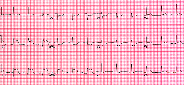
STEMI criteria: New ST elevation at the J point in two contiguous leads — at least 1 mm in limb leads, and at least 2 mm in V2–V3 for men aged 40 or over (2.5 mm under 40; 1.5 mm in women) and 1 mm in other chest leads.

Localising the infarct:

TerritoryECG leads → usual artery and what to watch for
Anterior / septalST elevation V1–V4 → LAD. Large territory: risk of heart failure, shock and VT.
LateralST elevation I, aVL, V5–V6 → LCx or a diagonal branch of the LAD.
InferiorST elevation II, III, aVF (reciprocal depression in I and aVL) → RCA (sometimes LCx). Watch for bradycardia, AV block and RV infarction.
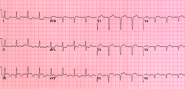
PosteriorST depression V1–V3 with tall R waves and upright T waves; ST elevation in posterior leads V7–V9 → LCx or RCA. Treat as STEMI.
Right ventricleST elevation in right-sided lead V4R, with an inferior STEMI → proximal RCA.

Other important ECG findings:

  • Hyperacute T waves: Tall, broad T waves that may come before ST elevation — the earliest sign of occlusion.
  • ST depression: Subendocardial ischaemia (NSTEMI) or a reciprocal change mirroring a STEMI elsewhere.
  • T-wave inversion: Ischaemia, or an evolving or reperfused infarct.
  • Pathological Q waves: An established infarct (dead muscle); may persist permanently.
  • Left bundle branch block: Hides ST changes. Use clinical judgement and the modified Sgarbossa criteria; get urgent senior or cardiology advice if the patient is unstable or has ongoing ischaemia.
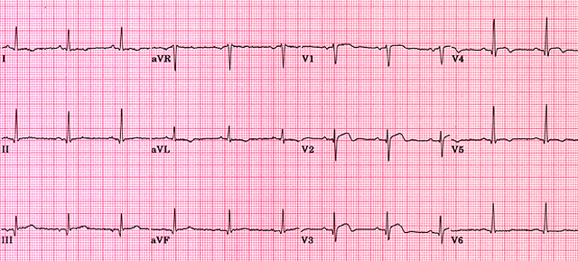
STEMI ECG ECG changes in ACS ACS ECG example Acute anterolateral MI Acute inferior MI Acute right ventricular MI Acute septal MI

⚠️ ECG Safety Pearl: A normal first ECG does not exclude ACS — repeat it. ST depression in V1–V3 may be a posterior STEMI: request posterior leads. With every inferior STEMI, record right-sided leads.

↑ Chapter index

1.16 Troponin

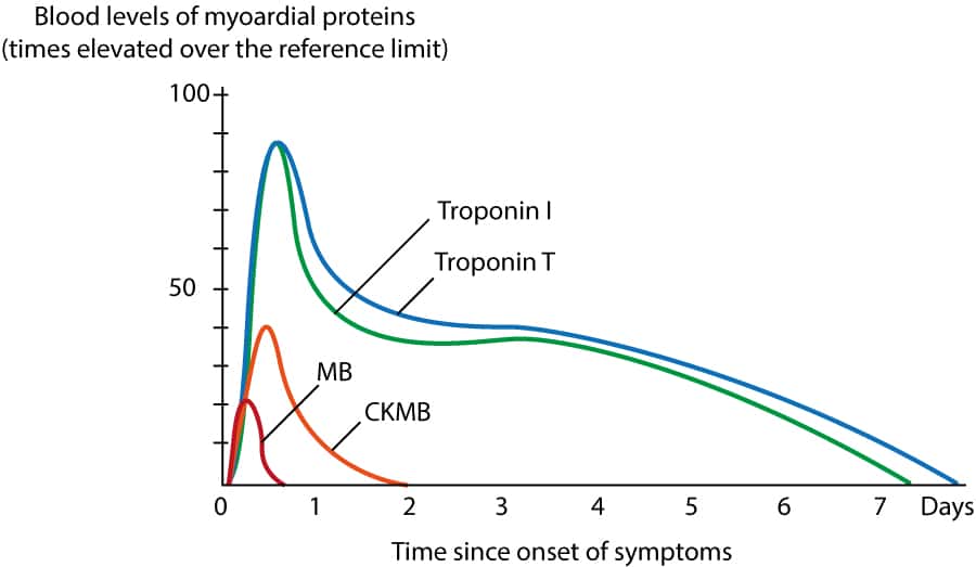
What it is: Troponin I and T are proteins in the contractile apparatus of heart muscle. They leak into the blood when heart muscle cells are damaged, so a raised troponin means myocardial injury.

How to use it:

  • High-sensitivity assays: Detect injury within 1–3 hours of symptom onset.
  • Abnormal threshold: Above the 99th percentile of the normal population for that particular assay.
  • Serial testing: A rise or fall between samples shows an acute process. A stable raised level suggests chronic injury (e.g. CKD, heart failure). Use the local validated pathway, commonly 0/1-hour or 0/3-hour sampling.
  • Very early samples: A sample taken soon after pain onset may be normal even in an evolving MI.

Causes of a raised troponin other than ACS:

  • Myocarditis.
  • Heart failure.
  • Pulmonary embolism.
  • Sepsis and critical illness.
  • Renal failure.
  • Fast arrhythmias.
  • Severe hypertension.
  • Takotsubo syndrome.
  • Cardiac contusion.
  • Aortic dissection.
  • Stroke and subarachnoid haemorrhage.
  • Strenuous endurance exercise.
Troponin kinetics Troponin interpretation

💡 Essential Teaching Point: Troponin tells you the heart muscle is injured, not why. A septic patient with a raised troponin does not usually need ACS treatment; they need treatment for sepsis.

↑ Chapter index

1.17 Definition and Types of MI

Universal definition: MI requires a rise and/or fall of troponin (at least one value above the 99th percentile) plus evidence of ischaemia — at least one of:

  • Ischaemic symptoms.
  • New ischaemic ECG changes.
  • New pathological Q waves.
  • New loss of viable muscle or a regional wall-motion abnormality on imaging.
  • Coronary thrombus on angiography or at autopsy.

Raised troponin without evidence of ischaemia is called myocardial injury, not infarction.

Types of MI:

  • Type 1: Plaque rupture or erosion with coronary thrombosis — the "classic" heart attack. Treat with antiplatelets, anticoagulation and angiography.
  • Type 2: A supply–demand mismatch without a new clot, e.g. severe anaemia, fast AF, hypoxia, hypotension or spasm. Treat the underlying cause; routine ACS antithrombotic treatment and urgent angiography are not automatically appropriate.
  • Type 3: Sudden cardiac death with features of ischaemia before troponin could be measured.
  • Type 4: Related to PCI, including stent thrombosis.
  • Type 5: Related to CABG.

💡 Clinical Pearl: Type 2 MI is very common in older inpatients (sepsis, anaemia, AF). Giving them dual antiplatelets and anticoagulation adds bleeding risk without treating the cause.

↑ Chapter index

1.18 Differential Diagnosis of Chest Pain

Life-threatening alternatives — consider these before giving antithrombotics:

  • Aortic dissection: Sudden, severe, tearing pain radiating to the back. Look for unequal arm pulses or blood pressures, a new aortic regurgitation murmur and neurological signs. Antiplatelets, anticoagulants and especially fibrinolysis can be fatal.
  • Pulmonary embolism: Pleuritic pain, breathlessness, tachycardia, hypoxia and risk factors for VTE.
  • Tension pneumothorax: Acute distress, absent breath sounds on one side, hypotension.
  • Oesophageal rupture (Boerhaave): Severe pain after vomiting, surgical emphysema and sepsis.

Other diagnoses:

  • Pericarditis: Sharp pain, worse lying flat and better sitting forward; widespread saddle-shaped ST elevation with PR depression.
  • Myocarditis: Chest pain and raised troponin after a viral illness, often in a young person.
  • Gastro-oesophageal reflux: Burning pain related to meals or lying down.
  • Musculoskeletal pain: Reproducible on palpation or movement.
  • Pneumonia: Fever, cough, pleuritic pain and focal chest signs.

⚠️ Safety Pearl: Before thrombolysis or anticoagulation, always ask: "Could this be a dissection?" Chest pain with syncope, neurological signs, unequal pulses or a new diastolic murmur needs urgent CT aortography first.

↑ Chapter index

1.19 Immediate Management

Use an ABCDE approach while working out which ACS pathway the patient needs.

First 10 minutes:

  • ABCDE assessment with heart rate, BP, oxygen saturations and respiratory rate.
  • 12-lead ECG immediately — if STEMI, activate the primary PCI pathway now.
  • IV access and cardiac monitoring.
  • Look for shock, pulmonary oedema and dangerous arrhythmias.

Bloods and imaging:

  • High-sensitivity troponin per local protocol.
  • FBC, U&E and creatinine (affects drug choice and doses).
  • Glucose and HbA1c, lipid profile.
  • Clotting if anticoagulation or procedures are planned.
  • Chest X-ray if pulmonary oedema or an alternative diagnosis is suspected.
  • Echo if unstable or a mechanical complication is suspected.

Initial drugs:

  • Aspirin 300 mg orally (chewed or dispersed) as soon as ACS is suspected, unless there is true allergy or suspected dissection.
  • GTN 400 micrograms sublingually for pain if systolic BP is above 90 mmHg. Avoid in RV infarction and after recent PDE-5 inhibitor use.
  • Analgesia: IV morphine 2.5–5 mg slowly, titrated to effect, with an antiemetic (e.g. metoclopramide 10 mg IV). Opioids can delay absorption of oral P2Y12 inhibitors, so use the lowest effective dose.
  • Oxygen: Only if hypoxic. Target saturations of 94–98%, or 88–92% if at risk of hypercapnia. Routine oxygen in normoxic patients gives no benefit.
  • Further antithrombotic treatment: A second antiplatelet and an anticoagulant are chosen according to the ACS type, bleeding risk and planned procedure (see the sections that follow).

⚠️ Clinical Pearl: Do not let blood tests delay treatment of a clear STEMI. Equally, do not reflexively give the full ACS drug package to every chest pain — think about dissection first.

↑ Chapter index

1.20 STEMI: Primary PCI

The aim in STEMI is to reopen the blocked artery as fast as possible. The preferred method is primary PCI: emergency angiography, followed by removing the clot and stenting the culprit artery.

NICE NG185 timing:

  • Within 12 hours of symptom onset: Offer primary PCI if it can be delivered within 120 minutes of the time fibrinolysis could have been given. In practice, paramedics transfer patients directly to a heart attack centre.
  • More than 12 hours: Consider angiography if there is ongoing ischaemia or cardiogenic shock.

Procedure points:

  • Radial access is preferred (fewer bleeding complications); drug-eluting stents are used.
  • Antiplatelet: Prasugrel with aspirin is preferred if the patient is not on an oral anticoagulant.
  • Anticoagulation: Unfractionated heparin during the procedure.
  • Multivessel disease without shock: Complete revascularisation of other significant narrowings is usually offered, during the same admission or soon after.

🚨 Time-Critical Principle: Every 30-minute delay to reperfusion increases mortality. The ECG and the story make the diagnosis — not the troponin.

↑ Chapter index

1.21 STEMI: Fibrinolysis

Fibrinolytic ("clot-busting") drugs activate plasminogen to plasmin, which dissolves fibrin in the clot. They are less effective than PCI and carry a risk of bleeding (including intracranial haemorrhage in about 1%), but they can be given anywhere.

Indications:

  • STEMI with symptom onset under 12 hours.
  • Primary PCI cannot be delivered within 120 minutes of the time fibrinolysis could be given (NICE NG185).
  • No contraindications.
  • Give as early as possible — ideally within 10 minutes of the diagnostic ECG, pre-hospital if available.

Fibrinolytic doses:

  • Tenecteplase (preferred): a single IV bolus over 5–10 seconds, weight-based. A half dose should be considered in patients aged 75 or over to reduce intracranial haemorrhage.
WeightTenecteplase dose
<60 kg30 mg
60–69 kg35 mg
70–79 kg40 mg
80–89 kg45 mg
≥90 kg50 mg
  • Reteplase: 10 units IV bolus, repeated once after 30 minutes.
  • Alteplase (accelerated 90-minute regimen): 15 mg IV bolus, then 0.75 mg/kg over 30 minutes (max 50 mg), then 0.5 mg/kg over 60 minutes (max 35 mg); total max 100 mg.

Drugs given with the fibrinolytic:

  • Aspirin: 300 mg loading, then 75 mg daily.
  • Clopidogrel: 300 mg loading if aged 75 or under; 75 mg (no loading) if over 75; then 75 mg daily. Ticagrelor and prasugrel are not used with fibrinolysis.
  • Enoxaparin (preferred anticoagulant), given at the same time as the fibrinolytic:
    • Under 75: 30 mg IV bolus, then 15 minutes later 1 mg/kg SC twice daily (max 100 mg for the first two SC doses).
    • 75 or over: No IV bolus; 0.75 mg/kg SC twice daily (max 75 mg for the first two doses).
    • Creatinine clearance below 30 mL/min: 1 mg/kg SC once daily.
    • Duration: Until revascularisation or for up to 8 days.
  • Unfractionated heparin (alternative): 60 units/kg IV bolus (max 4,000 units), then 12 units/kg/hour (max 1,000 units/hour), adjusted to an aPTT ratio of 1.5–2.0, for 24–48 hours.

Absolute contraindications — the risk of catastrophic bleeding is too high:

  • Previous intracranial haemorrhage, or stroke of unknown cause, at any time.
  • Ischaemic stroke within 6 months.
  • CNS tumour, damage or arteriovenous malformation.
  • Major trauma, surgery or head injury within 1 month.
  • GI bleeding within 1 month.
  • Known bleeding disorder (excluding menses).
  • Suspected aortic dissection.
  • Non-compressible punctures in the past 24 hours (e.g. liver biopsy, lumbar puncture).

Relative contraindications:

  • TIA within 6 months.
  • Oral anticoagulant therapy.
  • Pregnancy or within 1 week postpartum.
  • Refractory hypertension (systolic above 180 or diastolic above 110 mmHg).
  • Advanced liver disease.
  • Infective endocarditis.
  • Active peptic ulcer.
  • Prolonged or traumatic CPR.

After fibrinolysis:

  • Transfer to a PCI-capable centre.
  • Repeat the ECG at 60–90 minutes. Less than 50% ST resolution, ongoing pain or instability means the drug has failed: arrange urgent rescue PCI, not a second dose.
  • If reperfusion succeeds, perform coronary angiography within 2–24 hours.
  • Watch for bleeding. New headache, falling GCS or focal neurology suggests intracranial haemorrhage: stop anticoagulation, arrange urgent CT and seek haematology and neurosurgical advice.

💡 Clinical Pearl: Fibrinolysis is a bridge, not the end of treatment. Every patient should still go on to angiography.

↑ Chapter index

1.22 NSTEMI and Unstable Angina

In NSTEMI the artery is usually only partly blocked, so there is time to assess risk before deciding on angiography.

Initial treatment:

  • Aspirin 300 mg loading, then 75 mg daily.
  • Fondaparinux 2.5 mg SC daily for patients without high bleeding risk who are not having immediate angiography (NICE). Use unfractionated heparin instead if creatinine is above 265 µmol/L.
  • A second antiplatelet (P2Y12 inhibitor) — see Antiplatelet Therapy.
  • Anti-ischaemic treatment (GTN, beta blocker if no heart failure or shock) and pain relief.
  • High-intensity statin (atorvastatin 80 mg).
  • Serial ECGs and troponin; continuous monitoring if pain continues or the patient is unstable.

Risk stratification: Use the GRACE score, which combines age, heart rate, systolic BP, creatinine, Killip class (heart failure), cardiac arrest, ST changes and troponin. Assess bleeding risk at the same time.

Timing of angiography (NICE NG185):

  • Immediate: If clinically unstable — ongoing pain, haemodynamic instability, life-threatening arrhythmia or acute heart failure. These patients are managed like STEMI.
  • Within 72 hours: If the predicted 6-month mortality is above 3% (intermediate or higher risk).
  • Lower risk: Consider conservative management, with further testing guided by symptoms and evidence of ischaemia.

Revascularisation: PCI for suitable lesions; CABG for complex multivessel or left main disease; medical therapy alone may be best for some, especially frail patients.

💡 Clinical Pearl: NSTEMI is not a "small" heart attack. Long-term mortality is similar to STEMI because these patients are often older, with more widespread disease.

↑ Chapter index

1.23 Antiplatelet Therapy

Platelets drive coronary thrombosis, so blocking them is central to ACS treatment. Two pathways are targeted, and blocking both is more effective than blocking either alone. This is dual antiplatelet therapy (DAPT).

Aspirin irreversibly blocks cyclooxygenase-1, stopping thromboxane A₂ production and platelet aggregation.

  • Dose: 300 mg loading, then 75 mg daily for life.

P2Y12 inhibitors block the ADP receptor on platelets:

  • Clopidogrel: An irreversible prodrug needing liver activation, so its response varies.
    • Dose: 300 mg loading (600 mg before PCI), then 75 mg daily.
    • Used: With fibrinolysis, in high bleeding risk, and when the patient also needs an oral anticoagulant.
  • Ticagrelor: Reversible and direct-acting, with a faster, stronger effect.
    • Dose: 180 mg loading, then 90 mg twice daily for 12 months.
    • Beyond 12 months: 60 mg twice daily may be continued for up to 3 years in selected high-risk patients.
    • Side effects: Breathlessness and bradyarrhythmias.
  • Prasugrel: Potent and irreversible.
    • Dose: 60 mg loading, then 10 mg daily (5 mg daily if under 60 kg or aged 75 or over — generally avoid at 75 or over).
    • Contraindicated after previous stroke or TIA.

Which P2Y12 inhibitor? (NICE NG185):

  • STEMI with primary PCI: Prasugrel with aspirin if not on an oral anticoagulant; ticagrelor or clopidogrel as alternatives depending on bleeding risk.
  • NSTEMI or unstable angina having angiography: Prasugrel or ticagrelor. Give prasugrel only once the anatomy is known and PCI is planned.
  • Conservative management or high bleeding risk: Ticagrelor or clopidogrel.
  • Patient on an oral anticoagulant (e.g. for AF): Clopidogrel. Keep triple therapy (anticoagulant + aspirin + clopidogrel) as short as possible, often up to 1 month, then anticoagulant plus clopidogrel.

Duration: DAPT usually continues for 12 months after ACS (shorter if bleeding risk is high), then aspirin alone for life. Stopping DAPT early after a stent can cause stent thrombosis, a frequently fatal event. Consider a PPI for patients at risk of GI bleeding.

💡 Clinical Pearl: After ACS a patient takes aspirin plus one P2Y12 inhibitor — never two P2Y12 inhibitors together. Every antithrombotic added increases bleeding, especially in older, frail patients and those with CKD.

↑ Chapter index

1.24 Anticoagulation

Anticoagulants stop the coagulation cascade from building fibrin into the clot. They are used for the first few days of ACS and during PCI.

  • Fondaparinux (indirect factor Xa inhibitor):
    • Dose: 2.5 mg SC once daily for up to 8 days or until discharge.
    • Use: NICE's choice for NSTEMI/unstable angina without high bleeding risk when immediate angiography is not planned.
    • Avoid if creatinine clearance is below 20 mL/min.
    • During PCI: Extra unfractionated heparin must be given, because fondaparinux alone allows clots to form on catheters.
  • Unfractionated heparin (UFH): Acts via antithrombin. It is short-acting, easily adjusted and reversible with protamine, which makes it ideal around procedures and in renal failure.
    • Medical ACS: 60–70 units/kg IV bolus (max 5,000 units), then 12–15 units/kg/hour (max 1,000 units/hour), adjusted to an aPTT ratio of 1.5–2.5.
    • During PCI: 70–100 units/kg IV, guided by the ACT (50–70 units/kg if a GP IIb/IIIa inhibitor is used).
  • Enoxaparin (low-molecular-weight heparin):
    • NSTEMI/unstable angina: 1 mg/kg SC twice daily for 2–8 days.
    • STEMI under 75: 30 mg IV bolus plus 1 mg/kg SC, then 1 mg/kg SC twice daily (max 100 mg for the first two SC doses).
    • STEMI aged 75 or over: No bolus; 0.75 mg/kg SC twice daily (max 75 mg for the first two doses).
    • Creatinine clearance 15–30 mL/min: 1 mg/kg SC once daily.

Before anticoagulating, check:

  • Active bleeding and platelet count.
  • Renal function and weight.
  • Whether the patient already takes warfarin or a DOAC.
  • That combinations of antiplatelets and anticoagulants are not continued for longer than needed.

↑ Chapter index

1.25 MINOCA, SCAD and Coronary Embolism

MINOCA (Myocardial Infarction with Non-Obstructive Coronary Arteries):

  • What it is: The patient meets the criteria for MI, but angiography shows no significant narrowing. It is a working diagnosis, not a final one.
  • Possible causes: Plaque disruption without a lasting blockage, coronary spasm, embolism, spontaneous dissection and microvascular dysfunction.
  • Mimics: Myocarditis and takotsubo syndrome.
  • Key test: Cardiac MRI separates true infarction from myocarditis and takotsubo.
  • Treatment: Directed at the underlying cause.

Spontaneous coronary artery dissection (SCAD):

  • Mechanism: A tear or bleed within the artery wall compresses the true lumen.
  • Who gets it: Typically women under 60 without conventional risk factors, including in late pregnancy and after delivery. Associated with fibromuscular dysplasia and extreme physical or emotional stress.
  • Treatment: Usually conservative if stable, because the artery usually heals by itself and PCI risks extending the tear. Beta blockers are commonly used. PCI or CABG is reserved for ongoing ischaemia or instability.

Coronary embolism:

  • Sources: AF, prosthetic or diseased valves, infective endocarditis and LV thrombus.
  • Management: Find and treat the source. Long-term anticoagulation is usually needed.

💡 Clinical Pearl: A young woman with an MI in the weeks after childbirth has SCAD until proven otherwise.

↑ Chapter index

1.26 Arrhythmias After MI

Ischaemic and dying muscle is electrically unstable, so arrhythmias are commonest in the first 48 hours. Always correct potassium (aim above 4.0 mmol/L) and magnesium, and treat the ischaemia itself.

  • Ventricular fibrillation:
    • Immediate defibrillation and ALS: adrenaline 1 mg and amiodarone 300 mg after the third shock.
    • Urgent angiography after return of circulation.
    • VF within the first 48 hours does not by itself require an ICD.
  • Ventricular tachycardia:
    • Pulseless: ALS with defibrillation.
    • Pulse but adverse features (shock, syncope, ischaemia, heart failure): synchronised DC cardioversion (up to 3 attempts), then amiodarone 300 mg IV over 10–20 minutes.
    • Stable: Amiodarone 300 mg IV over 20–60 minutes.
    • Arrange urgent revascularisation.
  • Sinus bradycardia: Common and usually vagal in inferior MI. Treat only with adverse features: atropine 500 micrograms IV, repeated to a maximum of 3 mg. Review rate-limiting drugs.
  • AV block — inferior MI: Usually at the AV node and transient. Atropine if compromised; monitor.
  • AV block — anterior MI: Mobitz II or complete block means extensive damage below the node and a poor prognosis. Temporary pacing (transcutaneous, then transvenous), with adrenaline or isoprenaline as a bridge. Permanent pacing if the block persists.
  • Asystole: CPR and the non-shockable ALS pathway, with adrenaline 1 mg IV as soon as possible and every 3–5 minutes; treat the 4 Hs and 4 Ts. Consider pacing if P waves are present (ventricular standstill).

💡 Clinical Pearl: Heart block in inferior MI usually resolves; heart block in anterior MI signals a large infarct and usually needs pacing.

↑ Chapter index

1.27 Heart Failure and Cardiogenic Shock

Acute LV failure and pulmonary oedema:

  • Sit the patient upright; give oxygen to target saturations.
  • IV furosemide 40–80 mg (higher if already on a loop diuretic).
  • IV nitrate infusion if systolic BP allows.
  • CPAP if hypoxic or distressed.
  • Urgent echo and revascularisation.
  • Avoid routine opiates.

Cardiogenic shock occurs when the heart cannot pump enough blood to perfuse the organs. It usually follows a large anterior MI, and mortality is around 40–50%.

  • Features:
    • Systolic BP below 90 mmHg despite adequate filling, or the need for vasopressors.
    • Cold, clammy peripheries.
    • Confusion.
    • Oliguria.
    • Raised lactate.
    • Pulmonary oedema (the lungs may be clear if the RV is mainly affected).
  • Management:
    • Senior cardiology and critical care input immediately.
    • Emergency PCI of the culprit artery.
    • Bedside echo to exclude mechanical complications.
    • Noradrenaline for hypotension, with or without dobutamine for low output, under invasive monitoring.
    • Consider mechanical circulatory support (e.g. Impella, ECMO) in specialist centres.
    • Assess fluid needs individually rather than giving large volumes.
    • Avoid beta blockers and other negative inotropes.

⚠️ Clinical Pearl: Hypotension after MI is not always LV failure. Think RV infarction, papillary muscle rupture, septal rupture, tamponade and arrhythmia — echo distinguishes them.

↑ Chapter index

1.28 Right Ventricular Infarction

Mechanism: A proximal RCA occlusion can infarct the right ventricle as well as the inferior LV wall. A damaged RV cannot push enough blood through the lungs to fill the left ventricle, so LV output depends on adequate venous return (it is preload-dependent).

Clinical pattern: Inferior STEMI with the triad of hypotension, raised JVP and clear lungs, often with bradycardia or AV block.

Diagnosis: ST elevation in right-sided lead V4R; echo shows a dilated, poorly moving RV.

Management:

  • Urgent reperfusion.
  • Maintain preload: Cautious IV fluid boluses.
  • Avoid nitrates, diuretics and opiates — they reduce preload and can cause collapse.
  • Inotropes if hypotension persists despite fluid.
  • Pacing for AV block (keeping atrial and ventricular contraction in sequence helps the RV).

💡 Clinical Pearl: Inferior MI + low BP + high JVP + clear lungs = RV infarction. GTN may cause profound hypotension.

↑ Chapter index

1.29 Mechanical Complications

Dead muscle is soft and weakest about 3–5 days after MI, before scar forms. Mechanical complications are now less common thanks to early PCI, but they are frequently fatal. Suspect them with sudden deterioration, a new murmur or shock.

  • Papillary muscle rupture:
    • Presentation: Sudden severe mitral regurgitation, flash pulmonary oedema and a new pansystolic murmur (which may be quiet), usually within the first week.
    • Management: Urgent echo; diuretics and afterload reduction (IV nitrates if BP allows); intra-aortic balloon pump or other mechanical support as a bridge; emergency mitral valve surgery.
  • Ventricular septal rupture:
    • Presentation: A hole in the septum creates a left-to-right shunt, with a new harsh pansystolic murmur and shock.
    • Management: Urgent echo; immediate cardiothoracic referral; haemodynamic support (diuretics, vasodilators if BP allows, IABP or Impella) as a bridge to surgical repair; percutaneous closure in selected patients.
  • Free-wall rupture:
    • Presentation: Blood fills the pericardium, causing tamponade (hypotension, raised JVP, PEA arrest).
    • Management: Emergency echo; pericardiocentesis only as a temporary measure; immediate cardiac surgery. Often fatal.

🚨 Clinical Pearl: Sudden deterioration several days after MI with pulmonary oedema, shock or a new murmur needs an immediate echo and cardiothoracic referral.

↑ Chapter index

1.30 Pericarditis and Late Complications

Pericarditis:

  • Early post-MI pericarditis (days 1–3): Inflammation over the infarct, causing pleuritic pain and a friction rub. Usually self-limiting.
    • Treat with high-dose aspirin; colchicine may be added.
    • Avoid other NSAIDs and corticosteroids, which impair scar healing.
    • Exclude re-infarction and check for an effusion.
  • Dressler syndrome (weeks later): Immune-mediated pericarditis with fever and effusion.
    • High-dose aspirin plus colchicine 500 micrograms twice daily (once daily if under 70 kg) for about 3 months.
    • Echo to exclude effusion and tamponade.
    • Corticosteroids only if refractory.
    • Review anticoagulation if there is an effusion.

Late complications:

  • Ischaemic cardiomyopathy and heart failure:
    • Full heart failure therapy for reduced EF: ACE inhibitor/ARB or sacubitril/valsartan, beta blocker, MRA and SGLT2 inhibitor, plus diuretics for congestion.
    • Cardiac rehabilitation.
    • Consider an ICD or CRT if the EF remains 35% or less despite optimal therapy (reassess at least 40 days after MI).
  • LV aneurysm:
    • Suspect if ST elevation persists weeks after MI; confirm with echo or cardiac MRI.
    • Causes heart failure, thrombus and VT.
    • Heart failure therapy; anticoagulate if thrombus is present; surgery for refractory heart failure, angina or VT.
  • LV thrombus (risk of embolic stroke):
    • Anticoagulation (warfarin or DOAC) for 3–6 months with repeat imaging.
    • Keep triple therapy with antiplatelets as short as possible and give a PPI.
  • Recurrent ventricular arrhythmias: Optimise beta blockade, correct electrolytes and treat residual ischaemia; amiodarone if needed; electrophysiology referral and ablation; ICD for secondary prevention.
  • Recurrent MI: Treat as a new ACS, considering stent thrombosis; check adherence to DAPT.

↑ Chapter index

1.31 Secondary Prevention

Atherosclerosis affects all the arteries, so an MI marks a patient at high risk of another event. Secondary prevention reduces that risk and should be started before discharge. A simple way to remember the drugs is "5 drugs for life after MI", adjusted to the individual.

  • Antiplatelets:
    • Aspirin 75 mg daily for life.
    • Plus a P2Y12 inhibitor, usually for 12 months.
  • High-intensity statin:
    • Dose: Atorvastatin 80 mg daily; lower doses if there are interactions or intolerance.
    • Target (NICE NG238): LDL cholesterol 2.0 mmol/L or less, or non-HDL cholesterol 2.6 mmol/L or less.
    • Monitoring: Recheck lipids at 2–3 months.
    • If the target is not met: Add ezetimibe 10 mg daily, then consider other NICE-approved options (e.g. inclisiran, PCSK9 inhibitors).
  • ACE inhibitor:
    • Dose: Ramipril 2.5 mg twice daily once stable, titrated to 5 mg twice daily.
    • Monitoring: BP, renal function and potassium.
    • If not tolerated: Use an ARB.
  • Beta blocker:
    • When to start: Once haemodynamically stable.
    • Reduced EF: Continue indefinitely.
    • Preserved EF: NICE advises about 12 months; recent trials have questioned the benefit, so review continuation individually.
  • MRA (eplerenone):
    • Who: Patients with EF 40% or less plus heart failure or diabetes.
    • When: Start 3–14 days after MI.
    • Dose: 25 mg daily, increasing to 50 mg daily.
    • Monitoring: Potassium.
  • SGLT2 inhibitor: If there is HFrEF, diabetes or CKD.
  • Before discharge: Assess LV function by echo in all patients.

💡 Clinical Pearl: Revascularisation fixes one artery; secondary prevention protects all of them. Both are needed.

↑ Chapter index

1.32 Rehabilitation and Lifestyle

  • Cardiac rehabilitation: Offer to every patient. Structured exercise and education reduce cardiac death and readmission, but uptake is poor, so encourage it actively.
  • Smoking cessation: The single most effective lifestyle change — it substantially reduces the risk of death and further MI.
  • Exercise: Regular graded aerobic exercise, increasing gradually.
  • Diet: Mediterranean-style, low in saturated fat, high in fibre, fruit and vegetables.
  • Risk factors: Control blood pressure and diabetes, and optimise weight.
  • Psychological health: Depression and anxiety are common after MI and worsen outcomes — screen and support.
  • Driving: Group 1 (car) licence holders usually stop driving for 1 week after successful PCI, or 4 weeks otherwise; they do not need to notify the DVLA unless other conditions apply. Group 2 (bus and lorry) drivers must stop for at least 6 weeks and notify the DVLA.
  • Returning to normal life: Most people can return to work within weeks, depending on the job. Sexual activity can usually resume about 4 weeks after an uncomplicated MI.

↑ Chapter index

1.33 Drug Reference Table

🫀 Common adult doses (UK). Doses vary with indication, renal function, blood pressure, bleeding risk and local protocols. Always check the BNF.

DrugUsual adult dose
AspirinACS: 300 mg loading, then 75 mg daily for life.
Clopidogrel300–600 mg loading (600 mg before PCI), then 75 mg daily. With fibrinolysis: 300 mg if ≤75 years, 75 mg if >75.
Ticagrelor180 mg loading, then 90 mg twice daily for 12 months; 60 mg twice daily thereafter in selected patients.
Prasugrel60 mg loading, then 10 mg daily (5 mg if <60 kg or ≥75 years). Contraindicated after stroke/TIA.
Fondaparinux2.5 mg SC daily up to 8 days. Avoid if CrCl <20 mL/min. Extra UFH needed during PCI.
EnoxaparinNSTEMI: 1 mg/kg SC twice daily. STEMI: see Anticoagulation for age- and renal-adjusted regimens.
Unfractionated heparinMedical ACS: 60–70 units/kg IV bolus, then 12–15 units/kg/h (aPTT 1.5–2.5). PCI: 70–100 units/kg guided by ACT.
TenecteplaseSingle IV bolus 30–50 mg by weight band; consider half dose if ≥75 years.
GTN spray400 micrograms sublingually; repeat after 5 minutes. Call 999 if pain persists 5 minutes after the second dose.
Morphine2.5–5 mg IV slowly, titrated, with an antiemetic.
BisoprololAngina: 2.5–5 mg daily, usually 5–10 mg (max 20 mg). Target heart rate 55–60 bpm.
Amlodipine5 mg daily, increase to 10 mg if needed.
Diltiazem60 mg three times daily (twice daily in older people), max 360 mg/day. Prescribe MR by brand.
Verapamil80–120 mg three times daily. Never with a beta blocker.
Isosorbide mononitrate20–40 mg twice daily (asymmetric), or MR 25–60 mg each morning (max 120 mg).
Ivabradine5 mg twice daily (2.5 mg if ≥75), max 7.5 mg twice daily. Sinus rhythm, HR ≥70.
Nicorandil10 mg twice daily, usually 10–20 mg twice daily (max 30 mg twice daily). Watch for ulceration.
Ranolazine375 mg twice daily, then 500 mg, max 750 mg twice daily. Check QT and interactions.
AtorvastatinSecondary prevention: 80 mg daily. Primary prevention: 20 mg daily.
Ezetimibe10 mg daily when further LDL lowering is needed.
RamiprilPost-MI: 2.5 mg twice daily once stable, titrated to 5 mg twice daily.
EplerenonePost-MI LV dysfunction: 25 mg daily, increasing to 50 mg daily.

⚠️ Prescribing Pearls: Aspirin plus one P2Y12 inhibitor after ACS. Prasugrel is contraindicated after stroke or TIA. Never give nitrates with PDE-5 inhibitors or in RV infarction. Monitor potassium and creatinine with ACE inhibitors and eplerenone. Do not start beta blockers in shock or heart block.

↑ Chapter index

1.34 Spot the Diagnosis

ClueThink of
Predictable exertional chest tightness relieved by restStable angina
New or worsening angina at rest, normal troponinUnstable angina
ST elevation II, III, aVF with bradycardiaInferior STEMI (RCA)
Inferior STEMI, low BP, high JVP, clear lungsRV infarction
ST depression V1–V3 with tall R wavesPosterior STEMI
Early-morning rest pain, transient ST elevation, normal coronariesVasospastic angina
Angina in a woman, normal angiogram, positive stress testMicrovascular angina (INOCA)
Young woman, MI weeks after childbirthSCAD
Raised troponin in sepsis with fast AFType 2 MI / myocardial injury
Day 4 post-MI, flash pulmonary oedema, new murmurPapillary muscle rupture
Day 4 post-MI, shock, harsh pansystolic murmurVentricular septal rupture
Sudden PEA arrest days after MIFree-wall rupture with tamponade
Persistent ST elevation weeks after MILV aneurysm
Fever, pleuritic pain and effusion weeks after MIDressler syndrome
Tearing pain to back, unequal arm BPsAortic dissection

↑ Chapter index

1.35 Exam-style Scenarios

Decide your answer before reading on.

  • A 58-year-old man has 40 minutes of central chest pain. ECG shows ST elevation in V1–V4. The nearest PCI centre is 30 minutes away. Answer: Anterior STEMI. Give aspirin 300 mg and analgesia, and transfer for primary PCI (prasugrel loading in the cath lab if not on an anticoagulant). Fibrinolysis is not needed because PCI is available within 120 minutes.
  • A 72-year-old woman on a remote island has an inferior STEMI; the nearest PCI is 4 hours away. She had an ischaemic stroke 3 months ago. Answer: Ischaemic stroke within 6 months is an absolute contraindication to fibrinolysis. Arrange urgent transfer for primary PCI despite the delay.
  • Ninety minutes after tenecteplase, a patient still has pain and less than 50% ST resolution. Answer: Failed reperfusion. Arrange urgent rescue PCI; do not repeat the fibrinolytic.
  • A patient with an inferior STEMI becomes hypotensive after GTN. JVP is raised; lungs are clear. Answer: RV infarction. Stop nitrates, give IV fluid boluses, record V4R, and arrange urgent reperfusion.
  • An 80-year-old woman admitted with pneumonia and fast AF has a troponin rising from 60 to 90 ng/L and no chest pain. Answer: Probable type 2 MI or myocardial injury. Treat the sepsis and control the AF; do not start routine ACS DAPT and anticoagulation.
  • Four days after an anterior MI, a man suddenly becomes breathless and hypotensive with a new loud pansystolic murmur. Answer: Ventricular septal rupture (or papillary muscle rupture). Immediate echo and cardiothoracic referral, with mechanical support as a bridge to surgery.
  • A patient with stable angina on bisoprolol still has symptoms. Which drug should be added? Answer: A dihydropyridine CCB such as amlodipine — not verapamil, because of the risk of heart block.
  • A 34-year-old woman 3 weeks postpartum has chest pain and a raised troponin. Angiography shows a long, smooth narrowing of the LAD. Answer: SCAD. Treat conservatively if stable, with a beta blocker; avoid PCI unless there is ongoing ischaemia.

↑ Chapter index

1.36 Key Points

  • IHD is a mismatch between oxygen supply and demand; atherosclerosis is the usual cause.
  • Stable angina is reversible ischaemia; troponin does not rise.
  • ACS is plaque rupture plus clot; antiplatelets and anticoagulants target the clot.
  • MI often arises from plaques that were not severely narrowing the artery.
  • A normal ECG and a normal first troponin do not exclude ACS — repeat both.
  • STEMI needs reperfusion now; primary PCI within 120 minutes, otherwise fibrinolysis.
  • ST depression in V1–V3 may be a posterior STEMI.
  • Inferior MI: watch for heart block and RV infarction; avoid nitrates if the RV is involved.
  • Troponin means injury, not necessarily ACS; distinguish type 1 from type 2 MI.
  • Give oxygen only if hypoxic.
  • Aspirin plus one P2Y12 inhibitor for 12 months; never stop DAPT early after a stent.
  • A new murmur or shock days after MI needs an immediate echo.
  • Never combine a beta blocker with verapamil.
  • Secondary prevention: aspirin, P2Y12 inhibitor, atorvastatin 80 mg, ACE inhibitor and beta blocker, plus cardiac rehabilitation.
  • Exclude aortic dissection before giving antithrombotics or fibrinolysis.

↑ Chapter index

1.37 NICE and European Guidelines

↑ Chapter index

2. Heart Failure

🫀 Heart Failure (HF) is a clinical syndrome caused by structural or functional cardiac abnormalities that impair ventricular filling, ejection or both. It results in symptoms such as breathlessness, fatigue and exercise intolerance, often accompanied by signs of pulmonary or systemic congestion. Heart failure may develop gradually or present acutely with pulmonary oedema, hypotension or cardiogenic shock. Important: Heart failure does not necessarily mean that the left ventricular ejection fraction is reduced. Patients may have severe heart failure despite a normal or near-normal ejection fraction. ⚠️ Clinical priorities: Confirm the diagnosis, identify the underlying cause, assess congestion and perfusion, treat reversible precipitants and introduce evidence-based treatment.

2.01 🫀 Understanding Heart Failure

The heart must generate sufficient cardiac output to meet the metabolic requirements of the body while maintaining normal or near-normal filling pressures.

  • Cardiac output: Cardiac output = heart rate × stroke volume; Normal resting cardiac output is approximately 4–8 litres/minute, depending on body size and physiological conditions; Stroke volume depends on preload, afterload and myocardial contractility; Heart failure may result from impaired contraction, abnormal relaxation, excessive loading conditions or structural abnormalities.
  • Two major haemodynamic problems: Reduced forward flow: Inadequate cardiac output may cause fatigue, renal hypoperfusion, confusion and reduced exercise tolerance; Increased filling pressures: Elevated cardiac pressures cause pulmonary congestion, peripheral oedema, ascites and orthopnoea.

💡 Clinical Pearl: Many symptoms of heart failure result from increased cardiac filling pressures rather than reduced cardiac output alone. A patient with preserved cardiac output may still develop severe pulmonary oedema if left atrial pressure rises sufficiently.

2.02 ⚖️ Classification of Heart Failure

  • Heart Failure with Reduced Ejection Fraction (HFrEF): NICE classification: LVEF ≤40%; Usually involves impaired left ventricular systolic function; Common causes include previous myocardial infarction, dilated cardiomyopathy and myocarditis; Benefits substantially from disease-modifying pharmacological treatment.
  • Heart Failure with Mildly Reduced Ejection Fraction (HFmrEF): NICE classification: LVEF 41–49%; May represent partially recovered systolic dysfunction or a phenotype between HFrEF and HFpEF; Patients often benefit from treatments used for reduced ejection fraction.
  • Heart Failure with Preserved Ejection Fraction (HFpEF): NICE classification: LVEF ≥50%; Usually involves increased ventricular stiffness, impaired relaxation or abnormal ventricular-vascular coupling; Common in older adults with hypertension, obesity, diabetes, atrial fibrillation and chronic kidney disease; Diagnosis requires symptoms or signs of heart failure plus objective evidence supporting elevated filling pressures or cardiac structural or functional abnormalities.
  • Heart Failure with Improved Ejection Fraction (HFimpEF): Previously documented LVEF ≤40%, with subsequent improvement by at least 10 percentage points to more than 40%; Recovery may follow treatment of cardiomyopathy, tachyarrhythmia or myocardial ischaemia; Improved EF does not necessarily indicate permanent recovery; Continue disease-modifying treatment unless there is a compelling reason to change it.

📌 2026 Guideline Update: NICE retains the categories HFrEF ≤40%, HFmrEF 41–49% and HFpEF ≥50%. The 2026 ESC guidelines simplify classification to HFrEF with LVEF below 50% and HFpEF with LVEF ≥50%. For UK prescribing and examinations, recognise both systems and follow current NICE recommendations where appropriate.

2.03 📊 New York Heart Association (NYHA) Classification

  • Class I: No limitation of ordinary physical activity.
  • Class II: Comfortable at rest, but ordinary activity produces breathlessness, fatigue or palpitations.
  • Class III: Comfortable at rest, but less-than-ordinary activity produces symptoms.
  • Class IV: Symptoms occur at rest, and any physical activity increases discomfort.

NYHA classification assesses functional limitation, not ejection fraction. It is subjective but valuable for describing symptom burden, monitoring treatment response and selecting some specialist interventions.

2.04 🔬 Pathophysiology of Heart Failure

Heart failure initiates a series of compensatory mechanisms intended to maintain cardiac output and blood pressure. Initially beneficial, these adaptations become harmful when chronically activated.

  • Frank-Starling mechanism: Increased venous return stretches ventricular myocardial fibres; Within physiological limits, greater fibre stretch increases contractile force; In failing ventricles, further increases in preload produce relatively little improvement in stroke volume; Excessive preload increases ventricular filling pressures and congestion.
  • Sympathetic nervous system activation: Reduced effective arterial perfusion activates sympathetic responses; Noradrenaline increases heart rate, contractility and peripheral vascular resistance; Persistent activation increases myocardial oxygen demand and promotes arrhythmias; Chronic catecholamine stimulation contributes to myocardial injury and adverse ventricular remodelling.
  • Renin-angiotensin-aldosterone system (RAAS): Reduced renal perfusion stimulates renin release; Angiotensin II causes vasoconstriction and increases afterload; Aldosterone promotes sodium and water retention; Persistent activation contributes to myocardial fibrosis, ventricular remodelling and progressive dysfunction.
  • Vasopressin (ADH): Promotes renal water retention; Can contribute to dilutional hyponatraemia; Severe hyponatraemia is associated with advanced disease and adverse prognosis.
  • Natriuretic peptides: BNP and NT-proBNP increase in response to myocardial wall stress; BNP promotes natriuresis, diuresis and vasodilatation; The natriuretic peptide system counteracts RAAS and sympathetic activation; Despite this response, progressive heart failure often produces persistent sodium and water retention.
  • Ventricular remodelling: Chronic pressure overload promotes concentric hypertrophy; Chronic volume overload or myocardial injury may produce ventricular dilatation; Myocyte loss, interstitial fibrosis and altered ventricular geometry impair cardiac function; Mitral regurgitation may develop as a consequence of ventricular dilatation and papillary muscle displacement.

💡 MRCP Pearl: ACE inhibitors, beta blockers, MRAs and ARNIs modify the maladaptive neurohormonal response. Their benefit extends beyond reducing symptoms: they can slow or reverse remodelling and reduce mortality in selected heart failure populations.

2.05 🧬 Causes of Heart Failure

  • Ischaemic heart disease: Previous myocardial infarction; Extensive coronary artery disease; Chronic ischaemic myocardial dysfunction; Acute myocardial infarction causing severe LV dysfunction.
  • Hypertension: Chronic pressure overload produces LV hypertrophy; Myocardial fibrosis and impaired relaxation may cause HFpEF; Advanced disease can eventually cause systolic dysfunction.
  • Valvular heart disease: Aortic stenosis causes pressure overload; Aortic regurgitation causes LV volume overload; Mitral regurgitation causes left atrial and ventricular volume overload; Mitral stenosis produces increased left atrial and pulmonary venous pressure.
  • Cardiomyopathies: Dilated cardiomyopathy; Hypertrophic cardiomyopathy; Restrictive cardiomyopathy; Arrhythmogenic cardiomyopathy; Inherited or familial cardiomyopathies; Peripartum cardiomyopathy; Takotsubo syndrome.
  • Inflammatory and infiltrative disease: Myocarditis; Cardiac amyloidosis; Cardiac sarcoidosis; Haemochromatosis; Fabry disease.
  • Arrhythmias: Atrial fibrillation with rapid ventricular response; Persistent supraventricular tachycardia; High burden of ventricular ectopic beats; Severe bradyarrhythmias.
  • Toxic, endocrine and metabolic causes: Excess alcohol consumption; Anthracycline chemotherapy; Trastuzumab-associated cardiac dysfunction; Thyrotoxicosis or severe hypothyroidism; Thiamine deficiency; Severe chronic anaemia.

💡 Clinical Pearl: Every new diagnosis of heart failure should trigger a search for its cause. Identifying coronary disease, valve pathology, uncontrolled tachyarrhythmia or a specific cardiomyopathy may reveal a treatable or reversible mechanism.

2.06 🫁 Left-Sided Heart Failure

Left ventricular dysfunction increases LV end-diastolic pressure, which is transmitted backwards to the left atrium and pulmonary veins.

  • Symptoms: Exertional breathlessness. Orthopnoea; Paroxysmal nocturnal dyspnoea (PND); Nocturnal cough. Reduced exercise tolerance; Fatigue. Occasionally pink frothy sputum in severe pulmonary oedema.
  • Signs: Tachycardia; Basal inspiratory crackles; Third heart sound (S3), particularly in volume-overloaded dilated ventricles; Displaced apex beat in LV dilatation; Functional mitral regurgitation; Hypoxaemia in acute pulmonary oedema.
  • Why does orthopnoea occur?: Lying flat redistributes blood from the lower limbs and splanchnic circulation towards the thorax; Venous return increases; A failing LV cannot accommodate the additional preload without a substantial rise in filling pressure; Pulmonary venous congestion develops, producing breathlessness.
  • Why does PND occur?: Redistribution of peripheral oedema fluid occurs during sleep; Increased venous return and pulmonary congestion develop; Patients awaken after a period of sleep with severe breathlessness and frequently sit upright for relief.

2.07 🩺 Right-Sided Heart Failure

Right ventricular dysfunction causes increased systemic venous pressure and impaired pulmonary blood flow.

  • Symptoms: Dependent oedema; Abdominal distension; Early satiety; Right upper quadrant discomfort; Fatigue and reduced exercise tolerance.
  • Signs: Raised jugular venous pressure (JVP); Peripheral pitting oedema; Congestive hepatomegaly; Ascites; Tricuspid regurgitation; Right ventricular heave; Pleural effusions.
  • Important causes: Left-sided heart failure causing pulmonary hypertension; Pulmonary arterial hypertension; Chronic thromboembolic pulmonary hypertension; Chronic lung disease; Right ventricular myocardial infarction; Tricuspid or pulmonary valve disease; Right ventricular cardiomyopathy.

💡 Clinical Pearl: Right heart failure commonly develops secondary to left heart disease, but isolated right ventricular failure should prompt consideration of pulmonary hypertension, pulmonary embolic disease, RV infarction and primary RV pathology.

2.08 🩺 Examination of a Patient with Heart Failure

  • General examination: Assess respiratory distress, ability to speak and posture; Look for cyanosis, pallor, muscle wasting and frailty; Assess peripheral temperature and capillary refill; Look for peripheral oedema, including sacral oedema in bedbound patients.
  • Pulse and blood pressure: Tachycardia suggests sympathetic activation or another precipitant; An irregularly irregular pulse suggests atrial fibrillation; Hypotension may indicate advanced LV dysfunction or shock; Postural hypotension may occur with medication or volume depletion; A narrow pulse pressure may suggest low stroke volume.
  • Jugular venous pressure: An elevated JVP indicates raised right atrial pressure; Assess with the patient appropriately reclined and the neck relaxed; Observe the internal jugular venous pulsation and estimate its height above the sternal angle; The abdominojugular reflux may reveal elevated filling pressures; A prominent V wave suggests significant tricuspid regurgitation.
  • Cardiac examination: Displaced apex beat suggests ventricular enlargement; S3 occurs during rapid early diastolic filling and may indicate increased filling pressures in HFrEF; S4 may occur with a stiff ventricle in sinus rhythm, such as hypertensive heart disease; A pansystolic murmur may indicate functional mitral or tricuspid regurgitation; Consider aortic stenosis when there is an ejection systolic murmur and slow-rising carotid pulse.
  • Respiratory examination: Basal crackles suggest pulmonary congestion but are not specific; Pleural effusions may produce reduced breath sounds and stony dullness; Wheeze may occur in pulmonary oedema and has historically been termed cardiac asthma; The absence of crackles does not exclude chronic heart failure.
  • Abdominal examination: Congestive hepatomegaly. Ascites; Hepatic tenderness in acute congestion; Abdominojugular reflux.

💡 MRCP Examination Pearl: An elevated JVP is a useful sign of systemic congestion. An S3 supports ventricular dysfunction but is less sensitive. Always interpret these findings with the history, echocardiography and natriuretic peptides.

2.09 🧪 Investigations in Suspected Heart Failure

  • Blood investigations: FBC: Anaemia or infection; U&E and creatinine: Renal impairment and electrolyte disturbance; LFTs: Congestive hepatopathy or alternative liver disease; TSH: Thyroid dysfunction; HbA1c: Diabetes mellitus.
  • Continued: Serum calcium: Metabolic abnormalities; Lipid profile: Cardiovascular risk assessment; Ferritin and transferrin saturation: Iron deficiency; CRP and infection investigations where clinically indicated; Troponin if ACS, myocarditis or acute myocardial injury is suspected.
  • 12-lead ECG: Previous myocardial infarction or pathological Q waves; Atrial fibrillation; Left bundle branch block; Left ventricular hypertrophy; Conduction abnormalities; Tachyarrhythmias or bradyarrhythmias.

A completely normal ECG makes significant LV systolic dysfunction less likely, although it does not exclude HFpEF.

  • Chest X-ray: Cardiomegaly; Upper-lobe venous diversion; Interstitial pulmonary oedema; Kerley B lines; Perihilar or alveolar shadowing; Pleural effusions.

A normal chest X-ray does not exclude heart failure, especially HFpEF or previously treated chronic heart failure.

2.10 🧪 NT-proBNP and BNP

Natriuretic peptides are released in response to myocardial wall stress and provide important diagnostic and prognostic information.

  • Chronic suspected heart failure — NICE pathway: NT-proBNP below 400 ng/L: Heart failure is less likely in an untreated patient, although not excluded; NT-proBNP 400–2,000 ng/L: Refer for specialist assessment and echocardiography within 6 weeks; NT-proBNP above 2,000 ng/L: Refer urgently for specialist assessment and echocardiography within 2 weeks.
  • Acute suspected heart failure: BNP below 100 ng/L makes new acute heart failure unlikely; NT-proBNP below 300 ng/L makes new acute heart failure unlikely; These acute thresholds differ from the outpatient chronic heart failure pathway.
  • Factors increasing natriuretic peptides: Increasing age. Atrial fibrillation. Chronic kidney disease. Pulmonary embolism. Sepsis and critical illness. Pulmonary hypertension.
  • Factors reducing natriuretic peptides: Obesity. Established heart failure therapy. Some patients with HFpEF may have relatively low levels, particularly with obesity.

💡 MRCP Pearl: NT-proBNP is not a measurement of ejection fraction. A patient with HFpEF, AF and CKD may have markedly elevated NT-proBNP despite preserved LV systolic function. The chronic and acute NT-proBNP thresholds serve different clinical purposes and should not be interchanged.

2.11 🫀 Echocardiography in Heart Failure

Transthoracic echocardiography is central to confirming cardiac structural or functional abnormalities and identifying the heart failure phenotype.

  • Important measurements: LV ejection fraction. LV chamber dimensions; Regional wall motion abnormalities; LV hypertrophy. Left atrial size; Right ventricular size and function; Valvular stenosis and regurgitation; Estimated pulmonary artery pressure where measurable; Diastolic function and filling pressure indices.
  • Left ventricular ejection fraction: EF = (end-diastolic volume − end-systolic volume) ÷ end-diastolic volume × 100%; An EF of 30% indicates that 30% of the end-diastolic LV volume is ejected with each contraction; EF is influenced by loading conditions and ventricular geometry; It is not a direct measurement of total cardiac output or intrinsic myocardial contractility.
  • Diastolic dysfunction: Impaired relaxation may reduce early ventricular filling; Increased ventricular stiffness causes a disproportionate rise in pressure for a given filling volume; Important measurements include tissue Doppler e', E/e' ratio, left atrial size and tricuspid regurgitation velocity; No single echocardiographic measurement reliably establishes HFpEF in every patient.
  • Other investigations: Cardiac MRI: Useful for cardiomyopathy, myocarditis, scar, infiltrative disease and uncertain echocardiographic findings; Coronary CT or angiography: Consider when ischaemic aetiology is suspected and investigation would change management; Ambulatory ECG: Detects AF, tachycardia burden and other arrhythmias; Cardiopulmonary exercise testing: May clarify exercise limitation and assess advanced heart failure; Right-heart catheterisation: Reserved for selected patients with complex haemodynamics, pulmonary hypertension or advanced disease.

2.12 🩺 Diagnosing HFpEF

HFpEF is not simply breathlessness with a normal ejection fraction. Objective evidence of cardiac dysfunction or elevated filling pressures is required.

  • Common clinical context: Older age. Longstanding hypertension. Obesity. Type 2 diabetes. Atrial fibrillation. Chronic kidney disease. Left ventricular hypertrophy.
  • Supporting findings: Symptoms and signs of heart failure. Preserved LVEF; Elevated natriuretic peptides, although levels can be low in obesity; Left atrial enlargement. LV hypertrophy; Abnormal diastolic function measurements; Evidence of elevated filling pressures.
  • Important alternative diagnoses: Obesity-related breathlessness; COPD or interstitial lung disease; Anaemia. Deconditioning; Pulmonary hypertension; Severe valvular disease; Cardiac amyloidosis.

💡 Clinical Pearl: HFpEF is particularly important in geriatric medicine. Frailty, AF, CKD and multiple comorbidities are common, making diagnostic interpretation and treatment tolerability more challenging.

2.13 🚨 Acute Decompensated Heart Failure

Acute decompensated heart failure occurs when new or worsening cardiac dysfunction produces clinically significant congestion, impaired perfusion or both.

  • Common precipitating factors: Acute coronary syndrome; Atrial fibrillation with rapid ventricular response; Uncontrolled hypertension; Infection or sepsis; Medication non-adherence.
  • Continued: Excess sodium or fluid intake in susceptible patients; Renal dysfunction; Acute valvular dysfunction; Pulmonary embolism; Anaemia or thyrotoxicosis; NSAIDs or other fluid-retaining medications.

2.14 🩺 Haemodynamic Profiles in Acute Heart Failure

Patients can be classified clinically according to congestion (wet or dry) and peripheral perfusion (warm or cold).

  • Warm and wet: Congestion with relatively preserved perfusion; Commonest acute decompensated presentation; Diuretics are usually central to treatment.
  • Cold and wet: Congestion with impaired systemic perfusion; May have hypotension, cold extremities, oliguria and raised lactate; Requires urgent senior assessment and consideration of cardiogenic shock.
  • Cold and dry: Reduced perfusion without obvious congestion; May reflect overdiuresis, inadequate preload or severe low-output failure; Requires careful haemodynamic assessment rather than automatic fluid administration.
  • Warm and dry: Adequate perfusion without clinically significant congestion; Usually the desired state in stable chronic heart failure.

💡 MRCP Pearl: A patient with warm, oedematous extremities and preserved blood pressure usually has a different immediate treatment requirement from a patient with cold extremities, hypotension and a rising lactate. The second patient may have cardiogenic shock.

2.15 🚑 Acute Pulmonary Oedema

Acute cardiogenic pulmonary oedema results from a rapid increase in pulmonary capillary hydrostatic pressure, causing fluid accumulation within the pulmonary interstitium and alveoli.

  • Clinical features: Sudden severe breathlessness. Orthopnoea. Tachypnoea and increased work of breathing. Hypoxaemia. Widespread crackles. Diaphoresis and distress. Occasionally pink frothy sputum.
  • Immediate management — ABCDE: Assess airway, breathing, circulation and conscious level. Sit the patient upright when tolerated with legs dependent. Continuous pulse oximetry and cardiac monitoring. Obtain IV access. Record ECG and look for ACS or arrhythmia. Administer oxygen for hypoxaemia, using appropriate saturation targets. Check blood gas when respiratory failure, acidosis or poor perfusion is suspected. Obtain urgent senior medical and cardiology input.
  • IV loop diuretics: Give IV furosemide for congestive acute heart failure. A commonly used initial dose in a diuretic-naive adult is 20–40 mg IV, adjusted to the clinical situation. Patients already taking loop diuretics may require larger IV doses. Monitor urine output, blood pressure, renal function and electrolytes. Escalate diuretic treatment according to clinical response.
  • Non-invasive ventilation:CPAP or bilevel ventilation can reduce work of breathing and improve oxygenation. NICE does not recommend routine NIV in all cases of cardiogenic pulmonary oedema. Consider it promptly in patients with severe dyspnoea and acidaemia, or an inadequate response to initial treatment. Intubation may be required for persistent respiratory failure, exhaustion or reduced consciousness.
  • Nitrates: Not recommended routinely for all patients with acute heart failure. May be appropriate in selected patients with severe hypertension, concomitant myocardial ischaemia or regurgitant valve disease. Require careful blood pressure monitoring.
  • Other considerations: Do not routinely give morphine. Treat ACS, arrhythmia or hypertensive emergency when present. Assess for acute valvular lesions and mechanical complications. Consider critical care when oxygenation or haemodynamics remain unstable.

⚠️ Important: Not all acute pulmonary oedema results from large total-body fluid overload. Sudden hypertensive pulmonary oedema may be driven by rapid pressure elevation and fluid redistribution. These patients may require different haemodynamic management from those with marked peripheral oedema.

2.16 💉 Diuretics in Heart Failure

Diuretics relieve congestion by increasing renal sodium and water excretion.

  • Loop diuretics: Furosemide. Bumetanide. Act on the Na-K-2Cl cotransporter in the thick ascending limb of Henle. Effective for pulmonary and peripheral congestion. Can cause hypokalaemia, hyponatraemia, dehydration and renal dysfunction.
  • Thiazide-type diuretics: Metolazone may be used by specialists for resistant congestion. Produces sequential nephron blockade when combined with a loop diuretic. Can cause profound electrolyte depletion and volume loss. Requires close biochemical and fluid-balance monitoring.
  • Practical assessment of response: Daily weight. Urine output. Assess Oedema and JVP; Breathlessness and oxygen requirements; Creatinine, sodium and potassium; Fluid balance and haemodynamic stability.
  • Diuretic resistance: May result from impaired renal delivery of diuretic, intestinal oedema, reduced GFR or compensatory sodium reabsorption. Check adherence and medication interactions, including NSAIDs. Consider increased IV loop diuretic dosing. Sequential nephron blockade may be necessary under specialist supervision. Ultrafiltration is not routine and may be considered in confirmed resistant congestion

💡 Clinical Pearl: Diuretics improve congestion and symptoms, whereas foundational HFrEF treatments modify disease progression and improve outcomes. A patient who becomes euvolaemic on furosemide alone has not necessarily received adequate long-term heart failure treatment.

2.17 💊 Chronic HFrEF — The Four Foundational Drug Classes

The four foundational treatment classes are:

  • ACE inhibitor: Reduces RAAS-mediated vasoconstriction, sodium retention and remodelling.
  • Evidence-based beta blocker: Reduces adverse sympathetic activation and improves long-term ventricular function.
  • Mineralocorticoid receptor antagonist (MRA): Reduces aldosterone-mediated sodium retention, fibrosis and remodelling.
  • SGLT2 inhibitor: Reduces heart failure hospitalisation and cardiovascular events through renal and other cardiometabolic mechanisms.
  • NICE treatment approach: Offer all four classes to eligible patients with HFrEF. Do not necessarily wait for one drug to reach maximum dose before introducing another class. Choose the sequence and starting doses according to blood pressure, renal function, potassium, frailty and clinical stability. If symptoms persist despite maximally tolerated treatment, consider switching the ACE inhibitor to sacubitril/valsartan. When ACE inhibitor intolerance other than angioedema occurs, NICE supports using an ARNI with the other foundational classes. If ACE inhibitor-associated angioedema occurs, avoid ACE inhibitor and ARNI therapy and consider an ARB where clinically appropriate.

💡 Teaching Pearl: Combining different disease-modifying mechanisms is more effective than relying on one treatment class. Modern HFrEF care aims for early introduction of all suitable foundational therapies, followed by careful dose optimisation.

2.18 💊 ACE Inhibitors and ARBs

  • Mechanism: ACE inhibitors reduce formation of angiotensin II. They reduce vasoconstriction and aldosterone secretion. They lower ventricular loading conditions. They reduce adverse myocardial remodelling.
  • Common drugs: Ramipril. Enalapril. Lisinopril. Candesartan, an ARB alternative.
  • Benefits: Reduced mortality in HFrEF. Reduced hospitalisation. Improvement in symptoms and ventricular remodelling.
  • Adverse effects: Hypotension. Hyperkalaemia. Creatinine rise. Dry cough with ACE inhibitors. Angioedema, a potentially serious complication.
  • Monitoring: Measure renal function and potassium before treatment. Repeat 1–2 weeks after initiation or dose increase. Monitor blood pressure and postural symptoms. Once stable, repeat renal function and electrolytes every 3–6 months.

2.19 💊 Beta Blockers

Beta blockers reduce chronic sympathetic stimulation and are associated with improved survival in appropriate patients with HFrEF.

  • Evidence-based agents: Bisoprolol. Carvedilol. Metoprolol succinate modified-release. Nebivolol in selected patients.
  • Mechanism: Reduce heart rate. Reduce myocardial oxygen demand. Reduce arrhythmogenic sympathetic effects. Allow improved ventricular filling time. Promote favourable long-term remodelling.
  • Prescribing principles: Start at a low dose and increase gradually. Check ECG, heart rate and blood pressure. Introduce when haemodynamically stable rather than during active cardiogenic shock. Do not automatically withhold treatment solely because of COPD, peripheral vascular disease or older age. Avoid in clinically significant bradycardia or unpaced second- or third-degree AV block.

💡 Clinical Pearl: Beta blockers initially reduce contractility, but improve outcomes with sustained treatment. This explains why initiation during severe acute haemodynamic instability can be harmful despite their long-term benefits.

2.20 💊 Mineralocorticoid Receptor Antagonists

  • Drugs: Spironolactone. Eplerenone.
  • Mechanism: Block aldosterone receptors. Reduce sodium retention. Reduce myocardial fibrosis and remodelling. Improve outcomes in HFrEF.
  • Adverse effects: Hyperkalaemia. Renal dysfunction. Gynaecomastia with spironolactone. Menstrual or sexual adverse effects with spironolactone. Eplerenone is more selective for the mineralocorticoid receptor and causes fewer endocrine adverse effects.
  • Monitoring: Check potassium and renal function before treatment. Repeat 1–2 weeks after initiation and dose increases. Use caution with renal impairment or baseline hyperkalaemia. Avoid unnecessary potassium supplements and potassium-containing salt substitutes.

2.21 💊 SGLT2 Inhibitors

Sodium-glucose cotransporter-2 inhibitors were initially developed for diabetes but provide important benefits in heart failure, including in patients without diabetes.

  • Drugs Dapagliflozin 10 mg daily. Empagliflozin 10 mg daily.
  • Mechanisms: Inhibit sodium-glucose reabsorption in the proximal renal tubule. Promote glucose and sodium excretion. Produce modest osmotic diuresis and natriuresis. Influence renal haemodynamics and cardiometabolic pathways. Reduce heart failure admissions across the ejection fraction spectrum.
  • Adverse effects and cautions: Genital mycotic infections. Volume depletion in susceptible patients. Transient early reduction in eGFR. Rare euglycaemic diabetic ketoacidosis. Check kidney function and drug-specific initiation criteria. Provide advice about temporary interruption during severe acute illness, prolonged fasting or major surgery. Not routinely prescribed for heart failure in type 1 diabetes.

💡 MRCP Pearl: SGLT2 inhibitors provide heart failure benefits independent of glucose lowering. An important prescribing complication is euglycaemic DKA, which can occur despite a normal or only mildly elevated blood glucose concentration.

2.22 💊 Sacubitril/Valsartan (ARNI)

Sacubitril/valsartan combines an angiotensin receptor blocker with inhibition of neprilysin.

  • Mechanism: Sacubitril inhibits neprilysin, reducing breakdown of endogenous natriuretic peptides. Valsartan blocks angiotensin II AT1 receptors. The combination promotes natriuresis and vasodilatation and reduces maladaptive remodelling.
  • Benefits: Reduces heart failure hospitalisation in appropriate patients with HFrEF. Reduces cardiovascular mortality compared with ACE inhibitor treatment in selected populations. May improve symptoms and ventricular remodelling.
  • Important cautions: Do not prescribe together with an ACE inhibitor. Allow at least 36 hours after stopping an ACE inhibitor before starting sacubitril/valsartan. Contraindicated with a history of ACE inhibitor- or ARNI-related angioedema. Monitor blood pressure, potassium and renal function. May cause symptomatic hypotension. Should not be combined with another ARB.

💡 Clinical Pearl: Neprilysin inhibition increases beneficial endogenous vasoactive peptides. Concurrent ACE inhibition increases the risk of angioedema, which is why the 36-hour washout is essential.

2.23 💊 Management of HFmrEF

Under NICE classification, HFmrEF describes an ejection fraction of 41–49%.

  • NICE 2025 treatment: Consider an ACE inhibitor; Consider an evidence-based beta blocker; Consider an MRA; Consider an SGLT2 inhibitor; Use a loop diuretic when symptomatic congestion is present; Use an ARB when ACE inhibitor intolerance makes this appropriate.

Decisions should reflect the underlying mechanism, comorbidities, blood pressure, renal function and individual treatment tolerance.

2.24 💊 Management of HFpEF

Treatment focuses on reducing heart failure events, relieving congestion, managing comorbidities and identifying the underlying cardiac abnormality.

  • Recommended treatment approach: SGLT2 inhibitor: Consider dapagliflozin or empagliflozin, including in suitable patients without diabetes; MRA: NICE 2025 recommends considering an MRA, with careful monitoring of renal function and potassium; Loop diuretics: Use when fluid retention or congestion is present; Hypertension: Treat appropriately to reduce pressure overload and cardiovascular risk; Atrial fibrillation: Assess rate or rhythm control and anticoagulation requirements; Coronary disease: Investigate and manage when present; Obesity: Encourage appropriate weight management and exercise; Diabetes and CKD: Optimise treatment; Valvular disease: Consider intervention when clinically indicated.

💡 Clinical Pearl: Patients with HFpEF can be highly sensitive to both excessive fluid loading and excessive diuresis. A stiff ventricle requires adequate filling, but small increases in volume can cause large increases in filling pressure.

2.25 🩺 Heart Failure and Atrial Fibrillation

Atrial fibrillation and heart failure frequently coexist and can worsen each other.

  • Why AF worsens heart failure: Loss of organised atrial contraction reduces ventricular filling; Rapid ventricular rates shorten diastole; Persistent tachycardia can cause tachycardia-mediated cardiomyopathy; Irregular ventricular filling can reduce effective cardiac output.
  • Management: Determine whether AF is causing haemodynamic instability; Consider urgent electrical cardioversion for life-threatening instability caused by AF; Use appropriate rate control in stable patients; Beta blockers are often useful when tolerated; Digoxin may be helpful in selected patients with HFrEF; Consider rhythm-control strategies, including catheter ablation, when clinically appropriate; Assess stroke risk and anticoagulation using established AF guidance.
  • Important caution: Verapamil and diltiazem are generally avoided in HFrEF because of negative inotropic effects; Anticoagulation is not routinely indicated solely because LVEF is reduced in a patient in sinus rhythm.

2.26 🫀 Cardiac Devices

  • Implantable Cardioverter Defibrillator (ICD): Detects and treats life-threatening ventricular arrhythmias; Reduces sudden arrhythmic death in selected patients; Used for secondary prevention following certain sustained ventricular arrhythmias; May be appropriate for primary prevention in selected patients with persistently reduced EF despite optimal treatment; Patient selection depends on LV function, symptoms, arrhythmic risk, life expectancy and comorbidity.
  • Cardiac Resynchronisation Therapy (CRT): Coordinates ventricular contraction using appropriately positioned pacing leads; Particularly beneficial in selected patients with LV systolic dysfunction and electrical dyssynchrony; Greatest benefit is often seen with broad QRS complexes and left bundle branch block; Can improve symptoms, LV function and hospitalisation outcomes; CRT-P provides pacing alone; CRT-D combines resynchronisation and defibrillation.

NICE TA314 provides detailed device-selection criteria based on LVEF, NYHA class, QRS duration and bundle branch block morphology.

💡 MRCP Pearl: ICD therapy primarily prevents sudden arrhythmic death, whereas CRT addresses ventricular electrical dyssynchrony. They have different mechanisms and indications, although some patients require a combined device.

2.27 🧬 Specialist and Advanced Heart Failure Treatment

  • Ivabradine: Can reduce sinus rate in selected symptomatic HFrEF patients with elevated heart rate despite optimised therapy. Requires sinus rhythm and appropriate specialist assessment.
  • Digoxin: May be useful for worsening or severe HFrEF despite optimised treatment and for ventricular rate control in selected AF patients.
  • Hydralazine with nitrate: May be considered in selected patients who cannot tolerate RAAS-blocking therapies or in specific specialist populations.
  • Intravenous iron: Consider in eligible patients with HFrEF and iron deficiency.
  • Valve intervention: May be appropriate for severe aortic or mitral valve disease.
  • Revascularisation: Consider when appropriate in selected patients with ischaemic cardiomyopathy.
  • Ventricular assist devices: Mechanical circulatory support for carefully selected advanced cases.
  • Cardiac transplantation: Consider for suitable patients with advanced heart failure despite optimal therapy.

2.28 🩸 Iron Deficiency in Heart Failure

Iron deficiency is common in heart failure and may impair exercise capacity even without classical anaemia.

  • Mechanisms: Reduced dietary intake or gastrointestinal absorption. Chronic inflammation and altered iron handling. Blood loss. Functional iron deficiency, where iron stores are present but unavailable for effective cellular use.
  • NICE 2025 assessment: Check haemoglobin. Check ferritin. Check transferrin saturation (TSAT).
  • IV iron treatment: In HFrEF, NICE advises considering IV iron when haemoglobin is below 150 g/L and iron deficiency is present; Iron deficiency is defined for this recommendation by TSAT below 20% or ferritin below 100 ng/mL; Preparations include iron sucrose, ferric carboxymaltose and ferric derisomaltose; Benefits include improvement in exercise tolerance and quality of life in eligible patients; Investigate the underlying cause of iron deficiency, particularly possible gastrointestinal blood loss.

💡 MRCP Pearl: A normal haemoglobin does not exclude iron deficiency. Ferritin can be elevated as an acute-phase reactant, making transferrin saturation especially useful in inflammatory conditions.

2.29 🩺 Heart Failure and Renal Dysfunction

Heart failure and renal disease frequently coexist. The interaction between cardiac dysfunction and worsening renal function is termed cardiorenal syndrome.

  • Mechanisms: Reduced renal perfusion; Renal venous congestion; RAAS activation; Neurohormonal sodium retention; Pre-existing CKD; Medication-related haemodynamic changes.
  • Management principles: Assess whether the patient is congested, euvolaemic or intravascularly depleted; Persistent renal venous congestion may itself worsen kidney function; Do not automatically discontinue diuretics because of a modest creatinine rise in a patient who remains severely congested; Monitor potassium, creatinine and volume status carefully during RAAS inhibition; NICE recommends considering lower starting doses and smaller titration steps when eGFR is 45 mL/min/1.73 m² or lower; Consider renal specialist collaboration when eGFR is below 30 mL/min/1.73 m².

💡 Clinical Pearl: A deteriorating creatinine does not automatically indicate overdiuresis. In a patient with raised JVP, oedema and persistent congestion, high renal venous pressure may contribute substantially to impaired renal filtration.

2.30 🧪 Hyponatraemia and Electrolyte Disorders

  • Hyponatraemia: Often results from increased vasopressin-mediated water retention. May occur alongside marked fluid overload. Can also result from diuretics or other causes of sodium loss. Associated with more advanced heart failure and worse outcomes. Treatment depends on volume status, symptoms, severity and chronicity.
  • Hypokalaemia: Commonly caused by loop or thiazide-type diuretics. May provoke ventricular arrhythmias. Increases sensitivity to digoxin toxicity.
  • Hyperkalaemia: May result from renal dysfunction, ACE inhibitors, ARBs, ARNIs or MRAs. Can cause life-threatening conduction abnormalities. Requires careful biochemical monitoring and assessment of medication combinations.

2.31 🧬 Cardiac Amyloidosis

Cardiac amyloidosis is an infiltrative cardiomyopathy caused by extracellular deposition of misfolded proteins within myocardial tissue.

  • Important types: ATTR amyloidosis: Transthyretin-related disease, including wild-type disease in older adults; AL amyloidosis: Associated with monoclonal immunoglobulin light chains and requires urgent specialist evaluation.
  • Clinical clues: HFpEF or progressive heart failure in an older adult; Disproportionate ventricular wall thickening; Low-voltage ECG relative to ventricular wall thickness, although this is not universal; Conduction disease; Atrial fibrillation; Orthostatic hypotension; Carpal tunnel syndrome; Peripheral neuropathy or autonomic dysfunction.
  • Investigations: Echocardiography and strain imaging. Cardiac MRI. Serum free light chains plus serum and urine immunofixation to exclude AL amyloidosis. Bone scintigraphy with an appropriate tracer where ATTR is suspected. Specialist assessment for confirmation and amyloid subtype.

💡 MRCP Pearl: An older patient with HFpEF, thick ventricular walls, conduction disease and bilateral carpal tunnel syndrome should prompt consideration of ATTR cardiac amyloidosis. Excluding AL amyloidosis is essential because it requires different and potentially urgent treatment.

2.32 ⚠️ Cardiogenic Shock

Cardiogenic shock is a state of inadequate systemic tissue perfusion caused primarily by severe cardiac dysfunction.

  • Clinical findings: Hypotension or need for circulatory support. Cold extremities. Oliguria. Confusion or reduced consciousness. Raised lactate. Acidosis. Possible pulmonary oedema or systemic congestion.
  • Causes: Extensive acute myocardial infarction. Severe decompensated cardiomyopathy. Fulminant myocarditis. Severe ventricular arrhythmias. Acute mechanical complications of MI. Acute severe valve dysfunction.
  • Management: Immediate critical care and cardiology assessment. Identify and reverse the precipitating cause. Urgent coronary reperfusion if ACS is responsible. Bedside echocardiography. Careful fluid and haemodynamic assessment. Vasopressors or inotropes when indicated in monitored critical care. Consider specialist mechanical circulatory support in selected patients.

⚠️ Clinical Pearl: Cardiogenic shock may occur without marked hypotension, particularly in patients with pre-existing hypertension or compensatory vasoconstriction. Evidence of impaired organ perfusion is central to the diagnosis.

2.33 💊 Common Drugs and Adult Doses

These are typical adult starting doses and commonly used target or maintenance doses. They are educational examples, not substitutes for the BNF, prescribing information or local protocols.

Drug Usual Adult Dose
Furosemide PO 20–40 mg daily initially; titrate to congestion and response.
Furosemide IV Often 20–40 mg initially if diuretic-naive; larger doses may be needed with prior loop treatment.
Bumetanide Usually 0.5–1 mg daily initially; titrate to response.
Ramipril Start 1.25–2.5 mg daily in suitable patients, with lower doses for vulnerability. Titrate towards an evidence-based target such as 5 mg twice daily.
Enalapril 2.5 mg twice daily initially; target 10–20 mg twice daily.
Candesartan 4 mg daily initially; target 32 mg daily.
Bisoprolol 1.25 mg daily initially; increase gradually to 10 mg daily if tolerated.
Carvedilol 3.125 mg twice daily initially; target commonly 25 mg twice daily.
Spironolactone 25 mg daily commonly; sometimes 12.5 mg initially. Up to 50 mg daily in selected patients.
Eplerenone 25 mg daily initially; increase to 50 mg daily if tolerated.
Dapagliflozin 10 mg once daily.
Empagliflozin 10 mg once daily.
Sacubitril/valsartan 24/26 mg or 49/51 mg twice daily initially; target 97/103 mg twice daily.
Ivabradine Usually 5 mg twice daily initially; up to 7.5 mg twice daily when indicated.
Digoxin Often 62.5–125 micrograms daily in older or renally impaired patients; individualise dosing.

⚠️ Prescribing Safety: ACE inhibitors, ARNIs, ARBs and MRAs require potassium and renal monitoring. Beta blockers require heart-rate and haemodynamic assessment. SGLT2 inhibitors require consideration of renal function, acute illness and ketoacidosis risk. Sacubitril/valsartan must not be given concurrently with an ACE inhibitor. Observe the required 36-hour washout when switching from an ACE inhibitor.

2.34 🚫 Medications That Can Worsen Heart Failure

  • NSAIDs: Promote sodium retention, impair renal perfusion and reduce diuretic effectiveness.
  • Verapamil and diltiazem: Negative inotropic effects may worsen HFrEF.
  • Pioglitazone: Can cause fluid retention and aggravate heart failure.
  • Systemic corticosteroids: Some regimens may worsen fluid retention, depending on dose and mineralocorticoid activity.
  • Excessive IV fluids: May precipitate pulmonary oedema in susceptible patients.
  • Excess alcohol: Can cause or worsen cardiomyopathy.
  • Certain antiarrhythmics: May worsen systolic function or provoke arrhythmias.
  • Some cancer treatments: Can cause cardiomyopathy and require specialist monitoring.

2.35 🧑‍⚕️ Heart Failure in Older Adults

Heart failure is common in older adults and frequently coexists with frailty, cognitive impairment, CKD and multiple long-term conditions.

  • Important clinical considerations: HFpEF is common; Breathlessness may present as reduced mobility or functional deterioration; Fatigue, anorexia and falls may dominate the presentation; Delirium may be precipitated by hypoxia, infection, electrolyte disturbance or poor perfusion; Peripheral oedema may have multiple causes, including venous insufficiency and immobility; Orthostatic hypotension is common with vasodilators and diuretics; Renal impairment increases the risk of drug accumulation and electrolyte disturbance; Medication burden may compromise adherence.
  • Practical approach: Establish the cause and subtype of heart failure; Assess frailty, functional status, cognition and falls risk; Use evidence-based treatments where beneficial and tolerable; Start low and titrate thoughtfully, avoiding unnecessary therapeutic inertia; Consider treatment burden and patient priorities; Ensure clear follow-up and monitoring arrangements.

💡 Geriatric Medicine Pearl: Chronological age alone is not a reason to withhold effective heart failure treatment. However, frailty, postural hypotension, renal dysfunction, life expectancy and patient preferences influence the balance of benefit and harm.

2.36 🏃 Rehabilitation and Long-Term Management

  • Offer suitable patients a personalised, exercise-based cardiac rehabilitation programme; Support smoking cessation; Encourage a healthy, balanced diet.
  • Advise against excessive alcohol consumption; Individualise sodium and fluid advice; routine severe restriction is not appropriate for every patient; Monitor weight and symptoms where helpful.
  • Discuss vaccination in accordance with current national recommendations; Screen for depression and anxiety; Review medication adherence and practical barriers.
  • Provide clear instructions for deterioration.
  • Patient self-monitoring: Increasing breathlessness. Worsening orthopnoea; New or increasing peripheral oedema; Rapid weight gain consistent with fluid retention; Reduced urine output. Dizziness or symptomatic hypotension.

2.37 🏥 Discharge Planning After Acute Heart Failure

  • Before discharge: Confirm clinical stability and adequate decongestion; Identify the underlying cause and precipitating factors; Document ejection fraction and relevant echocardiographic findings; Review renal function and electrolytes; Optimise medication as far as clinically appropriate; Provide a clear medication and monitoring plan; Explain red flags and who to contact with worsening symptoms; Assess mobility, cognition, frailty and support at home; Arrange heart failure specialist follow-up.

NICE recommends clinical assessment by a member of the specialist heart failure team within 2 weeks after discharge following an acute heart failure admission.

2.38 🕊️ Advanced Heart Failure and Palliative Care

Advanced heart failure may involve persistent symptoms despite optimal treatment, repeated hospital admissions, progressive end-organ dysfunction and increasing dependence.

  • Features suggesting advanced disease: Persistent NYHA III–IV symptoms; Repeated decompensations; Recurrent hypotension; Progressive renal dysfunction; Inability to tolerate disease-modifying therapies; Severe frailty or cachexia; Frequent arrhythmias or device therapies.
  • Management principles: Assess whether advanced therapies are appropriate; Continue proportionate treatments that improve symptoms or outcomes; Address breathlessness, oedema, fatigue and anxiety; Discuss prognosis and treatment preferences sensitively; Consider advance care planning where appropriate; Review hospital admission preferences and resuscitation decisions; Discuss ICD deactivation when clinically appropriate and consistent with the patient's wishes; Involve palliative care alongside active cardiac management when beneficial.

💡 Clinical Pearl: Palliative care is not synonymous with stopping heart failure treatment. Diuretics, selected cardiac medicines and other interventions may provide meaningful symptom relief even when care priorities become predominantly comfort-focused.

2.39 📉 Prognosis

Prognosis varies considerably according to the underlying disease, response to treatment, comorbidities and frequency of decompensation.

  • Factors associated with poorer prognosis: Advanced NYHA class; Repeated hospitalisation; Low blood pressure with poor perfusion; Persistent congestion; Renal dysfunction.
  • Continued: Hyponatraemia; Elevated NT-proBNP; Severe right ventricular dysfunction; Significant arrhythmia burden; Cardiac cachexia and frailty.

Ejection fraction alone does not determine prognosis. Patients with HFpEF may experience substantial morbidity, recurrent admissions and mortality, particularly with advanced age and multimorbidity.

2.40 ✅ Key MRCP and Clinical Pearls

  • Heart failure is a clinical syndrome, not an echocardiographic measurement.
  • Normal ejection fraction does not exclude heart failure.
  • Elevated filling pressures explain many symptoms of HFpEF.
  • Orthopnoea results partly from redistribution of blood towards the thorax when supine.
  • An S3 suggests increased ventricular filling pressures, particularly in dilated HFrEF.
  • A raised JVP is an important sign of systemic venous congestion.
  • NT-proBNP is influenced by age, AF, renal function, obesity and treatment.
  • NICE chronic HF and acute HF use different NT-proBNP thresholds.
  • Diuretics relieve congestion but do not replace disease-modifying treatment.
  • HFrEF management should include the four foundational drug classes when appropriate.
  • SGLT2 inhibitors benefit heart failure patients even without diabetes.
  • Sacubitril/valsartan must not be combined with an ACE inhibitor.
  • Severe bradycardia, high-grade AV block and shock are important beta-blocker contraindications or precautions.
  • Renal dysfunction during decongestion does not automatically mean the patient is dehydrated.
  • Severe pulmonary oedema may be precipitated by acute hypertension without major total-body fluid overload.
  • A new diagnosis of heart failure requires an aetiological assessment.
  • Atrial fibrillation can both cause and result from heart failure.
  • Consider cardiac amyloidosis in an older adult with unexplained ventricular thickening and HFpEF.
  • Iron deficiency may worsen heart failure symptoms even without conventional anaemia.
  • Frailty changes treatment planning but does not automatically remove the benefits of evidence-based therapy.

2.41 🧑‍⚕️ Teaching Commentary

Heart failure is best understood as a syndrome of abnormal cardiac structure or function leading to inadequate forward flow, elevated filling pressures or both. Systolic dysfunction commonly involves impaired contraction and ventricular dilatation, while HFpEF frequently reflects a stiff ventricle that develops high filling pressures despite preserved ejection fraction. The neurohormonal response initially maintains blood pressure and perfusion, but chronic sympathetic and RAAS activation contribute to sodium retention, fibrosis and adverse ventricular remodelling. This explains why modern treatments target neurohormonal mechanisms rather than simply removing excess fluid. In acute presentations, the immediate clinical task is to establish whether the patient is congested, poorly perfused or both, while identifying reversible precipitants such as ACS, arrhythmia, infection or valve dysfunction. IV diuretics are central to the treatment of congestive decompensation, while respiratory support and specialist haemodynamic interventions may be needed in severe cases. Long-term treatment should be individualised according to ejection fraction, aetiology, renal function, blood pressure and comorbidity. The objective is not only to improve breathlessness and oedema but also to reduce hospitalisation, prevent progressive ventricular dysfunction and improve survival. For medical students and MRCP candidates, the most important distinction is between relief of congestion, treatment of the underlying disease and pharmacological modification of the processes that drive heart failure progression.

2.42 📌 NICE and European Guidelines

Part 3 of 12 · 61 topics

3. Cardiomyopathies and Myocardial Disease

🫀 Cardiomyopathies are disorders of the heart muscle that may cause heart failure, arrhythmia, syncope, thromboembolism or sudden cardiac death. The main phenotypes are dilated, hypertrophic, arrhythmogenic, restrictive and non-dilated LV cardiomyopathy. Some are inherited; others result from inflammation, infiltration, storage disease, metabolic disease or toxins.
Clinical priority: Define the phenotype, identify the cause, assess sudden-death risk, treat symptoms and protect family members.

🎯 Learning objectives: By the end of this chapter you should be able to (1) classify a cardiomyopathy by phenotype and then by cause; (2) choose the right first and second-line investigations; (3) recognise the specific diseases that have targeted treatments (ATTR and AL amyloid, sarcoid, Fabry, haemochromatosis, giant-cell and checkpoint-inhibitor myocarditis); (4) explain why obstructive HCM is managed differently from DCM; (5) outline sudden-death risk assessment, ICD/CRT principles and family screening.


3.01 Definition and Clinical Importance

Cardiomyopathy is structural or functional disease of the heart muscle that is not explained solely by coronary artery disease, abnormal loading conditions (hypertension, valve disease) or congenital heart disease. It may be genetic or acquired, and can present at any age from infancy to old age.

  • Why it matters: Cardiomyopathies are a leading cause of heart failure in younger adults, the commonest cause of heart transplantation, and an important cause of sudden cardiac death (SCD) in people under 35.
  • The diagnostic sequence: A morphological label such as "dilated" or "hypertrophic" is the beginning of a diagnosis, not the end. Work through phenotype → cause → genotype → risk → family.
  • Main priorities: Assess haemodynamic stability; establish the phenotype; identify treatable causes; assess arrhythmia and SCD risk; treat heart failure or obstruction; arrange genetic and family evaluation when indicated.
  • Ischaemic cardiomyopathy: Commonly used clinically for LV dysfunction due to coronary disease, but it is not a cardiomyopathy in the ESC phenotypic scheme; coronary disease must be excluded before a non-ischaemic diagnosis is made.
  • Typical presentations: Exertional dyspnoea, orthopnoea, oedema, chest pain, palpitations, exertional syncope, ventricular arrhythmia, incidental ECG or echo findings, or screening because a relative is affected.

💡 Clinical / MRCP Pearl: Unexpected syncope during exercise or a family history of sudden unexplained death is a major clue to inherited heart muscle disease and warrants urgent specialist risk assessment.

↑ Chapter index

3.02 Classification and Major Phenotypes

The 2023 ESC cardiomyopathy guideline uses a phenotype-first approach based mainly on imaging, followed by an aetiological and genetic diagnosis. The same gene can produce different phenotypes, and the same phenotype can have many causes.

  • Dilated cardiomyopathy (DCM): LV (or biventricular) dilatation with global systolic dysfunction not explained by coronary disease or abnormal loading.
  • Hypertrophic cardiomyopathy (HCM): Increased LV wall thickness not explained solely by loading conditions; obstructive or non-obstructive.
  • Arrhythmogenic right ventricular cardiomyopathy (ARVC): Predominantly RV dilatation/dysfunction with fibrofatty replacement and ventricular arrhythmia.
  • Non-dilated LV cardiomyopathy (NDLVC): Non-ischaemic LV scar or fatty replacement, or isolated global hypokinesia, without LV dilatation. Introduced in 2023; it absorbs many "left-dominant arrhythmogenic" cases.
  • Restrictive cardiomyopathy (RCM): Non-dilated ventricles with restrictive filling, raised filling pressures and biatrial enlargement; systolic function often preserved early.
  • Traits rather than cardiomyopathies: LV hypertrabeculation ("non-compaction") and isolated dilatation are described as morphological traits that need context.
  • Syndromes classified by mechanism: Takotsubo syndrome, peripartum cardiomyopathy, myocarditis and toxic, infiltrative or storage disease are described by cause, though each produces one of the phenotypes above.

💡 Clinical / MRCP Pearl: A normal or mildly reduced ejection fraction does not exclude an important cardiomyopathy; fibrosis and ventricular arrhythmia may precede pump failure, especially in NDLVC, ARVC and LMNA/FLNC disease.

↑ Chapter index

3.03 Epidemiology and Inheritance

Inherited cardiomyopathies are commoner than often assumed, and penetrance is typically incomplete and age-dependent.

  • HCM: About 1 in 500 adults have the phenotype; the commonest inherited cardiac disease. A sarcomeric variant is found in roughly 30–60% of probands.
  • DCM: Around 1 in 250–500; up to a third are familial when relatives are screened. Truncating TTN variants are the single commonest genetic cause.
  • ARVC: Roughly 1 in 2,000–5,000; often presents in adolescence or early adulthood, more commonly in males and athletes.
  • ATTR amyloid: Wild-type ATTR is common but under-recognised in men over 70 with HFpEF.
  • Autosomal dominant: Most sarcomeric, desmosomal and DCM genes (each child has a 50% risk of inheriting the variant).
  • X-linked: Fabry (GLA), Danon (LAMP2), Duchenne/Becker (DMD) — males usually more severe, females variably affected.
  • Autosomal recessive or mitochondrial: Friedreich's ataxia (FXN); Naxos disease (JUP); mitochondrial disease is maternally inherited.

💡 Clinical / MRCP Pearl: Male-to-male transmission excludes X-linked inheritance — a useful pedigree clue when distinguishing sarcomeric HCM from Fabry or Danon disease.

↑ Chapter index

3.04 Pathophysiology: Core Mechanisms

Different cardiomyopathies produce different haemodynamic and electrical problems; recognising the dominant mechanism guides treatment.

  • Systolic pump failure: Myocyte loss, sarcomeric dysfunction and remodelling reduce contractility and stroke volume (DCM, advanced disease).
  • Diastolic dysfunction: Hypertrophy, abnormal relaxation or infiltration reduce compliance, so small rises in volume cause large rises in filling pressure (HCM, RCM, amyloid).
  • Dynamic outflow obstruction: In obstructive HCM, septal hypertrophy and abnormal mitral–septal interaction produce a variable LVOT gradient and mitral regurgitation.
  • Electrical instability: Fibrosis, fibrofatty replacement and cell-to-cell uncoupling create re-entry circuits causing ventricular ectopy, NSVT, VT and VF.
  • Conduction disease: Infiltration or scar of the conduction system (sarcoid, amyloid, LMNA, myotonic dystrophy) causes AV block.
  • Neurohormonal remodelling: Sympathetic and RAAS activation drive fibrosis, chamber enlargement and functional valve regurgitation — the target of HFrEF therapy.
  • Thromboembolism: AF, low output, LV aneurysm or thrombus predispose to stroke and systemic embolism.

💡 Clinical / MRCP Pearl: In several inherited cardiomyopathies, preventing sudden arrhythmic death matters as much as treating symptoms or improving ejection fraction.

↑ Chapter index

3.05 Causes and Aetiological Clues

Look for treatable acquired causes while remembering that inherited susceptibility and an external trigger often coexist.

  • Sarcomeric: MYH7 and MYBPC3 (HCM); TTN truncating variants (DCM).
  • Desmosomal and arrhythmogenic: PKP2, DSP, DSG2, DSC2, JUP; non-desmosomal high-risk genes include LMNA, FLNC, PLN, RBM20 and DES.
  • Inflammatory/infective: Viral and immune myocarditis, giant-cell myocarditis, sarcoidosis, Chagas disease, checkpoint-inhibitor myocarditis.
  • Infiltrative/storage: ATTR and AL amyloid, haemochromatosis, Fabry, Danon, PRKAG2, glycogen storage disease.
  • Toxic: Alcohol, cocaine, methamphetamine, anabolic steroids, anthracyclines, trastuzumab, radiotherapy.
  • Haemodynamic/rate-related: Persistent tachyarrhythmia or high PVC burden; pregnancy-related (PPCM); catecholamine surge (takotsubo).
  • Endocrine/nutritional/neuromuscular: Thyroid disease, acromegaly, phaeochromocytoma, thiamine deficiency; Duchenne/Becker, myotonic dystrophy, Friedreich's ataxia.

💡 Clinical / MRCP Pearl: An apparently non-ischaemic DCM may have several contributors — for example a TTN variant unmasked by alcohol, chemotherapy or pregnancy ("two-hit" model).

↑ Chapter index

3.06 Symptoms and Red Flags

Presentations range from asymptomatic family-screening findings to cardiac arrest.

  • Heart failure: Exertional dyspnoea, orthopnoea, PND, fatigue, oedema, abdominal congestion, early satiety and reduced exercise tolerance.
  • Arrhythmia: Palpitations, light-headedness, syncope or documented VT; AF is common in HCM and amyloid.
  • Chest pain: Angina in HCM may reflect microvascular ischaemia or obstruction despite normal coronary arteries; acute pain suggests myocarditis or takotsubo.
  • Red flags: Exertional syncope, resuscitated arrest, sustained VT, sudden unexplained death in a relative under 50, new high-grade AV block in a younger adult, rapidly progressive heart failure and shock.
  • Extracardiac clues: Bilateral carpal tunnel and spinal stenosis (ATTR); macroglossia, periorbital purpura, nephrotic syndrome (AL); angiokeratomas and neuropathic pain (Fabry); ataxia (Friedreich); myotonia and cataracts (myotonic dystrophy); calf hypertrophy (Duchenne/Becker).

💡 Clinical / MRCP Pearl: Ask specifically about drowning, single-vehicle accidents, sudden infant death and "epilepsy" in relatives — an arrhythmic death may be hidden behind these descriptions.

↑ Chapter index

3.07 History and Examination

A targeted history and examination connect the symptoms to a phenotype and frequently suggest the cause.

  • History: Onset and tempo of breathlessness; exertional syncope; palpitations; recent viral illness; chemotherapy or radiotherapy; alcohol, stimulants and anabolic steroids; pregnancies; neuromuscular symptoms; travel or residence in Latin America.
  • Family history: A three-generation pedigree documenting sudden death, cardiomyopathy, AF, heart failure, pacemakers, ICDs, transplantation and unexplained accidents.
  • General: BP (including postural), pulse character and rhythm, JVP, oedema, ascites, cachexia, skin, eyes and muscles.
  • DCM: Displaced diffuse apex, S3, pansystolic murmur of functional MR, signs of congestion.
  • HCM: Brisk "jerky" carotid pulse, double apical impulse, S4, ejection systolic murmur at the left sternal edge that varies with manoeuvres.
  • RCM/amyloid: Raised JVP (may rise on inspiration — Kussmaul's sign), hepatomegaly, oedema with a relatively normal-sized heart; postural hypotension in amyloid.
  • Neurological/systemic: Peripheral or autonomic neuropathy, myopathy, ataxia, hearing loss, angiokeratoma, lymphadenopathy.

💡 Clinical / MRCP Pearl: Physical signs do not quantify sudden-death risk. A patient with few symptoms may have extensive fibrosis or a high-risk genotype.

↑ Chapter index

3.08 Baseline Investigations

Combine structural imaging, electrical assessment and targeted laboratory tests; avoid indiscriminate rare-disease panels.

  • ECG: Hypertrophy, repolarisation changes, Q waves, conduction delay, pre-excitation, AF, low voltage and ectopy.
  • Blood tests: FBC, U&E/eGFR, LFT, TSH, HbA1c, calcium, phosphate, magnesium, ferritin/transferrin saturation, CK, NT-proBNP, and troponin when injury is suspected.
  • Echocardiography: LV and RV size and function, maximal wall thickness, diastolic filling, valves, resting and provoked LVOT gradient, thrombus.
  • Ambulatory ECG: 24–48 hours (longer if symptoms are infrequent) for NSVT, AF, conduction disease and PVC burden.
  • Cardiac MRI: Volumes, function and tissue characterisation (LGE, T1/T2 mapping, ECV, T2*).
  • Coronary assessment: CT coronary angiography or invasive angiography when ischaemia is plausible.
  • Exercise testing / CPET: Functional capacity, BP response (abnormal in high-risk HCM), arrhythmias and transplant assessment.
  • Targeted tests: Serum free light chains plus serum and urine immunofixation (amyloid); bone scintigraphy (ATTR); alpha-galactosidase A and GLA testing (Fabry); FDG-PET (sarcoid); T. cruzi serology (Chagas); eosinophil count (Löffler).

💡 Clinical / MRCP Pearl: ECG, echo and CMR are complementary. The distribution of LGE on CMR is often the single most useful test for separating ischaemic scar from the various non-ischaemic diseases.

↑ Chapter index

3.09 ECG Patterns Worth Recognising

The ECG often suggests the phenotype, but a normal ECG does not exclude disease.

  • HCM: LVH voltage, deep narrow Q waves (inferolateral), lateral T-wave inversion; giant T-wave inversion in V2–V5 in apical HCM.
  • DCM: LBBB or nonspecific QRS widening, AF, ectopy; poor R-wave progression.
  • ARVC: T-wave inversion in V1–V3 beyond age 14 (without complete RBBB), prolonged terminal activation, epsilon wave; VT with LBBB morphology and often a superior axis.
  • Amyloid: Low limb-lead voltage despite thick walls (voltage–mass discordance), pseudo-infarct Q waves, AV block, AF.
  • Sarcoid / LMNA / myotonic dystrophy: PR prolongation, high-grade AV block, bundle-branch block, VT.
  • Fabry, Danon, PRKAG2: Short PR and/or pre-excitation with LVH.
  • Chagas: RBBB with left anterior fascicular block.
  • Myocarditis / takotsubo: ST elevation mimicking STEMI; takotsubo evolves to deep widespread T-wave inversion with QT prolongation.

💡 Clinical / MRCP Pearl: LVH with a short PR or delta wave should prompt consideration of a storage disease (Fabry, Danon, PRKAG2) rather than sarcomeric HCM.

↑ Chapter index

3.10 Echocardiography and Dynamic Testing

Echocardiography is the first-line imaging test; it establishes morphology and haemodynamic consequences.

  • DCM: Dilated LV with reduced EF (<50%), functional MR, RV dysfunction, possible apical thrombus.
  • HCM: Maximal segmental wall thickness, distribution of hypertrophy, systolic anterior motion (SAM) of the mitral valve, MR, LVOT gradient, apical aneurysm.
  • LVOT provocation: If the resting gradient is <50 mmHg in a symptomatic patient, measure with Valsalva, standing and, if needed, exercise echocardiography.
  • RCM: Non-dilated ventricles, large atria, restrictive mitral inflow (high E/A, short deceleration time), reduced tissue Doppler e′.
  • ARVC: RV dilatation, regional akinesia, dyskinesia or aneurysm; assess LV involvement.
  • Strain imaging: Reduced global longitudinal strain with relative apical sparing ("cherry-on-top") supports amyloid; serial GLS detects early chemotherapy toxicity.
  • Limitations: Echo is poor at tissue characterisation and may miss apical HCM, apical aneurysm and RV disease — CMR fills these gaps.

💡 Clinical / MRCP Pearl: Failing to provoke the LVOT gradient misses clinically important obstruction in about a third of HCM patients whose resting gradient is low.

↑ Chapter index

3.11 Cardiac MRI: Pattern Recognition

CMR is the reference standard for volumes and function and the best non-invasive test for tissue characterisation.

  • Ischaemic scar: Subendocardial or transmural LGE in a coronary territory.
  • Non-ischaemic DCM: Linear mid-wall septal LGE — a marker of fibrosis and arrhythmic risk.
  • HCM: Patchy mid-wall LGE in hypertrophied segments and at RV insertion points; extensive LGE (≥15% of LV mass) is a risk modifier.
  • Myocarditis: Oedema (T2) plus subepicardial or mid-wall LGE (T1-based), interpreted using updated Lake Louise criteria.
  • Amyloid: Diffuse subendocardial or transmural LGE, difficulty nulling myocardium, markedly raised native T1 and ECV.
  • Fabry: Low native T1 (sphingolipid) with basal inferolateral mid-wall LGE.
  • Sarcoid: Patchy multifocal LGE, often basal septum and RV side; FDG-PET shows active inflammation.
  • DSP/FLNC (NDLVC): Ring-like subepicardial LV LGE.
  • Iron overload: Myocardial T2* <20 ms; <10 ms indicates severe loading.

💡 Clinical / MRCP Pearl: Native T1 is high in amyloid but low in Fabry — useful when an older patient with LVH has an equivocal work-up.

↑ Chapter index

3.12 Genetic Testing and Family Screening

Cardiomyopathy care is family medicine as well as individual diagnosis. In England, testing is commissioned through the NHS Genomic Medicine Service and inherited cardiac conditions (ICC) clinics.

  • Who to test: Offer phenotype-directed panel testing to the affected individual (proband) with pre- and post-test counselling; the diagnostic yield is highest in HCM, ARVC and familial DCM.
  • Cascade testing: When a pathogenic or likely pathogenic variant is identified, offer predictive testing to at-risk relatives; those without the variant can usually be discharged from surveillance.
  • Variant of uncertain significance (VUS): Must not be used for predictive testing or to discharge relatives; classification may change with time.
  • Clinical screening: First-degree relatives receive ECG and echo (± CMR), typically starting in childhood for HCM and ARVC.
  • Surveillance intervals: Genotype-positive, phenotype-negative relatives are typically reviewed every 1–3 years in childhood and adolescence and every 3–5 years in adulthood, sooner for high-risk genes.
  • Implications of a result: Insurance (the UK Code on Genetic Testing and Insurance limits use of predictive results), reproductive options (including pre-implantation genetic testing) and psychological support.

💡 Clinical / MRCP Pearl: A negative gene panel does not exclude inherited disease, and an unaffected relative may develop disease later because penetrance is age-dependent.

↑ Chapter index

3.13 Endomyocardial Biopsy

Endomyocardial biopsy (EMB) is reserved for cases where histology changes urgent management.

  • Main indications: Unexplained new heart failure with haemodynamic compromise, ventricular arrhythmia or high-grade AV block not responding to treatment — especially suspected giant-cell, eosinophilic or sarcoid myocarditis.
  • Infiltration: Amyloid typing when non-invasive criteria are not met; storage disease when genetics are inconclusive.
  • Technique: Usually RV septal samples via the jugular or femoral vein; multiple samples reduce sampling error in patchy disease.
  • Analysis: Histology, immunohistochemistry, Congo red with mass spectrometry for amyloid, and viral PCR.
  • Risks: Perforation and tamponade, tricuspid injury, arrhythmia; complication rates are low in experienced centres.

💡 Clinical / MRCP Pearl: A negative biopsy does not exclude patchy diseases such as sarcoidosis; biopsy of an accessible extracardiac site (lymph node, skin) may give the diagnosis more safely.

↑ Chapter index

3.14 Dilated Cardiomyopathy (DCM)

DCM is LV or biventricular dilatation with global systolic dysfunction (LVEF <50%) not explained by coronary disease or abnormal loading. It is the commonest cardiomyopathy causing heart failure and the leading indication for heart transplantation.

  • Presentation: Progressive breathlessness, fatigue, orthopnoea, oedema, AF, ventricular ectopy, thromboembolism or acute decompensation; sometimes found incidentally.
  • Genetic: TTN truncating variants (commonest; often responds to therapy), LMNA (conduction disease, AF, high SCD risk), FLNC, RBM20, DSP, BAG3, PLN.
  • Acquired: Post-myocarditis, alcohol, chemotherapy, tachycardia, peripartum, endocrine and nutritional causes (each has its own section below).
  • Examination: Displaced apex, S3, functional MR murmur, raised JVP and oedema.
  • Investigations: Echo confirms the phenotype; exclude coronary disease; CMR for mid-wall LGE; Holter; TFTs, ferritin, CK; genetic testing if familial, young, conduction disease or arrhythmia.
  • Red flags for high-risk genotype: AV block or PR prolongation, early AF, raised CK or skeletal myopathy, ventricular arrhythmia out of proportion to EF, family SCD.

💡 Clinical / MRCP Pearl: DCM is not synonymous with irreversible disease. Many patients improve markedly with guideline therapy, control of tachycardia or removal of a toxin.

↑ Chapter index

3.15 DCM: Management and Prognosis

Separate relief of congestion from disease modification and arrhythmic protection.

  • Disease-modifying therapy: The four foundational HFrEF drug classes — ACE inhibitor (or sacubitril/valsartan), evidence-based beta blocker, MRA and SGLT2 inhibitor — introduced early and titrated.
  • Congestion: Loop diuretic titrated to the lowest dose that maintains euvolaemia.
  • Additional options: Ivabradine (sinus rhythm, heart rate ≥75/min, LVEF ≤35%), IV iron for iron deficiency, hydralazine/nitrate where ACE inhibitor/ARB/ARNI cannot be used.
  • Cause-specific: Rate or rhythm control of AF; ablation of high-burden PVCs; abstinence from alcohol; withdrawal of cardiotoxins; immunosuppression only for proven immune-mediated disease.
  • Devices: ICD and CRT by EF, QRS and NYHA class (NICE TA314); high-risk genotypes such as LMNA may justify an ICD with EF >35%.
  • Advanced disease: Early referral for transplant or LVAD assessment; palliative care in parallel with active treatment.
  • Recovered EF: Continue therapy — withdrawal frequently leads to relapse (TRED-HF).

💡 Clinical / MRCP Pearl: The UK TRED-HF trial showed that about 40% of patients with recovered DCM relapsed within 6 months of stopping treatment — "recovery" is usually remission.

↑ Chapter index

3.16 Hypertrophic Cardiomyopathy (HCM)

In adults, HCM is diagnosed with LV wall thickness ≥15 mm in any segment unexplained by loading conditions; ≥13 mm is sufficient in first-degree relatives of an affected patient or in a genotype-positive individual. In children, a z-score >2–2.5 is used.

  • Genetics: Usually autosomal dominant sarcomeric disease (MYBPC3, MYH7 commonest) with variable expressivity and incomplete penetrance.
  • Patterns: Asymmetric septal (commonest), apical (more common in Japanese and East Asian populations), concentric, mid-ventricular with apical aneurysm.
  • Obstructive HCM: LVOT gradient ≥30 mmHg at rest or with provocation, present in about two-thirds of patients.
  • Non-obstructive HCM: Symptoms from diastolic dysfunction, microvascular ischaemia and arrhythmia.
  • End-stage ("burnt-out") HCM: LVEF <50% with wall thinning and dilatation in a minority — high risk of heart failure and SCD.
  • Phenocopies to exclude: Hypertension, aortic stenosis, athlete's heart, ATTR amyloid, Fabry, Danon, PRKAG2, Friedreich's ataxia, Noonan syndrome (children), mitochondrial disease.

💡 Clinical / MRCP Pearl: Marked LVH in an older man with carpal tunnel syndrome, low ECG voltage or a raised troponin should not be labelled sarcomeric HCM until ATTR amyloid is excluded.

↑ Chapter index

3.17 HCM: Dynamic LVOT Obstruction

Obstruction in HCM is a dynamic haemodynamic phenomenon, not a fixed narrowing.

  • Mechanism: A narrowed outflow tract and abnormal mitral apparatus allow systolic flow to drag the anterior mitral leaflet towards the septum (SAM), narrowing the LVOT and causing posteriorly directed MR.
  • Thresholds: Peak gradient ≥30 mmHg defines obstruction; ≥50 mmHg is haemodynamically significant and the usual threshold for septal reduction or myosin inhibitors in symptomatic patients.
  • Worse with: ↓ preload (standing, Valsalva, dehydration, diuretics, nitrates), ↓ afterload (vasodilators, dihydropyridines, PDE5 inhibitors), ↑ contractility (inotropes, digoxin, exertion) and tachycardia.
  • Better with: ↑ preload (squatting, leg raising, fluids), ↑ afterload (handgrip, phenylephrine), ↓ contractility (beta blockers) and slower heart rates.
  • Consequences: Exertional angina, dyspnoea, presyncope, post-exertional or post-prandial syncope, MR and occasionally shock.
  • Acute hypotension: Give IV fluid and a pure alpha-agonist (phenylephrine or metaraminol), consider IV beta blocker; avoid inotropes and vasodilators.

💡 Clinical / MRCP Pearl: The HCM murmur gets louder on standing or Valsalva and softer on squatting or handgrip — the opposite of aortic stenosis.

↑ Chapter index

3.18 HCM: Clinical Features and Examination

Exertional symptoms, a family history and an unusual dynamic systolic murmur should prompt echocardiography.

  • Symptoms: Exertional dyspnoea (commonest), chest pain, presyncope, syncope, palpitations; many patients are asymptomatic.
  • Pulse: Brisk, "jerky" upstroke; may be bisferiens with obstruction.
  • Apex: Forceful, sustained, sometimes double impulse (palpable atrial contraction).
  • Heart sounds: S4 (absent in AF); reversed splitting of S2 with severe obstruction.
  • Murmurs: Harsh crescendo–decrescendo systolic murmur at the left sternal edge not radiating to the carotids; pansystolic MR murmur at the apex.
  • ECG: Abnormal in over 90% — LVH, Q waves, T-wave inversion, LA enlargement; giant T-wave inversion in apical HCM.
  • Imaging: Echo for gradient, SAM and MR; CMR for apical disease, aneurysm, LGE extent and phenocopies.

💡 Clinical / MRCP Pearl: Exertional syncope in HCM may be due to LVOT obstruction, an abnormal vascular response or VT. Treat it as a high-risk symptom until proven otherwise.

↑ Chapter index

3.19 HCM: Symptom Treatment and Septal Reduction

Management depends first on whether haemodynamically important LVOT obstruction is present.

  • First-line (obstructive): Non-vasodilating beta blocker (e.g. bisoprolol, propranolol), titrated to symptoms and heart rate.
  • Alternative: Verapamil or diltiazem if beta blockers are not tolerated; caution with very high gradients, hypotension or severe pulmonary hypertension.
  • Second-line: Add disopyramide (negative inotrope; QT and anticholinergic monitoring) or mavacamten, a cardiac myosin inhibitor (NICE TA913: add-on for symptomatic NYHA II–III obstructive HCM).
  • Septal reduction therapy: Surgical myectomy (preferred if concomitant mitral or other surgery needed) or alcohol septal ablation at an experienced centre, for gradient ≥50 mmHg and limiting symptoms despite medical therapy. Complete heart block requiring pacing is a recognised complication.
  • Avoid: Dehydration, high-dose diuretics, nitrates, dihydropyridines, ACE inhibitors and digoxin in symptomatic obstruction.
  • Non-obstructive HCM: Beta blocker or verapamil/diltiazem for angina or dyspnoea; cautious diuretics for congestion; HFrEF therapy if EF falls below 50%.
  • Advanced disease: Transplant assessment for refractory heart failure (including end-stage HCM).

💡 Clinical / MRCP Pearl: Obstruction can coexist with a normal or high EF. Giving a potent inotrope for hypotension in obstructive HCM worsens the gradient and reduces forward output.

↑ Chapter index

3.20 HCM: Sudden Death Risk and Atrial Fibrillation

Sudden-death prevention and thromboembolism prevention require separate, dedicated assessment.

  • Secondary prevention: ICD recommended after VF arrest or haemodynamically significant sustained VT.
  • HCM Risk-SCD (ESC): Estimates 5-year SCD risk from age, maximal wall thickness, LA diameter, maximal LVOT gradient, family history of SCD, NSVT and unexplained syncope. <4% low; 4–6% intermediate (ICD may be considered); ≥6% high (ICD should be considered). HCM Risk-Kids is used under 16.
  • Risk modifiers: Extensive LGE (≥15% of LV mass), LVEF <50%, apical aneurysm and multiple sarcomeric variants support an ICD in borderline cases.
  • Abnormal BP response: Failure of BP to rise during exercise is an additional adverse marker.
  • AF: Affects about a quarter of patients; poorly tolerated because the stiff ventricle depends on atrial contraction.
  • Anticoagulation: Oral anticoagulation (DOAC first-line) for clinical AF irrespective of CHA₂DS₂-VA score, unless contraindicated.
  • Rhythm control: Early cardioversion, amiodarone and catheter ablation are often valuable.

💡 Clinical / MRCP Pearl: An HCM patient with AF needs anticoagulation even with a stroke-risk score of zero — the score underestimates HCM-associated thromboembolism.

↑ Chapter index

3.21 HCM versus Athlete's Heart and Hypertension

A wall thickness of 13–16 mm is a "grey zone" where physiological adaptation and HCM overlap.

  • Favours athlete's heart: Symmetrical hypertrophy, LV end-diastolic diameter >55 mm, normal diastolic function (normal e′), no LGE, normal LA size for training load, VO₂max >50 mL/kg/min, regression after 6–12 weeks of detraining.
  • Favours HCM: Asymmetric or apical hypertrophy, small LV cavity, SAM or LVOT gradient, reduced e′, LGE, deep T-wave inversion in lateral leads, a family history or a pathogenic variant.
  • Ethnicity: Black athletes commonly have greater wall thickness (up to 15 mm) and anterior T-wave inversion in V1–V4 preceded by ST elevation, which can be physiological.
  • Sex: Wall thickness >12 mm is unusual in female athletes and should raise concern.
  • Hypertensive heart disease: Usually concentric and rarely exceeds 15 mm (higher in Black patients and dialysis); regresses with BP control. Marked asymmetry, SAM or a family history suggests coexisting HCM.
  • Aortic stenosis: Concentric LVH with a fixed valvular gradient; remember dual pathology with ATTR amyloid in older patients.

💡 Clinical / MRCP Pearl: Lateral T-wave inversion (V5–V6, I, aVL) is never a normal finding in an athlete and requires imaging including CMR.

↑ Chapter index

3.22 Arrhythmogenic Cardiomyopathy (ARVC)

ARVC is progressive fibrofatty replacement of predominantly RV myocardium, often caused by desmosomal gene variants, producing ventricular arrhythmia, RV dilatation and SCD in young people.

  • Presentation: Palpitations, exercise-induced syncope, frequent ectopy, VT or SCD — often before structural abnormalities are obvious; heart failure is a late feature.
  • Diagnosis: 2010 Task Force criteria (or the 2020 Padua criteria, which add LV involvement) combine imaging, tissue, ECG, arrhythmia and family/genetic findings into major and minor criteria.
  • ECG: T-wave inversion V1–V3 after age 14, epsilon wave, terminal activation delay; >500 PVCs/24 h is a minor criterion.
  • Genes: PKP2 commonest; DSP causes left-dominant disease; JUP causes Naxos disease (woolly hair and palmoplantar keratoderma).
  • Differentials: Idiopathic RVOT VT (normal ECG and imaging), cardiac sarcoidosis, Brugada syndrome.
  • Treatment: Beta blockers; amiodarone, sotalol or flecainide (with beta blocker) for arrhythmia; catheter ablation (often epicardial) for recurrent VT; ICD guided by the ARVC risk calculator or prior arrest/sustained VT.
  • Exercise: Avoid high-intensity and competitive sport — endurance exercise accelerates disease and arrhythmia in gene carriers.

💡 Clinical / MRCP Pearl: DSP-related cardiomyopathy may present with recurrent episodes of chest pain, troponin rise and myocarditis-like CMR ("hot phases") — think genetics in recurrent myocarditis.

↑ Chapter index

3.23 Non-dilated LV Cardiomyopathy (NDLVC)

NDLVC describes non-ischaemic LV scar or fatty replacement, with or without wall-motion abnormality, or isolated global hypokinesia without scar, in a non-dilated LV.

  • Causes: DSP, FLNC, LMNA, DES, PLN variants; post-myocarditis scar; sarcoidosis.
  • Imaging: CMR is essential — ring-like subepicardial or mid-wall LGE with normal or mildly reduced EF.
  • Importance: Malignant ventricular arrhythmias can occur despite preserved EF; EF-based ICD criteria underestimate risk.
  • Presentation: Palpitations, syncope, VT, family screening, incidental LGE on CMR performed for other reasons.
  • Management: Genetic testing, arrhythmia monitoring, genotype-specific ICD decisions, HF therapy if EF falls, and family screening.

💡 Clinical / MRCP Pearl: A young patient with VT, a near-normal echo and ring-like LGE on CMR has NDLVC until proven otherwise — not "idiopathic VT".

↑ Chapter index

3.24 LV Non-compaction (Hypertrabeculation)

Excessive LV trabeculation with deep intertrabecular recesses. The ESC 2023 guideline treats it as a trait ("hypertrabeculation") rather than a separate cardiomyopathy.

  • Imaging criteria: Non-compacted to compacted ratio >2.0 in systole on echo (Jenni) or >2.3 in diastole on CMR (Petersen); criteria are non-specific.
  • Physiological trabeculation: Common in athletes, pregnancy, sickle cell disease and people of Black African or Caribbean ancestry.
  • Pathological context: Matters when accompanied by LV dysfunction, dilatation, a family history or a pathogenic variant (e.g. MYH7, TTN), or with congenital heart or neuromuscular disease.
  • Complications: Heart failure, arrhythmia and thromboembolism occur according to the underlying LV function rather than trabeculation itself.
  • Management: Treat according to the coexisting phenotype; family screening if familial.

💡 Clinical / MRCP Pearl: "LVNC" on an imaging report should not lead to automatic anticoagulation, sports restriction or ICD implantation without other evidence of risk.

↑ Chapter index

3.25 Restrictive Cardiomyopathy (RCM)

RCM is abnormal myocardial stiffness with impaired ventricular filling and raised venous pressures, often with near-normal LV size and EF early in the disease. It is the least common phenotype but has many specific causes.

  • Presentation: Exercise intolerance, prominent right-sided congestion (raised JVP, hepatomegaly, ascites, oedema), AF and thromboembolism.
  • Echo/CMR: Biatrial enlargement, non-dilated ventricles, restrictive filling with reduced e′ and cause-specific tissue patterns.
  • Infiltrative causes: Amyloid (commonest in the UK), sarcoidosis.
  • Storage causes: Haemochromatosis, Fabry, glycogen storage disease.
  • Endomyocardial causes: Endomyocardial fibrosis, hypereosinophilic syndrome, radiation, carcinoid (right-sided valves), metastatic disease.
  • Idiopathic/familial: Sarcomeric variants (e.g. TNNI3) — rare, more aggressive in children.
  • Management: Careful diuresis, rate/rhythm control of AF, anticoagulation for AF, cause-specific therapy and early advanced-HF referral.

💡 Clinical / MRCP Pearl: Preserved EF in RCM does not mean normal output; small stroke volumes make these patients heart-rate dependent and intolerant of both over-diuresis and rate-slowing drugs.

↑ Chapter index

3.26 RCM versus Constrictive Pericarditis

Constrictive pericarditis is a curable mimic of RCM; the distinction is a classic examination and clinical problem.

  • History: Constriction follows previous cardiac surgery, radiotherapy, tuberculosis or pericarditis; RCM suggests amyloid, systemic disease or family history.
  • Examination: Both may show Kussmaul's sign; a pericardial knock suggests constriction; S3 and MR/TR murmurs are commoner in RCM.
  • Imaging: Pericardial thickening (>4 mm) or calcification on CT/CMR favours constriction — though up to a fifth have normal thickness.
  • Echo — constriction: Septal bounce, respiratory variation of mitral inflow >25%, preserved or increased medial e′, "annulus reversus", expiratory hepatic vein diastolic flow reversal.
  • Echo — restriction: Reduced e′ (typically <6–8 cm/s), little respiratory variation, marked biatrial enlargement.
  • Biomarkers: NT-proBNP is usually much higher in RCM than in constriction.
  • Catheterisation: Constriction shows equalised diastolic pressures and dynamic respiratory discordance of LV and RV systolic pressures; RCM shows concordance, LVEDP exceeding RVEDP by >5 mmHg and higher pulmonary pressures.
  • Treatment: Pericardiectomy for chronic constriction; cause-specific therapy for RCM.

💡 Clinical / MRCP Pearl: Ventricular interdependence (respiratory discordance) is the hallmark of constriction; it is absent in restriction because the problem is in the myocardium, not the pericardial "box".

↑ Chapter index

3.27 Ischaemic Cardiomyopathy

LV systolic dysfunction caused by coronary disease — the commonest cause of HFrEF in the UK. It is not a primary cardiomyopathy but must be excluded in every new presentation.

  • Mechanisms: Infarction with scar and remodelling, plus hibernating myocardium (chronically underperfused but viable).
  • Clues: Risk factors, previous MI, angina, regional wall-motion abnormality in a coronary territory, Q waves.
  • Investigations: CT coronary angiography or invasive angiography; CMR for subendocardial/transmural LGE and viability; stress imaging.
  • Revascularisation: CABG improves long-term survival in suitable patients (STICH/STICHES at 10 years); PCI did not reduce death or HF hospitalisation in stable ischaemic LV dysfunction (UK REVIVED-BCIS2 trial).
  • Treatment: Full HFrEF therapy, secondary prevention (antiplatelet, statin), ICD/CRT by NICE criteria.

💡 Clinical / MRCP Pearl: Mid-wall LGE indicates non-ischaemic disease even in a patient with coronary risk factors — the two can coexist, so match the scar pattern to the coronary anatomy.

↑ Chapter index

3.28 ATTR Cardiac Amyloidosis

Transthyretin amyloid cardiomyopathy (ATTR-CM) results from deposition of misfolded transthyretin, a liver-produced transport protein. It is either wild-type (age-related, mainly men over 70) or hereditary (variant TTR, autosomal dominant). It typically presents as HFpEF or conduction disease.

  • Red flags: HFpEF with LV wall ≥12 mm, intolerance of antihypertensives (previously hypertensive, now hypotensive), disproportionately high NT-proBNP and persistently raised troponin, AF, conduction disease, low-flow low-gradient aortic stenosis.
  • Extracardiac clues: Bilateral carpal tunnel syndrome (often 5–10 years earlier), lumbar spinal stenosis, spontaneous distal biceps tendon rupture, peripheral and autonomic neuropathy.
  • UK variants: V122I (p.Val142Ile) in about 3–4% of people of African-Caribbean ancestry; T60A (p.Thr80Ala) in people with Irish (especially north-west Irish) ancestry, with mixed cardiac and neuropathic disease.
  • Echo/CMR: Thick walls, biatrial enlargement, apical-sparing strain, diffuse LGE, high native T1 and ECV.
  • Non-biopsy diagnosis: Bone scintigraphy (DPD, PYP or HMDP) showing Perugini grade 2–3 myocardial uptake plus no monoclonal protein (normal serum free light chain ratio and negative serum and urine immunofixation) is diagnostic. Then perform TTR genotyping.
  • Disease-modifying therapy: TTR stabilisers — tafamidis (NICE TA984) and acoramidis (TA1121); TTR silencer — vutrisiran, a subcutaneous siRNA every 3 months (TA1115). Specialist-led, typically via the National Amyloidosis Centre network.
  • Supportive care: Loop diuretics; avoid or use cautiously beta blockers, ACE inhibitors and rate-limiting calcium channel blockers; anticoagulate AF regardless of risk score; pacing for conduction disease.

💡 Clinical / MRCP Pearl: Treatments slow progression rather than remove deposits, so outcomes depend on early recognition — ATTR is now a treatable cause of "HFpEF in an older man".

↑ Chapter index

3.29 AL Cardiac Amyloidosis

AL amyloidosis is deposition of monoclonal immunoglobulin light chains produced by a plasma-cell clone. Light chains are directly cardiotoxic, so cardiac AL is rapidly progressive and a haematological emergency.

  • Clinical features: Rapidly progressive restrictive heart failure, postural hypotension, nephrotic syndrome, peripheral and autonomic neuropathy, weight loss, macroglossia, periorbital purpura, hepatomegaly.
  • Investigations: Serum free light chains and ratio, serum and urine immunofixation, NT-proBNP, troponin, eGFR, urine albumin:creatinine ratio, bone marrow examination.
  • Confirm the type: Tissue biopsy (fat pad, bone marrow, kidney or heart) with Congo red apple-green birefringence and amyloid typing (immunohistochemistry or mass spectrometry).
  • Staging: Mayo staging uses NT-proBNP and troponin (± light-chain difference); very high NT-proBNP indicates very poor prognosis.
  • Treatment: Urgent plasma-cell-directed therapy — daratumumab with cyclophosphamide, bortezomib and dexamethasone (Dara-CyBorD) is standard first-line; autologous stem cell transplant in selected patients.
  • Supportive: Cautious diuresis; midodrine for postural hypotension; avoid digoxin (binds amyloid fibrils — toxicity) and poorly tolerated vasodilators.

💡 Clinical / MRCP Pearl: Positive bone scintigraphy does not exclude AL — some AL patients have uptake. Always complete the light-chain screen first; missing AL delays life-saving chemotherapy.

↑ Chapter index

3.30 Cardiac Sarcoidosis

Non-caseating granulomatous inflammation affects the myocardium and conduction system. Clinical cardiac involvement occurs in about 5% of patients with systemic sarcoidosis, but is found in about a quarter at autopsy; it can also occur in isolation.

  • Presentation: High-grade AV block in a younger adult, VT, syncope, heart failure or SCD.
  • Clues: Known pulmonary sarcoid, bilateral hilar lymphadenopathy, uveitis, erythema nodosum, lupus pernio, hypercalcaemia.
  • Imaging: CMR shows patchy multifocal LGE (basal septum, RV side, subepicardial); FDG-PET (after carbohydrate-free dietary preparation) shows active inflammation.
  • Tissue diagnosis: EMB has low sensitivity because disease is patchy; biopsy of extracardiac tissue (EBUS lymph node) plus typical cardiac imaging is usually accepted.
  • Treatment: Corticosteroids (e.g. prednisolone 30–40 mg daily, tapered) for active disease, with methotrexate as a steroid-sparing agent and anti-TNF therapy for refractory cases.
  • Devices: ICD rather than a simple pacemaker is generally preferred when pacing is needed, and recommended after sustained VT or with LVEF ≤35%.
  • Monitoring: Serial FDG-PET to guide immunosuppression; rhythm monitoring.

💡 Clinical / MRCP Pearl: Unexplained complete heart block under 60 should prompt CMR and screening for sarcoidosis, LMNA disease and Lyme carditis before a pacemaker is fitted.

↑ Chapter index

3.31 Haemochromatosis and Iron Overload

Myocardial iron loading causes initially restrictive then dilated cardiomyopathy with arrhythmia. It is one of the most reversible cardiomyopathies if treated early.

  • Causes: Hereditary haemochromatosis (HFE C282Y homozygosity, common in people of Northern European and Irish ancestry); transfusional iron overload (thalassaemia major, sickle cell disease, myelodysplasia).
  • Clinical clues: Diabetes, skin pigmentation, liver disease, arthropathy (2nd/3rd MCP joints), hypogonadism.
  • Investigations: Transferrin saturation and ferritin; HFE genotyping; CMR myocardial T2* (<20 ms indicates iron; <10 ms severe, high risk of heart failure).
  • Hereditary disease treatment: Venesection to deplete iron stores (target ferritin around 50 µg/L), then maintenance.
  • Transfusional overload: Chelation with desferrioxamine, deferasirox or deferiprone; intensified combination chelation for cardiac iron.
  • Heart failure care: Standard therapy plus arrhythmia management; cardiac function can recover substantially with iron removal.

💡 Clinical / MRCP Pearl: Deferiprone has the best myocardial iron penetration but causes agranulocytosis — weekly FBC monitoring is required.

↑ Chapter index

3.32 Anderson–Fabry Disease

X-linked lysosomal storage disorder caused by GLA variants and deficiency of alpha-galactosidase A, with accumulation of globotriaosylceramide (Gb3) in the heart, kidneys, nerves and vessels. It accounts for around 1% of patients labelled as HCM.

  • Cardiac: Concentric LVH (HCM phenocopy), short PR, conduction disease, AF, VT, chest pain.
  • Extracardiac: Childhood acroparaesthesia and neuropathic pain, angiokeratomas ("bathing-trunk" distribution), hypohidrosis, cornea verticillata, proteinuria and CKD, early stroke, hearing loss.
  • Females: Can be significantly affected because of X-inactivation; enzyme activity may be normal.
  • Diagnosis: Low alpha-Gal A activity in males; GLA genetic testing is essential in females; plasma lyso-Gb3 supports diagnosis and monitoring.
  • CMR: Low native T1 and basal inferolateral mid-wall LGE.
  • Treatment: Enzyme replacement therapy (agalsidase alfa or beta, pegunigalsidase alfa) or oral migalastat for amenable variants, through specialist lysosomal storage disorder centres.

💡 Clinical / MRCP Pearl: Cornea verticillata also occurs with amiodarone — check the drug history before attributing it to Fabry.

↑ Chapter index

3.33 Danon Disease

X-linked dominant glycogen/autophagic storage disease caused by LAMP2 variants.

  • Males: Massive LVH in adolescence, pre-excitation (WPW pattern), skeletal myopathy, raised CK, learning difficulties and retinopathy; rapid progression to end-stage heart failure by the 20s.
  • Females: Later onset, often DCM, with fewer extracardiac features.
  • Clues: Extreme wall thickness (often >30 mm), very high ECG voltages, raised CK and liver transaminases.
  • Management: ICD for SCD risk; early referral for transplantation; gene therapy is under investigation.

💡 Clinical / MRCP Pearl: A teenage boy with massive LVH, pre-excitation and a raised CK has Danon disease until proven otherwise.

↑ Chapter index

3.34 PRKAG2 Syndrome

Autosomal dominant glycogen storage cardiomyopathy caused by variants in the gamma-2 subunit of AMP-activated protein kinase.

  • Phenotype: LVH mimicking HCM, ventricular pre-excitation, progressive conduction disease and sinus node dysfunction.
  • Presentation: Palpitations, AF, syncope from AV block or rapidly conducted pre-excited AF.
  • Management: Pacing for conduction disease, ablation of accessory pathways where relevant, HF therapy, family screening.
  • Prognosis: Generally better than Danon disease but frequently requires a pacemaker.

💡 Clinical / MRCP Pearl: LVH + pre-excitation + progressive AV block in a young adult with autosomal dominant inheritance points to PRKAG2.

↑ Chapter index

3.35 Acute Myocarditis

Myocarditis is inflammation of the myocardium. Most viral cases recover, but a minority develop chronic DCM or life-threatening arrhythmia. The ESC 2025 guideline groups myocarditis and pericarditis as inflammatory myopericardial syndromes.

  • Causes: Viruses (parvovirus B19, HHV-6, enteroviruses such as Coxsackie B, SARS-CoV-2, influenza); mRNA vaccines (young males, within days of the second dose, usually mild); drugs (clozapine); autoimmune disease (SLE, sarcoid); bacterial (diphtheria, Lyme).
  • Presentation: Chest pain mimicking ACS, dyspnoea, palpitations, AV block, VT or cardiogenic shock (fulminant myocarditis), often after a viral prodrome.
  • Investigations: ECG, troponin, CRP, NT-proBNP, echo; coronary exclusion as needed; CMR using Lake Louise criteria (oedema plus non-ischaemic LGE).
  • High-risk features: Shock, LVEF <50%, sustained VT, high-grade AV block — admit to a centre with mechanical support and consider EMB.
  • Management: Monitoring, HF therapy for LV dysfunction, avoid NSAIDs if there is LV dysfunction, treat arrhythmias; immunosuppression only for biopsy-proven immune-mediated (virus-negative) disease.
  • Exercise: Restrict strenuous and competitive sport for several months (traditionally 3–6) until reassessment with ECG, Holter, echo and often CMR.

💡 Clinical / MRCP Pearl: Clozapine myocarditis usually occurs within the first 2 months; fever, tachycardia and a rising troponin or CRP during titration should prompt urgent review and stopping the drug.

↑ Chapter index

3.36 Giant-cell Myocarditis

A rare, rapidly fatal autoimmune T-cell myocarditis with multinucleated giant cells and myocyte necrosis.

  • Typical patient: Young to middle-aged adult, sometimes with thymoma, inflammatory bowel disease or other autoimmune disease.
  • Presentation: Rapidly progressive heart failure over days to weeks, refractory VT and high-grade AV block despite standard therapy.
  • Diagnosis: Endomyocardial biopsy — the key indication for EMB.
  • Treatment: Combined immunosuppression (high-dose corticosteroid plus ciclosporin or tacrolimus, ± anti-thymocyte globulin) improves survival.
  • Support: Temporary mechanical circulatory support, ICD and transplantation; recurrence in the transplanted heart is possible.

💡 Clinical / MRCP Pearl: Heart failure + VT + heart block that fails to respond to standard care within 1–2 weeks is the classic trigger for biopsy to look for giant-cell myocarditis.

↑ Chapter index

3.37 Immune Checkpoint Inhibitor Myocarditis

An immune-related adverse event of PD-1, PD-L1 and CTLA-4 inhibitors (e.g. pembrolizumab, nivolumab, ipilimumab). Uncommon (about 1%) but with mortality of up to a quarter to a half in fulminant cases.

  • Timing: Usually within the first 3 months; risk is highest with combination therapy.
  • Overlap syndrome: Frequently coexists with myositis and myasthenia-like features (ptosis, diplopia, bulbar weakness, respiratory muscle weakness); CK is often very high.
  • Investigations: Troponin (screening is often done before each early cycle), ECG (conduction disease, arrhythmia), echo (EF may be normal), CMR, EMB if uncertain.
  • Treatment: Stop the checkpoint inhibitor; IV methylprednisolone 500–1000 mg daily for 3–5 days, then oral steroid taper; second-line agents (e.g. abatacept, mycophenolate, ruxolitinib) for steroid-refractory disease.
  • Monitoring: Serial troponin and ECG — conduction disease can progress suddenly.

💡 Clinical / MRCP Pearl: A patient on immunotherapy with ptosis and weakness needs a troponin and ECG — the "myositis–myasthenia–myocarditis" triad has a high mortality.

↑ Chapter index

3.38 Eosinophilic (Löffler) Endomyocarditis

Eosinophil-mediated endomyocardial damage, typically in temperate climates, progressing through necrotic, thrombotic and fibrotic stages to a restrictive cardiomyopathy.

  • Causes: Hypereosinophilic syndrome (eosinophils >1.5 × 10⁹/L), EGPA, drug hypersensitivity (DRESS), parasitic infection, eosinophilic leukaemia.
  • Features: Heart failure, mural thrombus at the ventricular apices, embolic events, AV valve regurgitation.
  • Investigations: FBC film, stool and serology for parasites, ANCA, FIP1L1-PDGFRA testing, echo/CMR (apical obliteration, subendocardial LGE, thrombus), EMB.
  • Treatment: High-dose corticosteroids; imatinib for FIP1L1-PDGFRA-positive disease; anti-IL-5 therapy (mepolizumab) in selected cases; anticoagulation for thrombus; surgery for late fibrosis or valve disease.

💡 Clinical / MRCP Pearl: Always look at the eosinophil count in unexplained myocarditis or apical LV thrombus with normal wall motion.

↑ Chapter index

3.39 Endomyocardial Fibrosis

Endomyocardial fibrosis (EMF) is fibrous obliteration of the ventricular apices and inflow tracts, seen mainly in children and young adults in tropical Africa, South Asia and South America. It may represent the late stage of eosinophilic disease.

  • Features: Restrictive physiology, right-sided congestion with massive ascites, AV valve regurgitation, giant atria, AF and thromboembolism.
  • Imaging: Apical obliteration with preserved systolic function; calcified fibrosis.
  • Treatment: Diuretics, AF control and anticoagulation; surgical endocardiectomy with valve repair or replacement in selected patients.
  • UK relevance: Consider in migrants from endemic regions presenting with restrictive physiology.

💡 Clinical / MRCP Pearl: Apical obliteration on echo with a normal-sized LV and huge atria suggests endomyocardial disease rather than amyloid.

↑ Chapter index

3.40 Chagas Cardiomyopathy

Chronic infection with Trypanosoma cruzi, transmitted by triatomine ("kissing") bugs, blood transfusion, organ transplantation or vertically. It is the commonest cause of non-ischaemic cardiomyopathy in Latin America.

  • Course: Mild acute infection, then a long indeterminate phase; 20–30% develop cardiac disease decades later.
  • Cardiac features: RBBB with left anterior fascicular block, AV block, VT, LV apical aneurysm with thrombus, DCM and SCD.
  • GI disease: Megaoesophagus and megacolon.
  • Diagnosis: Two different positive serological tests in chronic disease.
  • Treatment: Benznidazole or nifurtimox in acute, congenital and early chronic disease (limited benefit in established cardiomyopathy); HF therapy, amiodarone, ICD and anticoagulation for aneurysm or thrombus.
  • UK relevance: Migrants from Latin America; UK blood services screen at-risk donors.

💡 Clinical / MRCP Pearl: RBBB + left anterior fascicular block + apical aneurysm in a patient from Bolivia or Brazil is Chagas disease.

↑ Chapter index

3.41 Takotsubo Syndrome

Takotsubo syndrome is acute, transient LV systolic dysfunction, usually extending beyond a single coronary territory, often triggered by emotional or physical stress. About 90% of patients are postmenopausal women.

  • Mechanism: Catecholamine-mediated myocardial stunning and microvascular dysfunction.
  • Triggers: Bereavement, conflict, acute illness, surgery, sepsis, subarachnoid haemorrhage, phaeochromocytoma; about a third have no identifiable trigger.
  • Patterns: Apical ballooning (commonest), mid-ventricular, basal ("inverted" — think phaeochromocytoma or SAH) and focal.
  • Diagnosis: InterTAK criteria; coronary angiography to exclude ACS (coexisting coronary disease does not exclude takotsubo); CMR to exclude myocarditis and infarction.
  • ECG: ST elevation initially, then deep widespread T-wave inversion with QT prolongation over days.
  • Complications: Acute HF, cardiogenic shock, dynamic LVOT obstruction, LV thrombus, torsades de pointes, free-wall rupture; in-hospital mortality is similar to ACS.
  • Treatment: Treat as ACS until clarified; then supportive HF care; avoid catecholamine inotropes (levosimendan or mechanical support if shocked); anticoagulate LV thrombus; avoid QT-prolonging drugs. Recurrence is around 1–2% per year.

💡 Clinical / MRCP Pearl: "Broken heart" is not benign — takotsubo can cause shock, LVOT obstruction and VT, and beta-agonist inotropes may worsen it.

↑ Chapter index

3.42 Peripartum Cardiomyopathy (PPCM)

PPCM is heart failure due to LV systolic dysfunction (LVEF usually <45%) arising towards the end of pregnancy or in the months after delivery (most within the first month), with no other identifiable cause.

  • Mechanism: A cleaved 16-kDa prolactin fragment with anti-angiogenic, cardiotoxic effects, often on a background of genetic susceptibility (e.g. TTN).
  • Risk factors: Pre-eclampsia and hypertension, multiple pregnancy, older maternal age, multiparity, African ancestry.
  • Presentation: Breathlessness, orthopnoea, oedema, palpitations or thromboembolism — easily mistaken for normal pregnancy symptoms.
  • Investigations: NT-proBNP, ECG, echo (EF, RV function, thrombus); exclude PE, pre-eclampsia, ACS (including SCAD) and pre-existing cardiomyopathy.
  • Treatment before delivery: Beta blocker (e.g. metoprolol), loop diuretic for congestion, hydralazine and nitrates for afterload reduction; avoid ACE inhibitors, ARBs, ARNIs, MRAs and SGLT2 inhibitors. Urgent pregnancy heart team input for timing and mode of delivery.
  • After delivery: Full HFrEF therapy (enalapril or captopril are compatible with breastfeeding); bromocriptine with prophylactic anticoagulation considered in specialist care.
  • Prognosis: About half recover LV function within 6–12 months; poor recovery is associated with LVEF <30%, LV dilatation and RV involvement.
  • Future pregnancy: High recurrence risk; pregnancy is advised against if LVEF has not normalised.

💡 Clinical / MRCP Pearl: Persistent breathlessness after delivery needs an NT-proBNP and echo, not reassurance that it is "physiological" or anxiety.

↑ Chapter index

Toxic cardiomyopathies are common and potentially reversible if exposure stops early.

  • Alcohol: DCM after prolonged heavy intake (typically >80 g/day for more than 5 years); often with AF ("holiday heart"), raised MCV and GGT.
  • Alcohol treatment: Complete abstinence (or marked reduction), thiamine supplementation, nutritional correction and HFrEF therapy; EF frequently improves within months.
  • Cocaine: Coronary vasospasm, MI, myocarditis, LVH and DCM; avoid beta blockers acutely in cocaine-related chest pain.
  • Methamphetamine: Increasingly recognised cause of severe DCM in young adults; partial reversibility with abstinence.
  • Anabolic-androgenic steroids: LVH, reduced systolic function, arrhythmia and premature coronary disease in bodybuilders.
  • Other drugs: Clozapine, high-dose 5-FU (vasospasm), some antiretrovirals, hydroxychloroquine (rare restrictive cardiomyopathy with conduction disease).

💡 Clinical / MRCP Pearl: Alcohol is a common "second hit" in genetically susceptible patients — consider TTN testing when alcohol-related DCM is severe or occurs with modest intake.

↑ Chapter index

Cancer therapy-related cardiac dysfunction (CTRCD) is managed in cardio-oncology services using the ESC 2022 cardio-oncology framework of baseline risk assessment, surveillance and early treatment.

  • Anthracyclines (doxorubicin, epirubicin): Dose-dependent myocyte injury; risk rises steeply above a cumulative doxorubicin dose of about 250 mg/m², with mediastinal radiotherapy, older age, pre-existing heart disease and childhood exposure. Damage may present years later.
  • HER2-targeted therapy (trastuzumab): Usually reversible LV dysfunction, not dose-related; treatment can often resume after recovery.
  • Other agents: Checkpoint inhibitors (myocarditis), VEGF inhibitors (hypertension, HF), proteasome inhibitors, CAR-T therapy (cytokine release syndrome), fluoropyrimidines (vasospasm).
  • Radiotherapy: Late restrictive cardiomyopathy, constrictive pericarditis, valve disease and premature coronary disease.
  • Surveillance: Baseline echo with GLS, troponin and NT-proBNP in moderate–high risk patients; serial imaging during treatment.
  • Treatment: ACE inhibitor/ARB and beta blocker for CTRCD; dexrazoxane can protect high-risk patients receiving anthracyclines; joint oncology decision on interruption.
  • Survivors: Childhood cancer survivors exposed to anthracyclines or chest radiotherapy need long-term cardiac follow-up.

💡 Clinical / MRCP Pearl: A fall in global longitudinal strain of more than 15% from baseline can identify toxicity before the ejection fraction falls.

↑ Chapter index

3.45 Tachycardia-mediated Cardiomyopathy

LV dysfunction caused by persistent tachyarrhythmia or frequent ectopy, which improves when the rhythm is controlled.

  • Causes: AF or flutter with rapid ventricular response, incessant atrial tachycardia, permanent junctional reciprocating tachycardia, frequent PVCs.
  • PVC-induced cardiomyopathy: Usually with a PVC burden >10% (risk increases above about 20%); outflow tract origin is common.
  • Diagnosis: Usually retrospective — EF recovers within weeks to months of restoring rate or rhythm control.
  • Treatment: Rhythm control preferred; catheter ablation of AF improves outcomes in HFrEF (CASTLE-AF); PVC ablation or antiarrhythmic drugs; HFrEF therapy during recovery.
  • Follow-up: Recurrent tachycardia can cause rapid decline; residual LGE on CMR suggests an underlying cardiomyopathy.

💡 Clinical / MRCP Pearl: When AF and LV dysfunction coexist, ask whether AF is the cause or the consequence — the distinction changes treatment priorities.

↑ Chapter index

3.46 Endocrine and Nutritional Cardiomyopathy

Uncommon but often fully reversible; screen selectively according to the clinical picture.

  • Thyrotoxicosis: High-output state, AF and tachycardia-mediated LV dysfunction; beta blocker and antithyroid therapy.
  • Hypothyroidism: Bradycardia, pericardial effusion, reduced contractility; reversible with levothyroxine (start low in heart disease).
  • Acromegaly: Biventricular hypertrophy then DCM, hypertension, arrhythmia; improves with GH control.
  • Phaeochromocytoma: Catecholamine cardiomyopathy, takotsubo-like (often inverted) patterns; alpha-blockade before beta-blockade.
  • Thiamine deficiency (wet beriberi): High-output failure with vasodilatation, oedema and lactic acidosis; seen in alcohol excess, malnutrition and bariatric surgery — rapid response to IV thiamine.
  • Other deficiencies: Selenium (Keshan disease), carnitine, severe hypocalcaemia and hypophosphataemia — correctable causes of LV dysfunction.

💡 Clinical / MRCP Pearl: Unexplained high-output heart failure with lactic acidosis in a malnourished or alcohol-dependent patient — give IV thiamine immediately.

↑ Chapter index

3.47 Duchenne and Becker Muscular Dystrophy

X-linked dystrophinopathies (DMD gene). With improved respiratory care, cardiomyopathy is now a leading cause of death in Duchenne muscular dystrophy.

  • Duchenne: Absent dystrophin; proximal weakness from early childhood, Gowers' sign, calf pseudohypertrophy, very high CK; DCM develops in adolescence.
  • Becker: Reduced or abnormal dystrophin; milder, later skeletal disease, but cardiomyopathy may be severe and disproportionate to weakness.
  • Female carriers: A proportion develop DCM and need cardiac surveillance.
  • Surveillance: Echo or (preferably) CMR at diagnosis and regularly from childhood; ECG and Holter.
  • Treatment: Early ACE inhibitor (often from about age 10 even with normal EF), beta blocker and MRA; ICD and CRT decisions are individualised.

💡 Clinical / MRCP Pearl: A man with DCM, mild proximal weakness and a raised CK may have Becker muscular dystrophy — check CK in every unexplained DCM.

↑ Chapter index

3.48 Myotonic Dystrophy

Myotonic dystrophy type 1 is an autosomal dominant CTG repeat expansion in DMPK, showing anticipation. Conduction disease and arrhythmia are the main cardiac threats.

  • Systemic clues: Myotonia (slow grip release), distal weakness, ptosis, frontal balding, cataracts, diabetes, testicular atrophy, hypersomnolence.
  • Cardiac features: Progressive conduction disease (PR prolongation, QRS widening, AV block), atrial arrhythmias, VT and SCD; LV dysfunction is less common.
  • Surveillance: At least annual ECG; Holter and echo periodically.
  • Management: Electrophysiology study and pacing are considered with PR >240 ms or QRS >120 ms; ICD if ventricular arrhythmia or LV impairment.
  • Anaesthesia: High risk of respiratory and arrhythmic complications.

💡 Clinical / MRCP Pearl: Sudden death in myotonic dystrophy is often due to heart block — an ECG with PR prolongation and wide QRS warrants urgent cardiology referral.

↑ Chapter index

3.49 Friedreich's Ataxia

Autosomal recessive GAA repeat expansion in FXN (frataxin), causing mitochondrial iron accumulation in neurons and myocytes.

  • Neurological: Progressive ataxia from adolescence, dysarthria, areflexia with extensor plantars, loss of proprioception, pes cavus, scoliosis.
  • Other features: Diabetes mellitus, hearing loss, optic atrophy.
  • Cardiac: Concentric LVH (HCM phenocopy) evolving to LV dilatation and systolic failure; AF and other arrhythmias.
  • Outcome: Cardiac disease is the commonest cause of death.
  • Management: Regular ECG and echo; standard HF and arrhythmia therapy; no proven cardiac disease-modifying therapy.

💡 Clinical / MRCP Pearl: A young adult with ataxia, diabetes and LVH on echo has Friedreich's ataxia — the "HCM" is part of the syndrome.

↑ Chapter index

3.50 Acute Presentation and Cardiogenic Shock

Manage unstable patients as an emergency while recognising phenotype-specific hazards.

  • Initial ABCDE: ECG, continuous monitoring, IV access, oxygen for hypoxaemia, bloods including troponin, lactate and NT-proBNP, and urgent focused echo.
  • Acute congestion: IV loop diuretic; nitrates if BP allows (not in obstructive HCM); CPAP for pulmonary oedema.
  • Cardiogenic shock: Early cardiology/ICU input; identify the mechanism — LV pump failure, RV failure, LVOT obstruction, acute valve lesion, arrhythmia or tamponade.
  • Arrhythmia: Synchronised DC cardioversion for unstable VT or AF; correct potassium and magnesium.
  • Obstructive HCM or takotsubo with LVOTO: Fluids, phenylephrine or metaraminol, consider beta blocker; avoid inotropes and vasodilators.
  • Fulminant myocarditis or PPCM: Early transfer to a centre with mechanical circulatory support (ECMO, Impella) and transplant capability.
  • Amyloid/RCM: Poorly tolerate both rapid diuresis and loss of atrial contraction; seek specialist input early.

💡 Clinical / MRCP Pearl: The same hypotension may need opposite treatments — inotropes for a dilated low-output ventricle, but fluids and vasoconstriction for an obstructed hypercontractile one. Echo before escalating.

↑ Chapter index

3.51 Heart Failure Treatment Across Cardiomyopathies

Treat the underlying cardiomyopathy and its heart-failure phenotype — one regimen does not fit all.

  • HFrEF (DCM, NDLVC with low EF, end-stage HCM): NICE NG106 foundational therapy — ACE inhibitor or sacubitril/valsartan, beta blocker, MRA, SGLT2 inhibitor; loop diuretic for congestion.
  • Obstructive HCM: Beta blocker or verapamil/diltiazem, then disopyramide or mavacamten; avoid vasodilators and over-diuresis.
  • Non-obstructive HCM with preserved EF: Rate-slowing drugs for symptoms; diuretics for congestion; SGLT2 inhibitors may help HFpEF symptoms.
  • RCM/amyloid: Loop diuretics (often with MRA); conventional neurohormonal drugs frequently cause hypotension; avoid digoxin in AL.
  • Arrhythmogenic disease: Arrhythmia management is central even before HF develops.
  • Treat secondary causes: Rate/rhythm control, alcohol abstinence, revascularisation where indicated, iron removal, disease-specific therapy.
  • Non-drug: Specialist HF nurse, cardiac rehabilitation, salt and fluid advice, daily weights, vaccination, iron deficiency correction.

💡 Clinical / MRCP Pearl: A beta blocker is a core drug in both DCM and obstructive HCM — but for different reasons: neurohormonal blockade in DCM, reducing contractility and lengthening diastole in HCM.

↑ Chapter index

3.52 Anticoagulation and Thromboembolism

Anticoagulation decisions in cardiomyopathy differ from standard AF practice.

  • Anticoagulate AF regardless of score: HCM and cardiac amyloidosis (ATTR and AL).
  • Other cardiomyopathies with AF: Use CHA₂DS₂-VA and bleeding risk in the usual way.
  • LV thrombus: Anticoagulate (warfarin or DOAC) typically for 3–6 months with repeat imaging.
  • High-risk structural features: HCM apical aneurysm, Chagas aneurysm, Löffler endomyocarditis and severe PPCM may warrant anticoagulation in sinus rhythm after specialist assessment.
  • Not routine: Low EF in sinus rhythm without thrombus — trials (WARCEF, COMMANDER-HF) showed no net benefit.
  • Before cardioversion: TOE in amyloid even if anticoagulated, because atrial thrombus is common.

💡 Clinical / MRCP Pearl: Amyloid patients may have left atrial appendage thrombus despite adequate anticoagulation — image before cardioversion.

↑ Chapter index

3.53 Ventricular Arrhythmias, ICD and CRT

Device decisions combine EF-based criteria with phenotype, genotype, CMR scar and arrhythmic history.

  • ICD secondary prevention: After VF/VT arrest or haemodynamically significant sustained VT without a reversible cause.
  • ICD primary prevention — EF-based: NICE TA314 uses LVEF ≤35%, NYHA class and QRS duration/morphology. In non-ischaemic DCM the mortality benefit is smaller (DANISH), particularly in older patients.
  • ICD primary prevention — disease-specific: HCM Risk-SCD; ARVC risk calculator; LMNA risk calculator (5-year risk ≥10%); FLNC, DSP and PLN variants with scar or arrhythmia; cardiac sarcoid.
  • Device type: Subcutaneous ICD in younger patients without pacing needs; wearable defibrillator as a bridge in myocarditis or PPCM while recovery is assessed.
  • CRT: HFrEF with LVEF ≤35% and broad QRS (especially LBBB ≥150 ms) despite optimal therapy, per NICE TA314.
  • Drugs and ablation: Beta blockers, amiodarone, sotalol; catheter ablation for recurrent monomorphic VT — it reduces shocks but does not replace the ICD.
  • Shared decision-making: Discuss shocks, complications, driving, frailty and deactivation at end of life.

💡 Clinical / MRCP Pearl: An ICD prevents arrhythmic death but does not treat the myocardial disease or prevent progressive pump failure.

↑ Chapter index

3.54 Common Adult Drugs and Doses

Educational adult examples only — always check the BNF, SmPC, renal function, interactions and local specialist protocols.

DrugCommon adult dose / indication
FurosemideCongestion: commonly 20–40 mg orally daily initially; acute overload often 20–40 mg IV if loop-diuretic-naive (at least the usual oral dose IV if already taking); adjust to response.
BisoprololHFrEF: 1.25 mg once daily, titrated every 2–4 weeks to 10 mg daily if tolerated. HCM: titrated to symptoms and heart rate.
CarvedilolHFrEF: 3.125 mg twice daily, titrated to 25 mg twice daily (50 mg twice daily if >85 kg).
RamiprilHFrEF: 1.25–2.5 mg daily, titrated to 10 mg daily (or 5 mg twice daily).
Sacubitril/valsartanHFrEF: 24/26 mg or 49/51 mg twice daily, titrated to 97/103 mg twice daily; stop ACE inhibitor at least 36 hours before.
SpironolactoneHFrEF: 25 mg once daily (12.5 mg if renal or potassium concerns), up to 50 mg daily; monitor potassium and eGFR.
Dapagliflozin / empagliflozinHeart failure: 10 mg once daily; sick-day rules; check eGFR thresholds.
IvabradineHFrEF in sinus rhythm with heart rate ≥75/min (NICE TA267): 5 mg twice daily, up to 7.5 mg twice daily (2.5 mg twice daily in older patients).
VerapamilSelected HCM: 40–80 mg three times daily initially, or modified-release; specialist titration. Avoid with significant LV systolic dysfunction.
DiltiazemSelected HCM: immediate-release 60 mg three times daily initially; modified-release preparations are not interchangeable.
DisopyramideObstructive HCM (specialist): 100 mg three to four times daily; total 400–600 mg daily; monitor QTc and anticholinergic effects.
MavacamtenObstructive HCM (specialist): 2.5–15 mg once daily; start only if LVEF ≥55%; CYP2C19 genotype guides dosing; mandatory echo surveillance; effective contraception required.
AmiodaroneArrhythmia: 200 mg three times daily for 1 week, twice daily for 1 week, then 200 mg daily; monitor TFT, LFT, lungs, eyes and skin.
ApixabanAF: 5 mg twice daily; 2.5 mg twice daily if two of: age ≥80, weight ≤60 kg, creatinine ≥133 µmol/L.
TafamidisATTR-CM: 61 mg once daily (free acid capsule); specialist.
AcoramidisATTR-CM: 712 mg (two 356 mg tablets) twice daily; specialist.
VutrisiranATTR-CM: 25 mg subcutaneously every 3 months; vitamin A supplementation; specialist.
BromocriptinePPCM (specialist): ESC regimen 2.5 mg once daily for 1 week (uncomplicated), or 2.5 mg twice daily for 2 weeks then once daily for 6 weeks (severe); always with at least prophylactic anticoagulation.
MethylprednisoloneCheckpoint-inhibitor or severe immune myocarditis (specialist): 500–1000 mg IV daily for 3–5 days, then oral prednisolone taper.

⚠️ Prescribing note: These are examples, not automatic prescriptions. Review contraindications, interactions, pregnancy, renal function and specialist advice — particularly for disopyramide, mavacamten, amiodarone and amyloid disease-modifying drugs.

↑ Chapter index

3.55 Monitoring and Safety

Cardiomyopathy therapy frequently requires serial ECG, biochemistry and echocardiography.

  • ACE inhibitor/ARB/ARNI/MRA: BP, potassium and eGFR before and 1–2 weeks after starting or increasing; a rise in creatinine of up to 30% is usually acceptable.
  • Beta blocker: Titrate when euvolaemic; check pulse, BP, AV block and congestion.
  • SGLT2 inhibitor: Volume status, genital infection, euglycaemic DKA; stop during acute illness or before major surgery.
  • Mavacamten: Serial echo for LVEF and gradient; interrupt if LVEF <50%; avoid interacting CYP2C19 and CYP3A4 drugs; may reduce hormonal contraceptive efficacy.
  • Disopyramide: QTc, urinary retention, glaucoma, dry mouth; may need pyridostigmine for anticholinergic effects.
  • Amiodarone: TFT and LFT 6-monthly, chest imaging if symptomatic, warfarin and digoxin interactions.
  • Vutrisiran: Vitamin A supplementation and eye symptoms (night vision).
  • Anticoagulation: Renal function, bleeding risk and adherence.

💡 Clinical / MRCP Pearl: Sacubitril/valsartan must never be combined with an ACE inhibitor — the 36-hour washout prevents angioedema.

↑ Chapter index

3.56 Pregnancy, Exercise, Driving and Family

Counselling should be tailored to diagnosis, genotype, symptom burden and measured risk.

  • Pregnancy: Pre-conception counselling for all women with significant cardiomyopathy. Severe LV dysfunction (LVEF <30% or NYHA III–IV) and previous PPCM with persisting impairment are very high risk (mWHO class IV) — pregnancy is advised against.
  • HCM in pregnancy: Usually well tolerated if asymptomatic; continue beta blockers (avoid atenolol); avoid dehydration and haemorrhage-related hypovolaemia at delivery.
  • Teratogenic drugs: Stop ACE inhibitors, ARBs, ARNIs, MRAs, SGLT2 inhibitors, mavacamten, amiodarone (where possible) and warfarin (first trimester) before conception.
  • Exercise: Regular moderate exercise is beneficial for most. HCM: individualised advice, with some low-risk patients able to compete after expert assessment. ARVC: avoid high-intensity and competitive sport. Myocarditis: restrict until reassessed.
  • Driving (DVLA): Patients must check current DVLA guidance. ICDs carry specific restrictions — Group 1: typically 6 months after a secondary-prevention ICD or appropriate shock and 1 month after a primary-prevention ICD; Group 2: permanently barred.
  • Family: Genetic counselling, cascade testing and screening of first-degree relatives; support from charities such as Cardiomyopathy UK and CRY.

💡 Clinical / MRCP Pearl: Sudden-death prevention is not achieved by blanket exercise bans; it depends on accurate phenotype- and genotype-specific risk stratification.

↑ Chapter index

3.57 Complications, Follow-up and Prognosis

The clinical course ranges from lifelong mild disease to progressive heart failure or sudden death.

  • Complications: AF, stroke, VT/VF, SCD, progressive HF, pulmonary hypertension, LV thrombus, apical aneurysm, conduction disease, endocarditis (obstructive HCM with MR).
  • Adverse prognostic factors: Previous arrest, unexplained syncope, extensive LGE, high-risk genotype, RV involvement, low EF, NYHA III–IV, high NT-proBNP and poor treatment response.
  • Follow-up: Clinical review, ECG and echo generally every 1–2 years in stable disease, sooner with symptoms; periodic Holter and CMR; reassess SCD risk at each review.
  • Variant reclassification: Revisit genetic results — a VUS may be upgraded or downgraded, changing family management.
  • Advanced disease: Timely referral for transplant or LVAD; palliative care for symptom control and advance care planning, including ICD deactivation discussions.
  • Prognosis: HCM has near-normal life expectancy with modern care; DCM outcomes have improved markedly with HFrEF therapy; untreated cardiac AL has the worst prognosis.

💡 Clinical / MRCP Pearl: A good follow-up plan addresses symptoms, electrical risk, genetics and family health — not just the latest ejection fraction.

↑ Chapter index

3.58 High-yield Comparisons for MRCP

Rapid "clue → diagnosis" discriminators for examinations and the acute take.

ClueThink of
Murmur louder on Valsalva/standing, softer on squattingObstructive HCM (SAM)
Large LV, low EF, S3, mid-wall septal LGENon-ischaemic DCM (consider TTN, alcohol, myocarditis)
DCM with AV block and early AFLMNA cardiomyopathy — ICD, not simple pacemaker
TWI V1–V3, epsilon wave, VT with LBBB morphologyARVC
Older man, HFpEF, bilateral carpal tunnel, low voltageATTR amyloid — light chains first, then DPD scan
Rapid restrictive HF, nephrotic syndrome, macroglossiaAL amyloid — urgent haematology
Complete heart block under 60, hilar nodesCardiac sarcoidosis
LVH, short PR, angiokeratomas, proteinuria, low T1Anderson–Fabry disease
Teenage boy, massive LVH, WPW, raised CKDanon disease
Diabetes, ataxia, LVHFriedreich's ataxia
Myotonia, cataracts, frontal balding, PR prolongationMyotonic dystrophy type 1
RBBB + LAFB, apical aneurysm, Latin American originChagas cardiomyopathy
Eosinophilia, apical thrombus, restrictive fillingLöffler endomyocarditis
Ptosis and myositis on immunotherapy, raised troponinCheckpoint-inhibitor myocarditis
Fulminant HF, VT and heart block unresponsive to treatmentGiant-cell myocarditis — biopsy
Apical ballooning after emotional stress, unobstructed coronariesTakotsubo syndrome
New HF in the last month of pregnancy or postpartum, EF <45%Peripartum cardiomyopathy
Raised JVP, pericardial knock, septal bounce, prior radiotherapyConstrictive pericarditis (not RCM)

💡 Clinical / MRCP Pearl: If the wall is thick, ask why; if the ventricle is dilated, ask why; if the ECG is malignant with a near-normal echo, still ask why.

↑ Chapter index

3.59 Exam-style Scenarios

Short clinical problems in MRCP style; cover the answer and decide first.

  • A 19-year-old footballer collapses during a match and recovers. ECG shows LVH with lateral T-wave inversion; echo shows a 24 mm septum with SAM. Answer: Obstructive HCM with unexplained syncope — stop competitive sport pending assessment, calculate HCM Risk-SCD (syncope and wall thickness push risk up; ICD likely), beta blocker, genetic testing and family screening.
  • A 79-year-old man has HFpEF, bilateral carpal tunnel release and a septum of 17 mm. DPD scan shows Perugini grade 3 uptake. What next? Answer: Serum free light chains with serum and urine immunofixation; if normal, ATTR-CM is confirmed without biopsy — then TTR genotyping and specialist referral for disease-modifying therapy.
  • A 38-year-old woman presents with complete heart block. CMR shows patchy basal septal LGE; chest CT shows bilateral hilar lymphadenopathy. Answer: Cardiac sarcoidosis — FDG-PET, EBUS node biopsy, corticosteroids, and an ICD rather than a simple pacemaker.
  • A 66-year-old woman develops chest pain after her husband's funeral. Anterior ST elevation; coronary angiography is unobstructed; ventriculography shows apical ballooning. She becomes hypotensive with a new systolic murmur. Answer: Takotsubo with dynamic LVOT obstruction — echo, fluids and a pure vasoconstrictor; avoid dobutamine.
  • A 31-year-old woman, 3 weeks postpartum and breastfeeding, is breathless with an LVEF of 28%. Answer: Peripartum cardiomyopathy — HFrEF therapy compatible with breastfeeding (e.g. enalapril, metoprolol), diuretics, consider bromocriptine with anticoagulation (stops lactation), wearable defibrillator, and counselling against further pregnancy unless EF normalises.
  • A 24-year-old runner has palpitations. ECG shows T-wave inversion in V1–V3; Holter shows 3,000 PVCs of LBBB morphology. Answer: Suspected ARVC — CMR, genetic testing, stop endurance exercise, beta blocker, ARVC risk calculator for ICD.
  • A 45-year-old man has DCM with LVEF 42%, first-degree AV block, AF and NSVT. His father died suddenly at 50. Answer: Likely LMNA cardiomyopathy — genetic testing and ICD consideration despite EF >35%; anticoagulate AF.
  • A 62-year-old man on nivolumab and ipilimumab develops ptosis, proximal weakness and a troponin of 900 ng/L four weeks after starting. Answer: Checkpoint-inhibitor myocarditis with myositis/myasthenia overlap — stop immunotherapy, IV methylprednisolone, ECG monitoring for heart block, involve oncology, neurology and cardiology.
  • A patient with known HCM is given sublingual GTN for chest pain and becomes profoundly hypotensive. Answer: Preload and afterload reduction worsened LVOT obstruction — lie flat with legs raised, IV fluid, phenylephrine; avoid inotropes.

💡 Clinical / MRCP Pearl: In MRCP questions the "next best test" is usually directed by the phenotype: provoked echo for obstruction, CMR for scar, light chains before scintigraphy, and Holter plus genetics for arrhythmic family disease.

↑ Chapter index

3.60 MRCP and Clinical Pearls

A concise checklist for written examinations, PACES and acute medical practice.

  • Cardiomyopathy is a phenotype — always search for the cause and the family implications.
  • A normal EF does not exclude HCM, amyloid, NDLVC or ARVC.
  • Exercise syncope and family sudden death require urgent arrhythmic risk evaluation.
  • HCM: ≥15 mm (≥13 mm in relatives); obstruction ≥30 mmHg; significant ≥50 mmHg.
  • HCM murmur ↑ with standing/Valsalva, ↓ with squatting/handgrip.
  • Non-vasodilating beta blockers are first-line for obstructive HCM; avoid nitrates, dihydropyridines and inotropes.
  • HCM or amyloid with AF: anticoagulate regardless of risk score.
  • HCM Risk-SCD ≥6% → ICD should be considered.
  • DCM may recover with tachycardia control, abstinence and HFrEF therapy — but do not stop treatment (TRED-HF).
  • LMNA, FLNC, DSP and PLN variants may justify an ICD with EF >35%.
  • ARVC: TWI V1–V3, epsilon wave, LBBB-morphology VT; restrict endurance sport.
  • Exclude AL (free light chains plus serum and urine immunofixation) before diagnosing ATTR non-invasively.
  • Avoid digoxin in AL amyloid; NICE recommends tafamidis, acoramidis and vutrisiran for eligible ATTR-CM.
  • Young adult with high-grade AV block: think sarcoid, LMNA, Lyme, myotonic dystrophy.
  • LVH + short PR/pre-excitation: think Fabry, Danon, PRKAG2.
  • Low native T1 = Fabry; high native T1/ECV = amyloid; low T2* = iron.
  • Takotsubo is reversible but can cause shock, LVOT obstruction and torsades.
  • PPCM: EF <45% late pregnancy/postpartum; avoid ACEi/ARB/ARNI/MRA/SGLT2i before delivery.
  • Constriction shows ventricular interdependence and preserved e′; restriction does not.
  • Never use a VUS to test or discharge relatives.

💡 Clinical / MRCP Pearl: Check CK in every unexplained cardiomyopathy — Becker dystrophy, Danon disease, desmin and LMNA disease and checkpoint-inhibitor myositis can all be uncovered by one blood test.

↑ Chapter index

3.61 Selected References and UK Resources

Check clinical guidance against current NICE appraisals, ESC recommendations and product information.

↑ Chapter index

Part 4 of 12 · 45 topics

4. Valvular Heart Disease

🫀 Valvular heart disease means one or more heart valves either fails to open properly (stenosis, an obstruction) or fails to close properly (regurgitation, a leak). The heart compensates for years, often silently, until it eventually fails.
Your four questions for every patient: Which valve, and is it stenosis or regurgitation? What caused it? How severe is it? Has it started to damage the heart, and does it need fixing?


4.01 What Is Valve Disease?

The four heart valves keep blood moving in one direction. Disease takes one of two forms:

  • Stenosis: The valve is narrowed and will not open fully. The chamber behind it must generate a higher pressure to push blood through — a pressure load.
  • Regurgitation (incompetence): The valve leaks when it should be closed, so blood flows backwards. The heart has to pump the same blood twice — a volume load.

A valve can be both stenosed and leaky (mixed disease), and several valves can be affected at once.

Primary versus secondary disease. In primary disease the valve itself is abnormal (e.g. calcified, prolapsing or infected). In secondary (functional) disease the valve leaflets are normal, but the surrounding chamber has enlarged and pulled them apart so they no longer meet — for example, MR from a dilated left ventricle. This distinction matters because secondary disease is treated mainly by treating the underlying heart.

The goals of management:

  • Identify the lesion.
  • Establish its cause and severity.
  • Detect damage to the heart.
  • Decide when to repair or replace the valve — before irreversible damage, but not unnecessarily early.

💡 Clinical Pearl: Murmur loudness does not reliably reflect severity. Severe AS with a failing ventricle and acute severe MR can both have quiet murmurs, because loudness depends on flow.

↑ Chapter index

4.02 Valves and the Cardiac Cycle

Working out when in the cardiac cycle blood flows across each valve lets you predict the timing of every murmur.

  • Aortic and pulmonary valves (semilunar) open in systole and close in diastole.
    • Stenosis obstructs flow in systole → ejection systolic murmur (AS, PS).
    • Regurgitation leaks in diastole → early diastolic murmur (AR, PR).
  • Mitral and tricuspid valves (atrioventricular) open in diastole and close in systole.
    • Stenosis obstructs flow in diastole → mid-diastolic murmur (MS, TS).
    • Regurgitation leaks in systole → pansystolic murmur (MR, TR).
  • Heart sounds: S1 is closure of the mitral and tricuspid valves; S2 is closure of the aortic (A2) and pulmonary (P2) valves.
  • Valve apparatus: The leaflets, annulus (ring), chordae tendineae, papillary muscles and the ventricle wall all work together. A fault in any part can cause a leak.
  • Added sounds: An ejection click after S1 suggests a bicuspid aortic valve or pulmonary stenosis. An opening snap after S2 suggests mobile rheumatic MS.

💡 Clinical Pearl: Time every murmur against the carotid pulse. A murmur that coincides with the pulse is systolic; one between pulses is diastolic. A diastolic murmur is always pathological.

↑ Chapter index

4.03 Pressure and Volume Overload

How the heart responds depends on whether the load is pressure or volume, and whether it develops slowly or suddenly.

  • Pressure overload (AS, hypertension):
    • The LV thickens with concentric hypertrophy to cope with the higher pressure.
    • This keeps up output but makes the ventricle stiff, impairs filling and raises oxygen demand.
    • Eventually fibrosis develops and the ventricle fails.
  • Volume overload (AR, MR):
    • The LV dilates (eccentric hypertrophy) to accommodate the extra volume.
    • Patients may feel well for years while contractility slowly declines.
    • This is why surgery is timed by ventricular size and function, not just symptoms.
  • The atria: MS and MR raise left atrial pressure and size, causing AF and pulmonary congestion.
  • The lungs and right heart: Long-standing high left-sided pressure causes pulmonary hypertension, then RV failure and secondary TR.
  • Acute versus chronic:
    • A sudden severe leak (acute MR or AR) hits a ventricle that has had no time to adapt. Pressure rises abruptly, causing pulmonary oedema and shock even though the heart looks a normal size.
    • Chronic disease allows years of adaptation before decompensation.

↑ Chapter index

4.04 Causes

  • Degenerative / calcific: The commonest cause in the UK. Age-related calcification causes AS and mitral annular calcification; it is more common with CKD and cardiovascular risk factors.
  • Congenital: Bicuspid aortic valve (1–2% of the population), pulmonary stenosis, Ebstein anomaly.
  • Rheumatic: Follows rheumatic fever (an immune reaction to streptococcal throat infection) years earlier. It classically causes MS, often with MR or aortic disease. Now uncommon in UK-born patients but still important in people from countries where rheumatic fever is common.
  • Infective endocarditis: Destroys or perforates leaflets, causing acute regurgitation, abscesses and emboli.
  • Functional / secondary:
    • LV dilatation pulls the mitral leaflets apart (secondary MR).
    • AF enlarges the atria and valve rings (atrial functional MR and TR).
    • RV dilatation causes TR.
  • Aortic root disease: Aneurysm, Marfan syndrome and dissection stretch the root, causing AR.
  • Other: Carcinoid syndrome, chest radiotherapy, drugs (historically ergot-derived dopamine agonists and fenfluramine), connective tissue disease and pacemaker leads crossing the tricuspid valve.

↑ Chapter index

4.05 History and Examination

Symptoms:

  • Exertional breathlessness, falling exercise tolerance and fatigue.
  • Angina.
  • Dizziness or blackouts on exertion.
  • Orthopnoea, PND and ankle swelling.
  • Palpitations.

Many older patients quietly reduce their activity to avoid symptoms, so ask specifically how far they can walk now compared with a year ago.

History:

  • Rheumatic fever, congenital heart disease, previous endocarditis or valve surgery.
  • Radiotherapy, AF, previous MI.
  • Family history (bicuspid valve, aortopathy).
  • Dental health, plans for pregnancy, current anticoagulation.

Examination: Work through the following systematically:

  • Pulse rate, rhythm and character (slow-rising, collapsing).
  • Blood pressure and pulse pressure.
  • JVP.
  • Apex position and character.
  • Thrills and heaves.
  • Heart sounds and added sounds.
  • Murmurs.
  • Lung bases, oedema and the liver.

Red flags needing urgent assessment:

  • Exertional syncope with a systolic murmur.
  • Angina or breathlessness on minimal exertion.
  • Pulmonary oedema or hypotension.
  • Fever with a new regurgitant murmur (endocarditis).
  • Sudden chest or back pain with a diastolic murmur (aortic dissection).

↑ Chapter index

4.06 Describing Murmurs

Describe every murmur in the same order: timing → site of maximal intensity → character → radiation → grade (and thrill) → effect of manoeuvres, then mention the heart sounds and pulse.

LesionMurmur and associated signs
Aortic stenosisHarsh ejection systolic, right upper sternal edge, radiating to the carotids. Slow-rising pulse, soft A2, heaving apex.
Aortic regurgitationHigh-pitched early diastolic, left sternal edge, best heard sitting forward in expiration. Collapsing pulse, wide pulse pressure.
Mitral stenosisLow-pitched mid-diastolic rumble at the apex, best with the bell in the left lateral position. Loud S1, opening snap, often AF.
Mitral regurgitationBlowing pansystolic at the apex, radiating to the axilla. Displaced apex, S3.
Tricuspid regurgitationPansystolic at the lower left sternal edge, louder on inspiration. Giant V waves in the JVP, pulsatile liver.
Pulmonary stenosisEjection systolic at the upper left sternal edge, louder on inspiration. RV heave, ejection click.
Pulmonary regurgitationEarly diastolic at the upper left sternal edge (Graham Steell murmur in pulmonary hypertension).

Grading: Systolic murmurs are graded 1–6.

  • Grade 4 or above has a palpable thrill.
  • Grade 6 is audible with the stethoscope just off the chest.

💡 MRCP Pearl: Right-sided murmurs get louder on inspiration and left-sided murmurs on expiration ("RILE"). It is a useful first clue to which side of the heart the lesion is on.

↑ Chapter index

4.07 Dynamic Manoeuvres

Changing how much blood returns to the heart (preload) or the resistance it pumps against (afterload) changes murmurs in predictable ways.

  • Inspiration: Increases venous return to the right heart, so right-sided murmurs get louder (e.g. TR — Carvallo's sign).
  • Handgrip: Raises afterload, so MR and AR murmurs get louder; the HCM murmur gets quieter.
  • Standing or Valsalva: Reduces preload. Most murmurs (including AS) get quieter. Two get louder or move: HCM gets louder, and the MVP click moves earlier.
  • Squatting or leg raising: Increases preload. The AS murmur may get louder; the HCM murmur gets quieter; the MVP click moves later.
  • Posture: Left lateral position for MS (with the bell); sitting forward in expiration for AR.
  • Limitations: Responses are unreliable in complex disease, low cardiac output or obesity. Echo decides.

💡 Clinical Pearl: If a systolic murmur gets louder on standing, think HCM. Almost every other murmur gets quieter.

↑ Chapter index

4.08 Investigations

  • ECG: AF, LVH or RVH, left atrial enlargement (P mitrale), conduction disease. A normal ECG does not exclude severe valve disease.
  • Bloods:
    • FBC, U&E and LFTs.
    • NT-proBNP if heart failure is suspected.
    • CRP and blood cultures if infection is possible.
    • Clotting if anticoagulated or before surgery.
  • Chest X-ray: Heart size, pulmonary congestion, valve calcification. It cannot grade severity.
  • Transthoracic echo (TTE): The key test. It shows the mechanism, leaflet motion, gradients, valve area, size of any leak, chamber size and function, and pulmonary pressure.
  • Transoesophageal echo (TOE): Gives a close-up view from the oesophagus behind the heart. Used for:
    • The mechanism of MR and planning repair.
    • Prosthetic valves.
    • Endocarditis and abscess.
    • Left atrial thrombus before procedures.
  • Cardiac CT:
    • Planning TAVI: sizing the valve ring and checking the access arteries.
    • Measuring the aorta.
    • Calcium scoring when the severity of AS is uncertain.
  • Cardiac MRI: Accurate ventricular volumes and regurgitant volumes when echo is inconclusive.
  • Exercise testing: In apparently asymptomatic severe AS, a supervised exercise test can reveal hidden symptoms. Never exercise-test a patient with symptomatic severe AS.
  • Dobutamine stress echo: For low-flow AS with reduced EF (see AS severity).

↑ Chapter index

4.09 Grading Severity on Echo

Severity is never decided by one number. Echo combines several measurements with the clinical picture.

  • Gradients depend on flow: The pressure gradient across a stenotic valve depends on how much blood goes through it. If the heart is weak and pumping little blood, a severely stenosed valve can show only a low gradient. Always check the stroke volume.
  • Regurgitation: The size of the colour jet alone is misleading, especially for eccentric jets that hug the wall. Severity combines:
    • Vena contracta (the width of the jet at its narrowest point).
    • Effective regurgitant orifice area (EROA).
    • Regurgitant volume and fraction.
    • Flow reversal in the veins or aorta.
    • Chamber size.
  • Size the chambers for the patient: Index chamber sizes to body surface area, especially in smaller patients.
  • Trends matter: Compare with previous scans; rapid progression changes management.
  • Mixed lesions: A leak increases flow across the same valve and exaggerates a stenotic gradient. Conversely, low output can hide severe stenosis.

↑ Chapter index

4.10 Referral, Surveillance and the Heart Team

NICE NG208 — who needs an echo?

  • Offer TTE to anyone with a suspected valve murmur plus symptoms, signs or an abnormal ECG, or an ejection systolic murmur with a soft or absent S2.
  • Urgent referral (within 2 weeks): A systolic murmur with exertional syncope. Also consider urgent referral for a murmur with severe breathlessness, or angina at rest or on minimal exertion.

When to intervene: Intervention is offered for symptomatic severe valve disease when appropriate. Asymptomatic severe disease may need earlier intervention if the ventricle starts to suffer or other high-risk features appear (thresholds are given under each valve).

Surveillance:

  • Asymptomatic severe disease suitable for intervention: Clinical review and echo every 6–12 months.
  • Moderate disease: Usually every 1–2 years.
  • Mild AS or MS: Every 3–5 years.
  • New symptoms at any stage mean prompt reassessment, not waiting for the next routine scan.

The Heart Team: Cardiologists, imaging specialists, cardiac surgeons and interventionists decide together. They weigh anatomy, operative risk, frailty, life expectancy, coronary disease, valve durability and — crucially — what the patient wants.

⚠️ Clinical Pearl: New symptoms in a patient with known severe stenosis are an indication for urgent review, not for bringing forward the annual echo.

↑ Chapter index

4.11 Aortic Stenosis

Mechanism: The aortic valve narrows, so the LV must generate a higher pressure to eject blood. It responds with concentric hypertrophy, which preserves output for years. However, the thick muscle is stiff, fills poorly and needs more oxygen than the coronary arteries can supply. Eventually fibrosis develops and the ventricle fails. The commonest valve disease requiring intervention in the UK.

Causes:

  • Degenerative calcification: Most cases over 70.
  • Bicuspid valve: Presents earlier, typically at 50–70.
  • Rheumatic disease: Usually with mitral disease.
  • Radiation.

Clinical pattern — the classic triad:

  • Exertional breathlessness: The stiff ventricle raises filling pressures.
  • Angina: The thick muscle needs more oxygen than the coronaries can deliver, even when they are normal.
  • Exertional syncope: The fixed obstruction prevents cardiac output rising during exercise while muscle blood vessels dilate, so blood pressure falls. Arrhythmias can also contribute.

Once symptoms appear, average survival without valve replacement is only 2–3 years.

Signs:

  • Pulse: Slow-rising, low-volume (pulsus parvus et tardus).
  • Blood pressure: Narrow pulse pressure.
  • Apex: Heaving, undisplaced.
  • Murmur: Harsh ejection systolic at the right upper sternal edge, radiating to the carotids. A late-peaking murmur suggests more severe disease.
  • Heart sounds: Soft or absent A2, possibly an S4. An ejection click suggests a mobile bicuspid valve.
  • Low output: In severe AS with a failing ventricle, the murmur may become quieter.

ECG: LVH with strain, left atrial enlargement, conduction disease or AF.

Differentials:

  • Aortic sclerosis: Thickened valve with a murmur but no obstruction; normal pulse and normal A2.
  • HCM: The murmur gets louder on standing.
  • Pulmonary stenosis and flow murmurs.

💡 Clinical Pearl: A soft A2 and a slow-rising pulse indicate severe AS. A loud murmur with a normal A2 is more often aortic sclerosis.

↑ Chapter index

4.12 AS: Severity and Low-flow States

SeverityEcho findings
MildPeak velocity 2.6–2.9 m/s; mean gradient <20 mmHg; valve area >1.5 cm²
ModeratePeak velocity 3.0–3.9 m/s; mean gradient 20–39 mmHg; valve area 1.0–1.5 cm²
SeverePeak velocity ≥4.0 m/s; mean gradient ≥40 mmHg; valve area ≤1.0 cm² (indexed ≤0.6 cm²/m²)
Very severePeak velocity >5 m/s — higher risk even without symptoms

Dimensionless index: The ratio of the velocity just below the valve to the velocity across it. A value below 0.25 supports severe AS when other measurements are uncertain.

Low-flow, low-gradient AS — an MRCP favourite: The valve area is ≤1.0 cm², but the mean gradient is below 40 mmHg because the heart is pumping too little blood (stroke volume index ≤35 mL/m²). There are two forms:

  • Classical (reduced EF): A weak LV cannot generate a high gradient.
    • Low-dose dobutamine stress echo increases flow.
    • If the gradient rises to severe levels while the valve area stays small, the AS is truly severe.
    • If the valve area increases, it was "pseudo-severe" — the valve only looked tight because flow was low.
  • Paradoxical (preserved EF): A small, thick-walled ventricle ejects a low stroke volume despite a normal EF. It is common in older women and hypertensive patients.
  • CT calcium score: Helps when uncertainty remains. Roughly >2,000 Agatston units in men or >1,200 in women supports severe AS.
  • Check the basics first: Confirm the measurements are technically correct (outflow tract diameter, Doppler alignment) and that blood pressure was controlled at the time of the scan.

💡 MRCP Pearl: A mean gradient of 25 mmHg does not exclude severe AS if the stroke volume is low. Ask first whether the ventricle can generate enough flow to produce a high gradient.

↑ Chapter index

4.13 AS: Management, SAVR and TAVI

No drug reverses a calcified stenosis. Valve replacement is the only definitive treatment.

  • Symptomatic severe AS: Prompt Heart Team referral for valve replacement.
  • Asymptomatic severe AS: Consider intervention if high-risk features are present. NICE NG208 lists:
    • Peak velocity above 5 m/s.
    • Valve area below 0.6 cm².
    • LVEF below 55%.
    • BNP or NT-proBNP more than twice the upper limit of normal.
    An exercise test that reveals symptoms or a fall in blood pressure also supports intervention (ESC).
  • Surgical aortic valve replacement (SAVR): Open surgery. Generally preferred for:
    • Younger patients.
    • Bicuspid valves with a dilated aorta.
    • Patients who also need bypass grafting or aortic surgery.
    • Anatomy unsuitable for TAVI.
  • Transcatheter aortic valve implantation (TAVI):
    • How it works: A biological valve on a stent is passed up from the femoral artery and expanded inside the old valve. There is no sternotomy and recovery is quick.
    • Who: Increasingly the default for older patients with suitable anatomy. ESC 2025 uses age 70 or over as one factor for tricuspid-valve AS; UK pathways individualise.
    • Drawbacks: Pacemaker requirement, leak around the valve (paravalvular leak), vascular complications and uncertain long-term durability.
  • Medical care while waiting:
    • Treat heart failure, AF, hypertension and coronary disease cautiously.
    • Diuretics relieve congestion, but too much preload reduction can cause hypotension.
    • Vasodilators are not absolutely forbidden but need care.
  • Balloon aortic valvuloplasty: A temporary bridge in unstable patients, not a lasting treatment.

💡 Clinical Pearl: Unexplained heart failure in an older patient with a soft ejection systolic murmur may be severe low-flow AS — request an echo before calling it "just a flow murmur".

↑ Chapter index

4.14 Aortic Regurgitation

Mechanism: Blood leaks back from the aorta into the LV in diastole. The LV then has to pump both the normal return from the lungs and the leaked blood. It dilates to cope and ejects a large stroke volume. This big volume ejected into the aorta, followed by rapid backflow, explains the wide pulse pressure and collapsing pulse.

Causes:

  • Leaflet disease: Bicuspid valve, rheumatic disease, endocarditis, degeneration.
  • Aortic root dilatation: Hypertension, bicuspid-associated aortopathy, Marfan syndrome, ankylosing spondylitis, syphilis (historical), aortic dissection.

Chronic AR: Often asymptomatic for years. Patients may notice a pounding heartbeat; later breathlessness, orthopnoea and angina develop.

Acute severe AR (endocarditis, type A dissection): The LV has had no time to dilate, so pressure rises abruptly, causing pulmonary oedema and shock. The classic peripheral signs and even the murmur may be absent. This is a surgical emergency.

Signs of chronic severe AR:

  • Pulse: Collapsing (water-hammer) pulse.
  • Blood pressure: Wide pulse pressure, e.g. 170/40.
  • Apex: Displaced and hyperdynamic.
  • Murmur: High-pitched early diastolic at the left sternal edge, best heard sitting forward in expiration. Often accompanied by a systolic flow murmur from the large stroke volume.
  • Austin Flint murmur: A mid-diastolic rumble at the apex. The regurgitant jet hits the mitral valve and partly closes it; it can be mistaken for MS.
  • Eponymous signs: Nodding head (de Musset), visible nail-bed pulsation (Quincke), pistol-shot femoral pulses. Classic, but neither sensitive nor specific.

ECG: LVH.

💡 Clinical Pearl: Chest pain radiating to the back with a new early diastolic murmur is aortic dissection until proven otherwise. Do not give anticoagulants or thrombolysis.

↑ Chapter index

4.15 AR: Assessment and Management

Echo: Shows the leaflets and aortic root, the size of the leak, LV size and EF. TOE or cardiac MRI help if the findings are unclear.

Signs of severe AR on echo:

  • Vena contracta >6 mm.
  • EROA ≥0.30 cm².
  • Regurgitant volume ≥60 mL per beat, or regurgitant fraction ≥50%.
  • Reversed flow throughout diastole in the descending aorta.
  • A dilated LV.

Management:

  • Symptomatic severe AR: Valve surgery, whatever the EF. The aortic root is repaired at the same time if dilated.
  • Asymptomatic severe AR (NICE NG208): Consider intervention if any of the following, which show the ventricle is starting to fail:
    • LVEF below 55%.
    • LV end-systolic diameter above 50 mm.
    • Indexed end-systolic diameter above 24 mm/m².
    ESC 2025 allows earlier surgery in selected low-risk patients. Do not mix NICE and ESC thresholds without Heart Team discussion.
  • Medical treatment: Treat hypertension (vasodilators such as ACE inhibitors or dihydropyridines) and heart failure while awaiting surgery. Drugs do not replace surgery when it is indicated.
  • Acute severe AR: Emergency echo and cardiothoracic surgery. An intra-aortic balloon pump is contraindicated, because it inflates in diastole and pushes more blood back through the leaking valve.

💡 Clinical Pearl: In chronic AR, surgery is timed by LV size and function, not just symptoms. Waiting for symptoms risks irreversible LV damage.

↑ Chapter index

4.16 Bicuspid Aortic Valve

Mechanism: The commonest congenital heart abnormality (1–2% of people). The aortic valve has two cusps instead of three. The abnormal flow wears the valve out faster, so AS develops 10–20 years earlier than in a normal valve, and AR can also occur. The aortic wall itself is often weak too.

Associations:

  • Dilatation of the ascending aorta (aortopathy), with a risk of dissection.
  • Coarctation of the aorta.
  • Turner syndrome.
  • Endocarditis.

Clinical pattern: An ejection click followed by a systolic murmur in a younger adult; AS presenting in the 50s–60s.

Assessment: Echo for the valve and the aorta, with CT or MRI if the aorta is not well seen. Consider screening first-degree relatives.

Management: The timing and type of valve replacement depends on age, aortic size and growth rate, and family history. Aortic surgery is sometimes needed at the same time. Lifelong surveillance of both the valve and the aorta is needed, even after valve replacement.

💡 Clinical Pearl: In a young adult with AS, look for coarctation: check for radio-femoral delay and measure blood pressure in both arms.

↑ Chapter index

4.17 Mitral Stenosis

Mechanism: The narrowed mitral valve obstructs flow from the left atrium to the LV in diastole. Pressure builds up in the left atrium and then the lungs, causing breathlessness and eventually pulmonary hypertension and right heart failure. The LV itself is usually normal and underfilled.

Causes:

  • Rheumatic heart disease: The classic cause — the commissures fuse and the leaflets thicken.
  • Mitral annular calcification: A non-rheumatic cause in older people and those with CKD.

Why heart rate matters so much: Blood can only cross the narrow valve during diastole. A fast heart rate shortens diastole, so left atrial pressure rises steeply. This is why AF, exercise, fever and pregnancy often unmask or worsen MS.

Clinical pattern:

  • Exertional breathlessness, orthopnoea and fatigue.
  • Palpitations from AF.
  • Haemoptysis from ruptured bronchial veins.
  • Embolic stroke from left atrial thrombus.
  • Right heart failure in advanced disease.
  • Malar flush (purplish cheeks) in severe, longstanding disease.

Signs:

  • Irregularly irregular pulse (AF).
  • Tapping apex beat (a palpable loud S1).
  • Loud S1 while the leaflets are still mobile.
  • Opening snap after S2 — the closer to S2, the more severe.
  • Low-pitched mid-diastolic rumble at the apex.
  • Signs of pulmonary hypertension: loud P2, RV heave, TR.

💡 MRCP Pearl: When a patient with MS develops AF, symptoms can worsen dramatically. Rate control alone often brings striking improvement.

↑ Chapter index

4.18 MS: Assessment and Management

Echo:

  • Commissural fusion and doming leaflets in rheumatic disease.
  • Valve area.
  • Mean gradient — strongly dependent on heart rate, so record the rate.
  • Left atrial size and pulmonary pressure.
  • Any MR and calcification.
  • Left atrial thrombus, on TOE before any procedure.

Severity: A valve area of 1.5 cm² or less is clinically significant MS and the threshold for considering intervention. A normal valve is 4–6 cm².

Medical treatment:

  • Diuretics: For congestion.
  • Rate control: Beta blockers or rate-limiting CCBs, to lengthen diastole for AF or sinus tachycardia.
  • Anticoagulation: Use warfarin (a vitamin K antagonist) — not a DOAC — for AF with moderate or severe rheumatic MS. Also anticoagulate for left atrial thrombus or previous embolism.

Intervention:

  • Percutaneous balloon mitral commissurotomy: A balloon is passed across the atrial septum and inflated to split the fused commissures. Preferred for symptomatic rheumatic MS with pliable, non-calcified leaflets, no left atrial thrombus and no more than mild MR.
  • Surgical repair or replacement: If the anatomy is unsuitable for balloon treatment.
  • Degenerative MS (annular calcification): Behaves differently — balloon treatment does not work. Needs specialist valve-centre review.

↑ Chapter index

4.19 Mitral Regurgitation

Mechanism: Blood leaks back into the left atrium in systole. The LV dilates to pump the extra volume, and the left atrium enlarges, leading to AF and pulmonary hypertension. Because the LV can empty easily into the low-pressure atrium, the EF looks higher than the true muscle function.

Three types — the distinction drives treatment:

  • Primary (degenerative) MR: The valve itself is abnormal — prolapse, a flail leaflet, chordal rupture, rheumatic disease or endocarditis. Fix the valve.
  • Ventricular secondary MR: A dilated or infarcted LV pulls the papillary muscles apart, so normal leaflets cannot meet. Treat the heart failure first.
  • Atrial secondary MR: Long-standing AF stretches the left atrium and the valve ring, even though the LV is normal. Treat the AF.

Acute severe MR (papillary muscle rupture after MI, chordal rupture, endocarditis):

  • A sudden leak into a small, non-compliant atrium causes flash pulmonary oedema and shock.
  • The murmur may be short or quiet because the pressures equalise quickly.

Clinical pattern: Chronic MR may cause no symptoms for years despite a big leak. Eventually breathlessness, fatigue and AF develop.

Signs:

  • Blowing pansystolic murmur at the apex radiating to the axilla. Posterior leaflet disease may radiate anteriorly instead.
  • Displaced, hyperdynamic apex.
  • Soft S1.
  • S3, reflecting large-volume filling.
  • AF.
  • Pulmonary crackles.

Differentials:

  • TR: Louder on inspiration, with giant V waves in the JVP.
  • VSD: Harsh pansystolic murmur at the lower left sternal edge, often with a thrill.

↑ Chapter index

4.20 MR: Echo Assessment

  • Mechanism first: Is the MR primary (prolapse or flail), ventricular secondary (tethered leaflets) or atrial secondary (dilated ring)? 3D TOE shows whether the valve can be repaired.
  • Severe primary MR:
    • EROA ≥0.40 cm².
    • Regurgitant volume ≥60 mL per beat.
    • Vena contracta ≥7 mm.
    • Supported by reversed systolic flow in the pulmonary veins and an enlarged LA and LV.
    Secondary MR is interpreted differently, so do not apply a single rigid cut-off.
  • Reading the EF in MR: The LV ejects both forwards into the aorta and backwards into the atrium, so the EF overestimates true function. An EF of 60% may already mean the ventricle is weakening.
  • If severity is unclear: Exercise echo, TOE or cardiac MRI.

💡 MRCP Pearl: In chronic severe MR, a "normal" EF is not reassuring. An EF below 60% signals LV dysfunction and is a trigger for surgery.

↑ Chapter index

4.21 Primary MR: Timing of Repair

  • Symptomatic severe primary MR: Refer for intervention. Repair is preferred to replacement when a durable repair is likely; it preserves LV function and avoids the problems of a prosthetic valve.
  • Asymptomatic severe primary MR (NICE NG208): Consider referral if any of the following:
    • LVEF below 60%.
    • LV end-systolic diameter above 45 mm (or indexed above 22 mm/m²).
    • Pulmonary artery systolic pressure above 60 mmHg on exercise.
  • Other triggers: New AF, pulmonary hypertension at rest, progressive enlargement of the LV or LA, rising symptoms.
  • ESC 2025 supports earlier repair in selected asymptomatic patients at expert centres where a durable repair is very likely (e.g. LV end-systolic diameter around 40 mm). NICE and ESC thresholds differ — do not merge them silently.
  • Surgery: Leaflet repair, artificial chords and an annuloplasty ring. Replacement (mechanical or biological) if repair is not feasible.
  • Transcatheter edge-to-edge repair (TEER): A clip is delivered via the femoral vein to pinch the leaflets together. For selected symptomatic patients at high surgical risk, after Heart Team review.

↑ Chapter index

4.22 Secondary MR

Secondary MR is a consequence of a diseased ventricle or atrium, so the first step is always to treat that underlying disease. The leak often improves when the heart failure is treated.

  • Ventricular secondary MR:
    • Optimise heart failure therapy first: ACE inhibitor or sacubitril/valsartan, beta blocker, MRA, SGLT2 inhibitor, and diuretics.
    • Consider CRT if eligible, and coronary revascularisation if appropriate.
  • Atrial secondary MR: Control the AF (rate or rhythm) and fluid status.
  • Mitral TEER: Consider if severe symptomatic MR persists despite optimal therapy, with suitable anatomy and LV size and function. Selection is by specialists.
  • Surgery: Usually only when other cardiac surgery (e.g. CABG) is being done.
  • Prognosis: Depends mainly on the underlying heart muscle disease.

💡 Clinical Pearl: Never "fix" secondary MR before optimising heart failure treatment. The valve is the victim, not the cause.

↑ Chapter index

4.23 Mitral Valve Prolapse

Mechanism: Floppy, thickened mitral leaflets (myxomatous degeneration) bulge back into the left atrium during systole. It is common (2–3% of people) and associated with Marfan and Ehlers–Danlos syndromes.

Clinical pattern: Often asymptomatic. The classic finding is a mid-systolic click followed by a late systolic murmur.

Manoeuvres: Standing or Valsalva makes the LV smaller, so the click comes earlier. Squatting delays it.

Complications:

  • Progressive MR, or sudden severe MR from chordal rupture (flail leaflet).
  • AF.
  • Endocarditis.
  • Rarely, ventricular arrhythmias.

Management: Uncomplicated MVP needs only echo follow-up. Treat significant MR as primary MR.

↑ Chapter index

4.24 Mitral Annular Calcification

Mechanism: Calcium is deposited in the ring around the mitral valve. It is common in older people and in CKD.

Effects: MR, degenerative (non-rheumatic) MS, or both. It is also associated with AF, conduction disease and stroke.

Management: Echo surveillance. The heavy calcium makes surgery and transcatheter procedures technically difficult, so refer significant disease to a specialist valve centre.

↑ Chapter index

4.25 Tricuspid Regurgitation

Mechanism: Blood leaks back from the RV into the right atrium in systole, raising venous pressure throughout the body. Most TR is secondary: the valve ring stretches because the right heart is enlarged by left-sided heart disease, pulmonary hypertension or AF.

Causes:

  • Secondary: Left heart failure, MS or MR, pulmonary hypertension, AF, RV infarction.
  • Primary: Infective endocarditis (especially in people who inject drugs), carcinoid syndrome, rheumatic disease, Ebstein anomaly, and pacemaker or ICD leads crossing the valve.

Clinical pattern: Right-sided congestion:

  • Swollen legs, ascites and abdominal fullness.
  • Early satiety and fatigue.
  • A pulsatile, enlarged liver.

Signs:

  • Raised JVP with giant V waves (a systolic pulsation).
  • Pulsatile liver.
  • RV heave.
  • Pansystolic murmur at the lower left sternal edge that gets louder on inspiration (Carvallo's sign). Very severe "wide-open" TR may have almost no murmur.

Echo:

  • RV size and function, and ring dilatation.
  • Pacemaker lead interference.
  • Pulmonary pressure.
  • Severity: vena contracta ≥7 mm, EROA ≥0.40 cm², and reversed systolic flow in the hepatic veins in severe TR.

Management:

  • Treat the cause; use diuretics for congestion; manage heart failure and AF.
  • Refer before the RV, kidneys or liver are irreversibly damaged.
  • Surgical repair is often done at the same time as left-sided valve surgery; isolated surgery in selected patients.
  • Transcatheter repair or replacement for selected high-risk patients in expert centres.

💡 Clinical Pearl: Giant V waves and a pulsatile liver strongly suggest severe TR, but always confirm with echo.

↑ Chapter index

4.26 Tricuspid Stenosis

Mechanism: Obstruction of flow from the right atrium to the RV in diastole raises systemic venous pressure and limits cardiac output. It is rare.

Causes: Rheumatic disease (almost always with mitral disease), carcinoid syndrome and congenital abnormalities.

Clinical pattern: Fatigue, oedema, an enlarged liver and ascites.

Signs:

  • Raised JVP with giant A waves in sinus rhythm (the atrium contracts against the narrowed valve).
  • A low-pitched mid-diastolic murmur at the lower left sternal edge, louder on inspiration.

Management: Diuretics and rhythm control. Symptomatic severe TS usually needs intervention, typically surgery.

↑ Chapter index

4.27 Pulmonary Stenosis

Mechanism: Obstruction to RV outflow causes RV pressure overload, then RV hypertrophy, and eventually RV failure.

Causes: Usually congenital. Associated with Noonan syndrome, tetralogy of Fallot, congenital rubella and carcinoid disease.

Signs:

  • Ejection systolic murmur at the upper left sternal edge, louder on inspiration.
  • Ejection click.
  • RV heave.
  • Soft, delayed P2 in severe disease.
  • Prominent A waves in the JVP.

Assessment: Echo measures the peak velocity and gradient, and the RV response.

Management: Balloon pulmonary valvuloplasty for significant valvular PS. Surgery for dysplastic valves or complex anatomy.

↑ Chapter index

4.28 Pulmonary Regurgitation

Mechanism: A leaky pulmonary valve causes RV volume overload and progressive RV dilatation.

Causes:

  • After surgical repair of tetralogy of Fallot (the commonest significant cause in adults).
  • Pulmonary hypertension.
  • Endocarditis.
  • Carcinoid disease.

Signs: Early diastolic murmur at the upper left sternal edge. In pulmonary hypertension this high-pitched murmur is called the Graham Steell murmur.

Management: Treat the cause and monitor RV size and function, usually with cardiac MRI. Pulmonary valve replacement (surgical or transcatheter) before irreversible RV dilatation or arrhythmias, particularly after Fallot repair.

↑ Chapter index

4.29 Carcinoid Heart Disease

Mechanism: Neuroendocrine (carcinoid) tumours that have spread to the liver release serotonin directly into the hepatic veins and on to the right heart. Serotonin causes plaque-like fibrosis of the right-sided valves, making them thick, retracted and stiff. The lungs inactivate serotonin, so left-sided valves are usually spared, unless there is a lung tumour or a patent foramen ovale.

Clinical pattern:

  • Carcinoid syndrome: Flushing, diarrhoea and wheeze.
  • Cardiac involvement: Right heart failure from TR and pulmonary stenosis.

Diagnosis:

  • Raised urinary 5-HIAA.
  • Echo shows thickened, immobile tricuspid and pulmonary leaflets.

Management:

  • Control hormone release with somatostatin analogues (e.g. octreotide).
  • Diuretics for congestion.
  • Valve surgery in selected patients, coordinated with the neuroendocrine tumour team.

Other unusual causes of valve thickening:

  • Radiotherapy: Multivalve fibrosis and calcification years after mediastinal radiotherapy (e.g. for Hodgkin lymphoma).
  • Serotonergic drugs: Historically pergolide, cabergoline and fenfluramine.

💡 Clinical Pearl: Flushing and diarrhoea with right heart failure and a TR murmur is carcinoid heart disease.

↑ Chapter index

4.30 Mixed and Multiple-valve Disease

  • Mixed disease: Stenosis and regurgitation of the same valve, each distorting the measurement of the other.
  • Multiple valves: Common in rheumatic disease (mitral plus aortic), and after radiation or with degenerative disease.
  • How lesions mask each other:
    • Severe MR reduces forward flow, lowering the AS gradient and making AS look milder.
    • AR increases flow across a stenotic aortic valve, exaggerating the gradient.
    • MS reduces forward flow and can hide coexisting severe AS.
  • Assessment: Combine symptoms, gradients, leak volumes, stroke volume, ventricular and pulmonary responses, and multimodality imaging.
  • Management: The Heart Team decides which lesion is causing the problem, whether to treat lesions together or in stages, and how much intervention is reasonable in frail patients.

↑ Chapter index

4.31 Acute Valve Emergencies

General approach:

  • ABCDE, ECG and monitoring, oxygen if hypoxic, IV access and bloods.
  • Blood cultures if infection is possible.
  • Urgent echo.
  • Avoid giving large fluid volumes or vasoactive drugs until the haemodynamics are known.

Specific emergencies:

  • Acute severe MR (papillary muscle rupture after MI, chordal rupture, endocarditis):
    • Presentation: Flash pulmonary oedema and shock; the heart is a normal size and the murmur may be quiet.
    • Management: Urgent echo and cardiothoracic referral; afterload reduction and mechanical support as a bridge to surgery.
  • Acute severe AR (type A dissection, endocarditis):
    • Presentation: Hypotension, pulmonary oedema and a short, soft diastolic murmur.
    • Management: Emergency imaging and surgery. Avoid an intra-aortic balloon pump.
  • Prosthetic valve thrombosis:
    • Presentation: Acute breathlessness, shock or embolism. Often there is a history of low INR or missed warfarin; muffled prosthetic clicks may be noted.
    • Management: Urgent echo, fluoroscopy or CT, and valve-centre management (surgery or thrombolysis).
  • Infective endocarditis:
    • Presentation: Fever, a new or changing murmur, emboli or heart failure.
    • Management: Take three sets of blood cultures before antibiotics whenever it is safe to do so; urgent echo; endocarditis team.
  • Decompensated severe AS (shock or pulmonary oedema):
    • Management: Senior cardiology input; urgent TAVI, surgery or bridging balloon valvuloplasty.

⚠️ Safety Pearl: Sudden pulmonary oedema with a new murmur after MI is a mechanical complication until proven otherwise. In acute regurgitation the murmur can be deceptively quiet.

↑ Chapter index

4.32 Surgery versus Transcatheter Treatment

ValveOptions
Aortic stenosisSAVR: Open surgery; allows bypass grafting, other valve or aortic root surgery at the same time; mechanical valve possible in younger patients. TAVI: No sternotomy, rapid recovery; consider pacemaker need, paravalvular leak, coronary access and durability. ESC 2025 favours TAVI for suitable tricuspid AS in patients aged 70 or over.
Mitral regurgitationSurgical repair preferred for repairable primary MR. TEER (clip) for selected high-risk primary MR or persistent secondary MR.
Mitral stenosisBalloon commissurotomy for favourable rheumatic anatomy; surgery otherwise.
Tricuspid regurgitationSurgical repair (often with left-sided surgery); transcatheter repair or replacement for high-risk severe TR before advanced RV failure.
Pulmonary stenosisBalloon valvuloplasty for valvular PS.

The Heart Team's decision takes into account:

  • Frailty and comorbidity.
  • Expected valve lifespan versus patient life expectancy.
  • Endocarditis risk and coronary anatomy.
  • Whether future procedures (e.g. a valve-in-valve TAVI) will be possible.
  • Whether the patient can take anticoagulation safely.
  • The patient's own priorities.

↑ Chapter index

4.33 Mechanical and Biological Valves

FeatureMechanicalBiological (tissue)
DurabilityLifelongDegenerates, typically after 10–20 years (faster in the young and in CKD)
AnticoagulationLifelong warfarin (VKA)Usually not needed long term unless there is another reason (e.g. AF)
Usually chosen forYounger patients who can take warfarin safelyOlder patients, women planning pregnancy, high bleeding risk, poor adherence
ExaminationAudible metallic clickNo click; normal heart sounds

Other points:

  • TAVI valves are biological. They can thrombose, leak around the edge, degenerate and become infected, so they need imaging follow-up.
  • Choosing a valve also depends on occupation, lifestyle, bleeding risk and whether a later valve-in-valve procedure is feasible.
  • After mitral repair with an annuloplasty ring, short-term antithrombotic treatment may be needed according to local protocol.

💡 Clinical Pearl: A muffled or absent mechanical valve click in a breathless patient suggests valve thrombosis — check the INR and arrange an urgent echo.

↑ Chapter index

4.34 Anticoagulation and Valves

  • Mechanical valves:
    • Lifelong warfarin (VKA). DOACs (apixaban, rivaroxaban, dabigatran, edoxaban) must never be used — they caused excess valve thrombosis and stroke in trials.
    • INR target: Depends on valve type, position and patient risk. Typically about 2.5 for aortic and 3.0 for mitral mechanical valves; always use the target set by the cardiology team.
  • Moderate or severe rheumatic MS with AF: Warfarin, not a DOAC.
  • Other native valve disease with AF (including AS, MR and bioprosthetic valves after the early period): Anticoagulate as for ordinary AF; DOACs are acceptable.
  • Surgical bioprosthetic valve: Early warfarin or antiplatelet therapy depending on valve position, rhythm, bleeding risk and local policy; long-term anticoagulation only if there is another indication such as AF.
  • TAVI:
    • No other indication for anticoagulation: Single antiplatelet therapy (usually aspirin 75 mg daily). Routine dual antiplatelet therapy is not needed.
    • AF or another indication: Continue the anticoagulant alone; avoid adding antiplatelets unless there is a specific reason such as recent PCI.
  • Patient education:
    • Signs of bleeding and of valve thrombosis.
    • Drug and food interactions with warfarin.
    • Planning around dental work and surgery.
    • Pregnancy planning.
    • What to do about missed doses.
    Never stop mechanical-valve anticoagulation without an individual plan.

💡 MRCP Pearl: Mechanical valve + DOAC is a dangerous prescribing error. Even if the patient also has AF and was previously on a DOAC, a mechanical valve needs warfarin.

↑ Chapter index

4.35 Prosthetic Valve Complications

  • Valve thrombosis:
    • Effect: Obstruction (high gradient, stuck leaflet) or leak; presents with acute breathlessness, shock or embolism.
    • Action: Check the INR history.
  • Pannus: Fibrous tissue slowly growing over a mechanical valve, causing gradual obstruction. TOE, CT or fluoroscopy distinguish it from thrombus.
  • Structural valve deterioration: Biological valves gradually stiffen (stenosis) or tear (regurgitation).
  • Paravalvular leak:
    • Effect: A leak around the sewing ring causes heart failure or haemolytic anaemia (red cells shredded by the jet).
    • Warning sign: A new severe leak suggests endocarditis or valve dehiscence.
  • Prosthetic valve endocarditis: High risk of abscess, dehiscence and emboli. Managed by the endocarditis team, often with surgery.
  • Investigations:
    • Compare with the post-operative baseline echo.
    • TOE or 3D echo, gated CT and fluoroscopy.
    • Blood cultures, FBC, LDH and haptoglobin (for haemolysis).

↑ Chapter index

4.36 Infective Endocarditis

High-risk patients:

  • Prosthetic valves, including TAVI.
  • Previous endocarditis.
  • Some congenital heart disease.

Good dental and skin hygiene matters more than antibiotics before procedures.

When to suspect it:

  • Fever with a new or changing murmur.
  • Positive blood cultures.
  • Heart failure.
  • Embolic events (stroke, splinter haemorrhages, Janeway lesions).
  • Immune phenomena (Osler nodes, Roth spots, haematuria).
  • Back pain (discitis).

Work-up:

  • Three sets of blood cultures from separate sites before antibiotics, if the patient is stable enough to wait.
  • TTE, and usually TOE, especially with prosthetic valves or suspected complications.
  • Diagnosis is made using the modified Duke criteria.

Antibiotic prophylaxis:

  • NICE CG64: Antibiotic prophylaxis is not recommended routinely for dental or other procedures.
  • High-risk patients: Individual decisions follow UK implementation advice (SDCEP) and shared decision-making, not a blanket rule.

Patient advice: Seek prompt review for unexplained persistent fever, keep good oral hygiene, and get dental and skin infections treated early.

Treatment: Prevention advice is different from treating established endocarditis, which needs prolonged IV antibiotics guided by cultures and early surgical assessment.

💡 Clinical Pearl: "Fever + murmur = endocarditis until proven otherwise." Do not give oral antibiotics to a febrile patient with a prosthetic valve before taking blood cultures.

↑ Chapter index

4.37 Valve Disease in Pregnancy

Why pregnancy is a stress test: Blood volume rises by about 50%, heart rate and cardiac output rise, and there are large fluid shifts during labour and after delivery. Stenotic lesions tolerate this poorly; regurgitant lesions usually tolerate it better.

High-risk lesions:

  • Severe symptomatic MS or AS.
  • Severe pulmonary hypertension.
  • Severe regurgitation with LV dysfunction.
  • Mechanical valves.

Before conception: Joint cardiology and obstetric review, with valve intervention first where indicated.

Rheumatic MS: Worsened by the rising heart rate and volume. Beta blockers and diuretics help; balloon commissurotomy can be performed in pregnancy for severe disease.

Mechanical valves: The hardest problem.

  • Warfarin crosses the placenta and can cause embryopathy, especially in weeks 6–12.
  • Low-molecular-weight heparin carries a higher risk of valve thrombosis for the mother.
  • Specialist anticoagulation planning throughout pregnancy is essential.

Drugs to avoid: ACE inhibitors, ARBs, ARNIs and others are contraindicated in pregnancy — check each drug.

↑ Chapter index

4.38 Non-cardiac Surgery

  • Assess before surgery:
    • Valve severity, symptoms, LV and RV function, and pulmonary pressure.
    • The urgency and size of the operation.
    • Anticoagulation.
  • Severe symptomatic AS: High risk of hypotension and ischaemia under anaesthesia; consider valve treatment before elective major surgery.
  • MS: Avoid tachycardia and fluid overload, both of which raise left atrial pressure.
  • Regurgitant lesions: Usually better tolerated. Anaesthetic goals depend on whether the lesion is acute or chronic and on ventricular function.
  • Mechanical valves: Interrupting warfarin and bridging with heparin is a specialist decision, balancing valve thrombosis against bleeding.
  • Practical: In the notes, record the lesion, the date of the most recent echo, functional class, the anticoagulation plan and the cardiology and anaesthetic plan — not just "murmur".

↑ Chapter index

4.39 Medical Treatment: AF and Heart Failure

Drugs relieve symptoms and treat complications, but they do not fix a severe structural lesion. Intervention is usually needed.

  • Heart failure: Diuretics for congestion; standard HFrEF therapy for a weak ventricle.
  • AF: Common with MS, MR and TR. Rate control relieves symptoms, especially in MS. Anticoagulation depends on the valve lesion (warfarin for mechanical valves and moderate or severe rheumatic MS) and the usual AF risk assessment.
  • AS: Treat hypertension and congestion gently, avoiding sudden drops in blood pressure.
  • AR and MR: Vasodilators and heart failure drugs can help, but symptom relief does not mean the lesion has improved.
  • Endocarditis: Antibiotics treat the infection, but urgent surgery may be needed for acute severe regurgitation, abscess, uncontrolled infection or a high embolic risk.
  • Shared decisions: Consider quality of life, frailty and patient preferences. Age alone should not exclude a patient from intervention.

↑ Chapter index

4.40 Drug Reference Table

These are typical adult doses for treating symptoms, AF, hypertension or heart failure in valve disease. They do not correct the valve lesion. Adjust for renal function, heart rate, blood pressure and pregnancy, and check the BNF.

DrugUsual adult dose / purpose
Furosemide (oral)20–40 mg once daily initially for congestion; titrate to response, renal function and electrolytes.
Furosemide (IV)20–40 mg IV if diuretic-naive in acute congestion; higher if already on a loop diuretic. Monitor BP, urine output and U&E.
Bumetanide0.5–1 mg orally daily initially; titrate to congestion.
BisoprololRate control or HFrEF: 1.25 mg daily initially in HFrEF, titrated to 10 mg daily if tolerated.
Digoxin62.5–125 micrograms daily (older or renally impaired patients) for AF rate control; monitor level and renal function.
RamiprilHypertension or HFrEF: 1.25–2.5 mg daily initially, titrated to 5 mg twice daily; check BP, potassium and creatinine.
Spironolactone12.5–25 mg daily for HFrEF or congestion; check potassium and renal function.
Dapagliflozin / empagliflozin10 mg once daily for eligible heart failure, with or without diabetes; sick-day rules.
WarfarinNo fixed dose — titrate to the individual INR target for mechanical valves or rheumatic MS with AF.
ApixabanAF with non-mechanical valve disease: 5 mg twice daily; 2.5 mg twice daily only if two of: age ≥80, weight ≤60 kg, creatinine ≥133 µmol/L. Never for mechanical valves.
Aspirin75 mg daily as single antiplatelet therapy after TAVI if no anticoagulation is needed.
AmoxicillinNot routine endocarditis prophylaxis. Any prophylaxis in high-risk patients follows individual assessment and UK implementation advice.

⚠️ Safety: No DOACs with mechanical valves. Warfarin dosing is INR-led. Rate-limiting drugs can cause bradycardia and hypotension. Diuretics and RAAS blockers need blood monitoring.

↑ Chapter index

4.41 Spot the Diagnosis

ClueThink of
Ejection systolic murmur to carotids, slow-rising pulse, soft A2Aortic stenosis
Exertional syncope in an older person with a systolic murmurSevere AS — urgent echo
Early diastolic murmur, collapsing pulse, wide pulse pressureAortic regurgitation
Chest pain to the back plus new early diastolic murmurAortic dissection with AR
Loud S1, opening snap, mid-diastolic rumble, AFMitral stenosis (rheumatic)
Apical pansystolic murmur to axilla, displaced apexMitral regurgitation
Mid-systolic click and late systolic murmurMitral valve prolapse
Pansystolic murmur louder on inspiration, giant V wavesTricuspid regurgitation
Systolic murmur louder on standingHCM (not AS)
Young adult with AS, radio-femoral delayBicuspid valve with coarctation
Flushing, diarrhoea, TR and pulmonary stenosisCarcinoid heart disease
Fever, new murmur, splinter haemorrhagesInfective endocarditis
Mechanical valve, breathless, muffled clicks, low INRProsthetic valve thrombosis
Anaemia with raised LDH after valve surgeryParavalvular leak with haemolysis

↑ Chapter index

4.42 MRCP Traps

  • Murmur intensity: Severe AS with low output and acute severe MR or AR can have quiet murmurs.
  • Low gradient: Does not exclude severe AS in a low-flow state — check the stroke volume and EF.
  • EF in MR: An EF below 60% in chronic severe MR already means LV impairment.
  • Tachycardia in MS: Shortens filling time; rate control is a key treatment.
  • AS treatment: Symptomatic severe AS needs valve replacement; drugs cannot remove calcium.
  • Acute versus chronic: A normal-sized heart does not exclude acute severe regurgitation.
  • Anticoagulation: Mechanical valve → warfarin for life. Rheumatic MS with AF → warfarin. Never a DOAC with a mechanical valve.
  • Valve choice: Biological avoids warfarin but wears out; mechanical lasts but needs warfarin.
  • Right heart: Massive TR may have little murmur but severe congestion; refer before liver and kidney damage.
  • Endocarditis prophylaxis: Not routine for dental work (NICE CG64); good oral health matters most.
  • Exertional syncope with a systolic murmur: Urgent echo within 2 weeks, not a routine appointment.
  • Multiple lesions: MR can mask AS gradients — assess the whole circulation.
  • IABP: Contraindicated in significant AR.

↑ Chapter index

4.43 Exam-style Scenarios

Decide your answer before reading on.

  • An 82-year-old man has two episodes of dizziness while walking uphill. He has an ejection systolic murmur radiating to the carotids and a soft S2. Answer: Likely severe AS with exertional syncope. Urgent echo and specialist referral within 2 weeks; if severe, Heart Team review for TAVI or SAVR. Do not exercise-test him.
  • A 74-year-old woman with heart failure and EF 30% has AS with a valve area of 0.8 cm² but a mean gradient of only 22 mmHg. Answer: Classical low-flow, low-gradient AS. Arrange a low-dose dobutamine stress echo to distinguish true severe from pseudo-severe AS; consider a CT calcium score.
  • A 32-year-old woman from Pakistan, 28 weeks pregnant, becomes increasingly breathless with new AF. There is a mid-diastolic rumble at the apex. Answer: Rheumatic MS unmasked by pregnancy and tachycardia. Rate control (beta blocker), cautious diuretics, anticoagulation with LMWH in pregnancy, and specialist review; balloon commissurotomy if severe and refractory.
  • A 55-year-old man with asymptomatic severe primary MR from a flail posterior leaflet has an EF of 58% and an LV end-systolic diameter of 47 mm. Answer: Refer for mitral valve repair: EF below 60% and LVESD above 45 mm both meet the NICE criteria despite no symptoms.
  • A patient with a mechanical mitral valve and AF asks to switch from warfarin to apixaban. Answer: No. DOACs are contraindicated with mechanical valves; continue warfarin to the target INR (typically about 3.0 for mitral).
  • A 45-year-old man develops sudden severe breathlessness and hypotension. He has had fever for 2 weeks. There is a short early diastolic murmur, but his pulse pressure is not wide. Answer: Acute severe AR from endocarditis. Blood cultures, urgent echo and emergency cardiothoracic referral. Avoid an intra-aortic balloon pump.
  • A man with liver metastases from a small-bowel tumour has flushing, ankle oedema and a pansystolic murmur louder on inspiration. Answer: Carcinoid heart disease with TR. Check urinary 5-HIAA and do an echo; start a somatostatin analogue; refer to the cardiac and neuroendocrine teams.

↑ Chapter index

4.44 Key Points

  • Stenosis = pressure load (hypertrophy); regurgitation = volume load (dilatation).
  • Time every murmur against the carotid pulse; diastolic murmurs are always pathological.
  • AS triad: breathlessness, angina, exertional syncope. After symptoms appear, survival without replacement is 2–3 years.
  • Severe AS: peak velocity ≥4 m/s, mean gradient ≥40 mmHg, valve area ≤1.0 cm² — but watch for low-flow states.
  • TAVI is increasingly used for older patients; SAVR for younger patients, bicuspid valves and combined surgery.
  • Chronic AR and MR: operate on LV size and function, not just symptoms.
  • MS: heart rate is everything — rate control, warfarin for AF, balloon commissurotomy for suitable valves.
  • Primary MR: repair rather than replace; secondary MR: treat the heart failure first.
  • Right-sided murmurs get louder on inspiration.
  • Mechanical valve = lifelong warfarin; never a DOAC.
  • Fever + murmur = blood cultures before antibiotics.
  • Antibiotic prophylaxis for dental work is not routine (NICE CG64).

↑ Chapter index

4.45 NICE and ESC Guidelines

📌 Guideline note: NICE NG208 sets UK referral and intervention thresholds; the 2025 ESC/EACTS guideline gives more recent European procedural advice. Where their numbers differ, follow local Heart Team policy.

↑ Chapter index

Part 5 of 12 · 45 topics

5. Cardiac Arrhythmias and Conduction Disorders

❤️ Cardiac Arrhythmias — Causes, ECG Diagnosis and Management
A comprehensive, compact guide for UK medical students, the UKMLA, foundation/IMT doctors and MRCP candidates. Arrhythmias arise from abnormal impulse formation, abnormal conduction, or both; management is determined by haemodynamic stability, ECG morphology, the underlying cause and thromboembolic/sudden-death risk.
⚠️ First principle: An unstable tachyarrhythmia generally requires urgent electrical treatment; life-threatening bradycardia requires the resuscitation pathway and possible pacing. Prescribing examples are adult educational doses: follow current RCUK, BNF and local specialist protocols.

5.01 Cardiac Conduction System and Normal Rhythm

The sinoatrial (SA) node initiates impulses that spread across the atria, pass through the atrioventricular (AV) node and His–Purkinje system, and depolarise the ventricles. Normal sinus rhythm has a P wave before each QRS complex, a constant PR interval and an appropriate rate.

  • SA node: Primary pacemaker; intrinsic automaticity is altered by autonomic tone, drugs and metabolic disease.
  • AV node: Slow, calcium-dependent conduction creates the PR interval and limits ventricular transmission during rapid atrial rhythms.
  • His–Purkinje system: Rapid ventricular activation produces a narrow QRS, usually under 120 ms; bundle branch disease or ventricular origin widens the QRS.
  • ECG intervals: PR normally 120–200 ms; QRS below 120 ms; QT varies with heart rate and must be corrected (QTc).
  • Automaticity, triggered activity and re-entry: The three major mechanisms responsible for tachyarrhythmias.

💡 Clinical Pearl: A narrow QRS suggests ventricular activation through the normal His–Purkinje system; a broad QRS may arise from VT, bundle branch block, pacing or pre-excitation.

↑ Chapter index

5.02 Mechanisms of Arrhythmia

  • Increased automaticity: Enhanced spontaneous pacemaker firing; examples include sinus tachycardia and some focal atrial tachycardias.
  • Abnormal automaticity: Latent pacemaker tissue develops spontaneous depolarisation during ischaemia or electrolyte disturbance.
  • Triggered activity: Early afterdepolarisations occur with prolonged repolarisation and may initiate torsades de pointes; delayed afterdepolarisations can follow catecholamine excess or digoxin toxicity.
  • Re-entry: An impulse circulates repeatedly around tissue with different conduction speeds and refractory periods; causes AVNRT, AVRT, typical atrial flutter and many monomorphic VTs.
  • Substrate plus trigger: Scar, fibrosis, hypertrophy or an accessory pathway creates susceptibility; ectopy, ischaemia, electrolyte abnormalities or adrenergic stimulation precipitate the episode.

💡 Clinical Pearl: Monomorphic VT after myocardial infarction is commonly scar-related re-entry, whereas torsades is linked to abnormal repolarisation and early afterdepolarisations.

↑ Chapter index

5.03 Causes and Reversible Triggers

  • Cardiac disease: Acute coronary syndrome, old MI scar, heart failure, cardiomyopathy, myocarditis, valvular disease, congenital heart disease and postoperative inflammation.
  • Physiological stress: Fever, sepsis, pain, hypovolaemia, haemorrhage, hypoxia, pulmonary embolism, anaemia and pregnancy.
  • Endocrine/metabolic: Thyrotoxicosis, electrolyte disturbance (K, Mg, Ca), hypoglycaemia, acidosis and severe renal impairment.
  • Drugs/toxins: Beta agonists, theophylline, digoxin, sympathomimetics, alcohol, cocaine, amphetamines, excess caffeine and medications prolonging QT.
  • Bradycardia causes: Age-related conduction fibrosis, inferior MI, AV nodal blockers, hyperkalaemia, hypothyroidism, raised intracranial pressure and sleep-related vagal tone.
  • Inherited disease: Channelopathies (long QT, Brugada, CPVT), inherited cardiomyopathy and familial conduction disease.

💡 Clinical Pearl: Treat the cause as well as the rhythm. Sinus tachycardia in sepsis is often a compensatory response; cardioverting or simply slowing it may harm the patient.

↑ Chapter index

5.04 Clinical Presentation and Red Flags

  • Symptoms: Palpitations, skipped beats, dizziness, presyncope, syncope, fatigue, reduced exercise tolerance, chest discomfort or dyspnoea.
  • Haemodynamic compromise: Shock, severe or ongoing hypotension with syncope, myocardial ischaemia, acute pulmonary oedema, or immediately post-ROSC.
  • High-risk history: Syncope during exertion or while supine, family history of sudden death under 40, known cardiomyopathy, congenital heart disease or prior ventricular arrhythmia.
  • Examination: ABCDE, pulse regularity, BP, oxygen saturation, JVP, signs of heart failure, murmurs, perfusion and neurological status.
  • History details: Abrupt versus gradual onset/offset; duration; frequency; exertional triggers; medicines; alcohol/stimulants; previous ECG; personal and family history.
  • Immediate action: Monitor ECG, obtain 12-lead ECG where possible, gain IV access, correct reversible causes and summon senior/resuscitation help for instability.

💡 Clinical Pearl: Syncope associated with a palpitation immediately beforehand, exertion, structural heart disease or an abnormal ECG deserves urgent cardiac assessment.

↑ Chapter index

5.05 Stepwise ECG Interpretation

  • Rate: Estimate the ventricular and atrial rate; compare them if P waves can be identified.
  • Regularity: Regular or irregular R–R intervals; irregularly irregular suggests AF, but irregular broad-complex rhythms have additional dangerous causes.
  • QRS duration: Narrow under 120 ms or broad 120 ms and above; initially presume a broad-complex tachycardia is VT until reliably established otherwise.
  • Atrial activity: Sinus P waves, flutter waves, absent discrete P waves, retrograde P waves or AV dissociation.
  • PR and QTc: Look for AV block, pre-excitation (short PR with delta wave) and prolonged QT.
  • Morphology: Capture/fusion beats and AV dissociation favour VT; compare with an old ECG for pre-existing bundle branch block.
  • Patient first: Do not delay emergency electrical treatment to complete the ECG analysis in a patient with adverse features.

💡 Clinical Pearl: A ventricular rate around 150/min with an apparently regular narrow QRS should prompt a search for 2:1 atrial flutter, not just “SVT”.

↑ Chapter index

5.06 Investigations and Rhythm Monitoring

  • Baseline: 12-lead ECG during symptoms, FBC, U&E, magnesium, calcium, thyroid tests when indicated, and troponin if ACS or injury is suspected.
  • Echocardiography: Assess structural heart disease, LV/RV function, valves and atrial enlargement.
  • 24–48-hour Holter: Useful when episodes occur daily; quantify ventricular ectopic burden and correlate symptoms.
  • Patch/event recorder: Use for less frequent palpitations over days or weeks; select duration according to symptom frequency.
  • Implantable loop recorder: Useful for infrequent unexplained syncope or suspected intermittent high-risk arrhythmia.
  • Exercise ECG or imaging: Consider exertional symptoms, exercise-triggered arrhythmia or suspected ischaemia, after excluding contraindications.
  • Cardiac MRI / electrophysiology: For suspected myocarditis, scar, inherited cardiomyopathy or specialist mechanism assessment.
  • Family assessment: Consider genetic counselling, targeted testing and cascade screening for inherited arrhythmia syndromes.
↑ Chapter index

5.07 Sinus Tachycardia

  • ECG: Regular rhythm, sinus P wave before each QRS, usually rate above 100/min with gradual onset and resolution.
  • Causes: Exercise, anxiety, pain, pyrexia, sepsis, haemorrhage, dehydration, anaemia, PE, hypoxia, thyrotoxicosis, stimulants and medication withdrawal.
  • Assessment: Find the precipitant, examine perfusion and volume status, review ECG and blood results.
  • Management: Treat the underlying cause: fluids when hypovolaemic, oxygen when hypoxaemic, antibiotics where indicated and analgesia. Do not routinely cardiovert or suppress the compensatory sinus rate.
  • Inappropriate sinus tachycardia: Persistent symptomatic sinus tachycardia without an appropriate trigger is a diagnosis of exclusion; consider specialist management.

💡 Clinical Pearl: Sinus tachycardia is usually a sign, not a primary rhythm disorder.

↑ Chapter index

5.08 Sinus Bradycardia

  • ECG: Sinus P wave before each QRS at a rate below 60/min; can be normal during sleep and in trained athletes.
  • Causes: Vagal tone, beta blockers, verapamil/diltiazem, digoxin, hypothyroidism, hypothermia, inferior MI and sinus node dysfunction.
  • Assess: Symptoms, haemodynamics, medication history, electrolytes, ECG, thyroid function and evidence of reversible disease.
  • Management: Asymptomatic physiological bradycardia often needs no intervention; symptomatic or unstable cases follow the bradyarrhythmia algorithm.
  • Persistent symptomatic disease: Consider specialist assessment for permanent pacing where symptoms correlate with bradycardia.
↑ Chapter index

5.09 Atrial and Ventricular Ectopic Beats

  • Premature atrial complexes (PACs): Early atrial P wave, often with different morphology, followed by normal or sometimes aberrant QRS; may predispose to AF.
  • Premature ventricular complexes (PVCs): Early broad QRS without a preceding conducted P wave, usually followed by a compensatory pause.
  • Triggers: Caffeine, alcohol, sympathetic stimulation, electrolyte disturbance, myocardial ischaemia and structural heart disease.
  • Investigations: ECG, ambulatory monitor to quantify ectopic burden, echocardiography if frequent/symptomatic or abnormal ECG; consider MRI if structural disease is suspected.
  • Management: Reassurance for benign low-burden ectopy, treat triggers; beta blocker may improve symptoms; catheter ablation for selected highly symptomatic or high-burden PVCs.
  • PVC cardiomyopathy: A sustained high PVC burden (often around 10% or more) can contribute to reversible LV dysfunction; interpret alongside ventricular function and morphology.

💡 Clinical Pearl: A PVC is not automatically dangerous. The important questions are burden, symptoms, LV function, morphology and evidence of structural heart disease.

↑ Chapter index

5.10 Supraventricular Tachycardia: General Approach

  • Definition: Tachyarrhythmia arising above the ventricles; includes AVNRT, AVRT, atrial tachycardia, flutter and AF, although “SVT” often refers to regular paroxysmal narrow-complex tachycardia.
  • ECG: Regular narrow QRS in typical AVNRT/AVRT, with P waves frequently hidden or retrograde; abrupt onset and termination.
  • Stable regular narrow-complex SVT: Modified Valsalva first; if unsuccessful, monitored adenosine 6 mg IV rapid bolus, then 12 mg, then 18 mg as in RCUK March 2026 algorithm, when appropriate.
  • After failed adenosine: Consider specialist-guided verapamil or beta blocker if suitable, or synchronised cardioversion.
  • Unstable patient: Synchronised cardioversion with appropriate sedation/anaesthesia if conscious and time permits.
  • Contraindications / cautions: Adenosine can provoke bronchospasm; avoid in pre-excited AF or an irregular broad-complex rhythm; use caution with heart transplant and relevant drug interactions.

💡 Clinical Pearl: Adenosine briefly blocks AV nodal conduction. It may terminate AV node-dependent re-entry but often only unmasks flutter waves or atrial activity in atrial tachycardia.

↑ Chapter index

5.11 AV Nodal Re-entrant Tachycardia (AVNRT)

  • Mechanism: Re-entry within or adjacent to the AV node through dual fast and slow pathways; the commonest regular paroxysmal SVT in adults.
  • ECG: Regular narrow-complex tachycardia, often 150–250/min, with buried or very short-RP retrograde P waves; pseudo-r' in V1 or pseudo-S in inferior leads may occur.
  • Clinical features: Sudden palpitations, sometimes neck pounding from simultaneous atrial and ventricular contraction; usually haemodynamically tolerated.
  • Acute management: Vagal manoeuvres then adenosine if stable; synchronised cardioversion if unstable.
  • Prevention: Avoid triggers where relevant, consider beta blocker or non-dihydropyridine calcium blocker in suitable patients; slow-pathway catheter ablation is highly effective.
↑ Chapter index

5.12 AV Re-entrant Tachycardia and Wolff–Parkinson–White

  • Mechanism: An accessory atrioventricular pathway permits re-entry involving atria and ventricles; orthodromic AVRT generally has a narrow QRS, antidromic AVRT a broad QRS.
  • WPW pattern: Short PR interval, delta wave (slurred initial QRS upstroke), broad QRS when manifest pre-excitation is present.
  • WPW syndrome: Pre-excitation associated with symptomatic tachyarrhythmia.
  • Major risk: AF conducted rapidly down an accessory pathway may degenerate to VF; ECG may be an extremely rapid irregular broad-complex tachycardia with variable QRS morphology.
  • Stable orthodromic AVRT: Vagal manoeuvres/adenosine may be used when diagnosis is secure and pre-excited AF has been excluded, with monitoring and defibrillation available.
  • Pre-excited AF: Urgent specialist help; electrical cardioversion if unstable; procainamide may be considered in selected stable patients.
  • Avoid in pre-excited AF: Adenosine, beta blockers, verapamil, diltiazem, digoxin and IV amiodarone can be dangerous; do not use AV nodal blockade.
  • Definitive management: Electrophysiological evaluation and accessory pathway ablation, particularly with symptomatic or high-risk pathways.

💡 Critical Safety Pearl: Irregular, very fast, wide-complex tachycardia in WPW is a potential pre-arrest rhythm: the main error is giving an AV nodal blocker.

↑ Chapter index

5.13 Focal Atrial Tachycardia

  • Mechanism: An ectopic atrial focus drives the atria independently of the sinus node; may arise from automaticity, triggered activity or micro-re-entry.
  • ECG: Atrial rate often 100–250/min; abnormal P-wave axis or shape; possible warm-up/cool-down and variable AV conduction.
  • Causes: Structural disease, postoperative scarring, pulmonary disease, catecholamine excess and digoxin toxicity.
  • Management: Treat cause; rate control with beta blocker or suitable calcium blocker; adenosine may identify atrial activity but frequently does not terminate the mechanism; catheter ablation for recurrent symptomatic episodes.
  • Clinical risk: Incessant atrial tachycardia may cause tachycardia-induced cardiomyopathy.
↑ Chapter index

5.14 Atrial Fibrillation: Causes, Types and ECG

  • Mechanism: Disorganised atrial activation, often initiated by pulmonary-vein triggers in an abnormal atrial substrate; loss of effective atrial contraction and irregular AV conduction.
  • ECG: No consistent discrete P waves; irregularly irregular R–R intervals, usually narrow QRS unless aberrancy or pre-excitation.
  • Phenotypes: First-diagnosed, paroxysmal (terminates within 7 days), persistent (over 7 days or requiring termination) and permanent (shared decision not to restore sinus rhythm).
  • Causes: Hypertension, coronary disease, heart failure, valvular disease, obesity, OSA, hyperthyroidism, alcohol, infection, postoperative state and ageing.
  • Clinical impact: Palpitations, fatigue, dyspnoea, stroke/systemic embolism, loss of atrial contribution to filling and tachycardia-mediated cardiomyopathy.
  • Investigations: ECG confirmation, renal function, thyroid tests, FBC, echocardiography when structural assessment changes management, and assessment for triggers.
  • Framework: Address comorbidities and risks, prevent stroke, choose rate/rhythm control, and reassess regularly.

💡 Clinical Pearl: Atrial fibrillation is both a rhythm problem and a long-term thromboembolic risk state; restoring sinus rhythm does not automatically remove the indication for anticoagulation.

↑ Chapter index

5.15 AF: Stroke Risk and Anticoagulation

  • UK stroke score: NICE NG196 uses CHA2DS2-VASc: heart failure 1, hypertension 1, age ≥75 = 2, diabetes 1, stroke/TIA/thromboembolism 2, vascular disease 1, age 65–74 = 1, female sex 1.
  • NICE treatment threshold: Offer a DOAC when CHA2DS2-VASc ≥2 and bleeding risk is acceptable; consider for men with a score of 1. Do not anticoagulate solely for female sex.
  • Bleeding risk: Use ORBIT under NICE to identify and modify bleeding risk; a high score prompts intervention and shared decision-making, not automatic withholding.
  • Anticoagulant choice: DOAC usually preferred; use warfarin/VKA for mechanical prosthetic valves or moderate–severe rheumatic mitral stenosis. Check renal function using Cockcroft–Gault CrCl for drug dosing.
  • Cardioversion timing: NICE guidance has historically allowed consideration of rhythm control for AF of definite onset under 48 h; ESC 2024 recommends a more cautious 24-hour threshold before elective cardioversion without ≥3 weeks of therapeutic anticoagulation or TOE exclusion of thrombus.
  • After cardioversion: Anticoagulation for at least 4 weeks in most patients and indefinitely when stroke-risk criteria justify it, regardless of apparent sinus rhythm.
  • Not substitutes: Aspirin is not an adequate alternative to oral anticoagulation for stroke prevention in AF.

💡 MRCP Pearl: NICE CHA2DS2-VASc and ESC CHA2DS2-VA scores are not identical. UK practice should use the local NICE pathway; distinguish emergency cardioversion for instability from elective cardioversion planning.

↑ Chapter index

5.16 AF: Rate Control

  • Objective: Reduce symptoms and avoid tachycardia-mediated cardiomyopathy; a resting rate below roughly 110/min is a reasonable initial lenient target for many stable patients, tightened if symptoms persist.
  • First-line in suitable patients: Oral beta blocker (for example bisoprolol) or rate-limiting calcium blocker (diltiazem/verapamil) when LV systolic function permits.
  • HFrEF: Use appropriately titrated beta blocker when stable; digoxin can be useful, especially when sedentary or BP limits other agents. Do not use verapamil/diltiazem in reduced EF or acutely decompensated HF.
  • Digoxin limitations: Less effective during exertion or high sympathetic tone; monitor renal function, potassium, interacting drugs and toxicity.
  • Acute AF with decompensated HF: Seek senior input. Avoid non-dihydropyridine calcium blockers; electrical cardioversion for instability, with carefully selected digoxin or amiodarone in specialist settings.
  • Refractory rate control: Specialist consideration of rhythm control or AV node ablation with pacing.
↑ Chapter index

5.17 AF: Rhythm Control and Cardioversion

  • When to consider: Persistent symptoms despite good rate control, new-onset AF, tachycardia cardiomyopathy, selected HF patients, patient preference or unsuitable rate-control strategy.
  • Electrical cardioversion: Synchronised shock under appropriate sedation or anaesthesia; immediate if life-threatening instability due to the rhythm.
  • Pharmacological cardioversion: Flecainide in suitable patients without structural/ischaemic disease; amiodarone can be considered when structural heart disease is present; choose with cardiology expertise.
  • Antiarrhythmic maintenance: Selected use of beta blockers, flecainide, dronedarone, sotalol or amiodarone according to heart disease, QT, renal function and interactions.
  • Catheter ablation: Pulmonary vein isolation for symptomatic AF in selected patients; especially relevant for recurrent paroxysmal AF and some AF-related cardiomyopathy.
  • Stroke prevention: Continue or stop long-term anticoagulation based on stroke risk and patient factors—not just ablation success or a normal follow-up ECG.
  • Emergency nuance: Life-threatening unstable AF may require immediate cardioversion before the usual elective anticoagulation pathway; anticoagulate as soon as clinically appropriate.
↑ Chapter index

5.18 Atrial Flutter

  • Mechanism: Typical flutter is a macro-re-entrant circuit around the tricuspid annulus through the cavotricuspid isthmus.
  • ECG: Sawtooth flutter waves, often prominent in II, III and aVF; atrial rate near 300/min; 2:1 conduction produces ventricular rate near 150/min.
  • Causes: Structural heart disease, chronic lung disease, postoperative atrial scar and AF-related atrial disease.
  • Acute treatment: Assess instability and provide synchronised cardioversion if required; otherwise careful rate control, sometimes difficult to achieve.
  • Long-term: Cavotricuspid isthmus ablation is highly effective for typical flutter.
  • Anticoagulation: Assess stroke risk as for AF; after flutter ablation, subsequent AF risk remains important.

💡 Clinical Pearl: Regular narrow-complex tachycardia at approximately 150/min should trigger the question: “Is this atrial flutter with 2:1 block?”

↑ Chapter index

5.19 Multifocal Atrial Tachycardia (MAT)

  • ECG: Irregular atrial tachycardia with ≥3 distinct P-wave morphologies, variable P–P and PR intervals, and an identifiable isoelectric baseline.
  • Causes: Severe COPD, hypoxia, infection, theophylline, electrolyte disturbance or other acute illness.
  • Management: Correct hypoxia and triggers, replace magnesium/potassium as needed; selected rate control if tolerated, usually guided by the underlying pulmonary and cardiac state.
  • Key distinction: Unlike AF, discrete P waves of different shapes are present; routine AF cardioversion and anticoagulation are not indicated solely for MAT.
↑ Chapter index

5.20 Broad-Complex Tachycardia: Differential

  • Most important: VT must be assumed unless another diagnosis is confidently established, particularly in older patients, with previous MI or cardiomyopathy.
  • Other causes: SVT with aberrant bundle branch conduction, pre-excited AVRT, ventricular pacing, severe hyperkalaemia or sodium-channel blocker toxicity.
  • VT clues: AV dissociation, capture beats, fusion beats, extreme axis, concordance across chest leads, and atypical bundle branch morphology.
  • Regular broad complex: Treat as monomorphic VT when uncertain; expert ECG interpretation and cardioversion planning.
  • Irregular broad complex: Consider pre-excited AF, AF with bundle branch block, polymorphic VT and artefact; identify dangerous drug contraindications.
  • Unstable: Synchronised cardioversion for organised tachycardia with a pulse; unsynchronised defibrillation for VF/pulseless VT or when synchronisation cannot be achieved in a life-threatening polymorphic rhythm.

💡 Critical Safety Pearl: Do not rely on the patient being awake and talking to rule out VT. Stable monomorphic VT is often initially well tolerated.

↑ Chapter index

5.21 Monomorphic Ventricular Tachycardia

  • Definition / ECG: At least 3 consecutive ventricular beats, usually broad and regular with a uniform QRS shape, often rate above 100/min.
  • Causes: Old MI scar, dilated/arrhythmogenic cardiomyopathy, myocarditis, inherited disease or idiopathic outflow tract VT.
  • Clinical presentation: Palpitations, presyncope, syncope, chest pain, hypotension and potentially cardiac arrest.
  • Management if unstable: Immediate synchronised cardioversion; use sedation/anaesthesia if conscious and feasible; correct ischaemia and electrolytes.
  • Stable monomorphic VT: RCUK March 2026 favours synchronised cardioversion for most; if sedation risks are high, expert-guided procainamide 10–15 mg/kg IV over 20 min (maximum 1 g), or amiodarone 300 mg IV over 10–60 min followed by 900 mg over 24 h when procainamide unsuitable.
  • Prevention: Investigate scar or cardiomyopathy, optimise HF/ischaemia therapy, assess ICD indications and consider catheter ablation for recurrent VT.

💡 Clinical Pearl: Scar-related VT after MI may be haemodynamically stable, but recurrence and sudden-death risk require specialist evaluation.

↑ Chapter index

5.22 Polymorphic VT and Torsades de Pointes

  • Polymorphic VT: Changing QRS morphology and axis; may be associated with acute ischaemia or prolonged QT.
  • Torsades: Polymorphic VT occurring in a prolonged-QT setting with characteristic twisting of QRS polarity around baseline.
  • Causes of acquired QT prolongation: Low potassium, magnesium or calcium; bradycardia; QT-prolonging drugs; renal/hepatic drug accumulation.
  • Immediate response: If pulseless or unstable with unsynchronisable rhythm, defibrillate according to ALS; stop offending medicines and urgently correct electrolytes.
  • RCUK 2026 torsades regimen: Magnesium sulfate 8 mmol IV over 10 min; consider isoprenaline or temporary overdrive pacing for pause-dependent recurrent torsades with expert input.
  • Avoid: Amiodarone and other QT-prolonging antiarrhythmics in long-QT-related torsades.
  • Differentiate: Polymorphic VT with normal QT from acute ischaemia may require urgent coronary reperfusion and different antiarrhythmic strategy.

💡 Clinical Pearl: Torsades is a type of polymorphic VT, but not every polymorphic VT is torsades. The QT interval matters.

↑ Chapter index

5.23 Ventricular Fibrillation and Pulseless VT

  • Recognition: Cardiac arrest without a pulse; VF is chaotic electrical activity, while pulseless VT has organised fast ventricular complexes but no effective circulation.
  • Treatment: Call 2222 in hospital; CPR, defibrillator pads and immediate unsynchronised shocks for shockable rhythm, with 2-minute CPR cycles.
  • Defibrillation: Follow device-specific biphasic recommendations; RCUK generally specifies first shock ≥150 J for most biphasic waveforms, with escalation when appropriate.
  • Adrenaline: 1 mg IV/IO after the third shock for shockable arrest, then every 3–5 min.
  • Amiodarone: 300 mg IV/IO after three shocks; 150 mg after five shocks for recurrent/persistent VF/pVT. Lidocaine is an alternative where specified.
  • Reversible causes: Four Hs and four Ts: hypoxia, hypovolaemia, hypo/hyperkalaemia or metabolic disturbance, hypothermia; tension pneumothorax, tamponade, toxins, thrombosis (coronary/pulmonary).
  • After ROSC: Treat cause, assess for coronary occlusion, manage oxygenation, haemodynamics, temperature and neurological care.
↑ Chapter index

5.24 PEA and Asystole

  • PEA: Electrical activity without a palpable pulse or effective cardiac output; non-shockable.
  • Asystole: Absent detectable ventricular electrical activity; check leads, gain and distinguish fine VF where uncertain.
  • Treatment: CPR, oxygenation/ventilation, IV/IO access and adrenaline 1 mg as soon as possible then every 3–5 min.
  • Do not shock: PEA and true asystole are non-shockable; defibrillation is not indicated.
  • Reversible causes: Search actively for four Hs and four Ts, especially hypoxia, hypovolaemia, hyperkalaemia, pulmonary embolism and tamponade.
  • P waves without QRS: Ventricular standstill can sometimes respond to pacing; identify on the tracing and seek specialist help.
↑ Chapter index

5.25 RCUK 2026 Tachyarrhythmia Algorithm

  • Initial: ABCDE, monitor ECG/BP/SpO2, 12-lead ECG if feasible, IV access, treat reversible causes and give oxygen if hypoxaemic.
  • Life-threatening features: Shock, syncope with severe/ongoing hypotension, myocardial ischaemia, severe HF with pulmonary oedema, or immediately post-ROSC.
  • Unstable tachyarrhythmia: Synchronised cardioversion up to 3 attempts; appropriate sedation/anaesthesia when conscious. If unsuccessful give amiodarone 300 mg IV over 10–20 min OR procainamide 10–15 mg/kg (maximum 1 g) over 20 min and repeat shock.
  • Stable regular narrow-complex: Vagal manoeuvres, then adenosine 6 mg → 12 mg → 18 mg by rapid IV bolus when no pre-excitation; then specialist-guided beta blocker/verapamil or cardioversion.
  • Stable irregular narrow-complex: Usually AF: select rate control based on EF, anticoagulation status and clinical context.
  • Stable regular broad-complex: Treat as VT; synchronised cardioversion preferred; monitored antiarrhythmic option if sedation/anaesthesia presents excessive risk.
  • Stable irregular broad-complex: Consider pre-excited AF and polymorphic VT; obtain expert advice, avoid AV nodal blockers if pre-excitation is suspected.
  • Sinus tachycardia: Not covered by the peri-arrest tachyarrhythmia treatment pathway: correct the underlying cause rather than giving antiarrhythmics or cardioversion.

💡 Resuscitation Pearl: Electrical treatment depends on whether the patient has a pulse and whether the shock can be safely synchronised. Unstable organised VT with a pulse is cardioverted; VF/pulseless VT is defibrillated.

↑ Chapter index

5.26 RCUK 2025 Bradyarrhythmia Algorithm

  • Initial: ABCDE, monitor, 12-lead ECG, IV access, identify reversible causes and review rate-slowing medicines.
  • Adverse features: Shock, syncope, myocardial ischaemia or severe heart failure; assess severity rather than treating the heart rate alone.
  • Atropine: 500 micrograms IV; repeat every 3–5 min as needed to a maximum cumulative 3 mg for symptomatic bradycardia when appropriate.
  • When atropine is ineffective: Consider isoprenaline infusion (starting 5 micrograms/min) or adrenaline infusion (2–10 micrograms/min) in a monitored setting while arranging pacing.
  • High-degree block with broad QRS: Do not give atropine: it is unlikely to be effective and may worsen block; expedite pacing.
  • Cardiac transplant: Avoid atropine; aminophylline 100–200 mg slow IV may be considered with specialist advice.
  • Pacing: Transcutaneous pacing as a bridge to urgent transvenous pacing when instability persists.
  • Additional causes: If beta blocker or calcium blocker toxicity suspected, seek toxicology advice; glucagon and other specific treatment may be relevant.
  • Ongoing risk: Even after initial improvement, high-grade AV block, asystolic pauses or recent asystole merit monitoring and definitive pacing assessment.
↑ Chapter index

5.27 Cardioversion, Defibrillation and Sedation

  • Synchronised cardioversion: Shock timed to the R wave, avoiding delivery during the T wave; for unstable tachyarrhythmia with a pulse.
  • Initial energy in RCUK guidance: AF: maximum defibrillator output is reasonable; atrial flutter/regular PSVT: 70–120 J; VT with a pulse: 120–150 J, increasing if needed.
  • Defibrillation: Unsynchronised shock for VF, pulseless VT or critical polymorphic VT that cannot be safely synchronised.
  • Sedation: Conscious cardioversion requires appropriately skilled anaesthesia/sedation and airway monitoring; do not allow avoidable delay in an immediately life-threatening situation.
  • After shock: Reassess rhythm, pulse, BP and clinical response; treat underlying trigger and decide on recurrence prevention.
↑ Chapter index

5.28 Sinus Node Dysfunction and Tachy–Brady Syndrome

  • Mechanism: Degeneration or fibrosis of the SA node/atrial tissue produces inappropriate bradycardia, sinus pauses or chronotropic incompetence.
  • Tachy–brady: Alternating AF or atrial tachyarrhythmias and symptomatic sinus pauses, sometimes after termination of AF.
  • Features: Fatigue, dizziness, exercise intolerance, presyncope or syncope.
  • Investigations: ECG, symptom-correlated Holter or loop recording, medicine review and echo as indicated.
  • Management: Remove reversible rate-limiting factors; permanent pacemaker when symptoms are attributable to significant bradycardia/pauses.
  • AF treatment: Pacing may permit necessary rate-control medication, but does not itself prevent AF-related stroke.
↑ Chapter index

5.29 First-Degree and Second-Degree AV Block

  • First-degree AV block: PR over 200 ms with every P wave conducted; usually observation unless symptoms, marked PR prolongation or underlying progressive disease.
  • Mobitz I (Wenckebach): Progressive PR lengthening followed by a dropped QRS; usually AV nodal, often transient/vagal, but clinical context matters.
  • Mobitz II: Fixed PR in conducted beats with sudden non-conducted P waves; often infranodal and more likely to progress to complete block.
  • 2:1 AV block: Every second P wave blocked; cannot reliably distinguish Mobitz I from II on that strip alone, so assess QRS, context and conduction system disease.
  • Causes: Age-related fibrosis, ischaemia, myocarditis, medications, electrolyte disturbance and infiltrative disease.
  • Management: Assess symptoms and reversibility; Mobitz II and high-grade AV block usually need urgent specialist assessment and pacing, even if temporarily stable.

💡 Clinical Pearl: The important distinction is level of block and risk of progression, not simply the measured ventricular rate.

↑ Chapter index

5.30 Complete Heart Block and High-Grade Block

  • Complete AV block: Atria and ventricles beat independently; no P–QRS relationship, often with a slow escape rhythm.
  • Narrow escape: Often junctional and may be relatively more stable.
  • Broad escape: Often ventricular and may be unreliable, especially in acute anterior MI.
  • Symptoms: Syncope (Stokes–Adams attacks), fatigue, hypotension, chest pain, heart failure or cardiac arrest.
  • Acute management: Monitor closely, correct reversible causes, follow RCUK bradyarrhythmia pathway and prepare for transcutaneous/transvenous pacing when unstable.
  • Definitive management: Permanent pacemaker for persistent or non-reversible high-grade or complete AV block in appropriate patients.
  • MI distinction: Inferior MI often produces transient nodal block; anterior MI with infranodal block suggests extensive septal injury and worse prognosis.
↑ Chapter index

5.31 Bundle Branch and Bifascicular Block

  • Right bundle branch block: QRS ≥120 ms with rsR' or related morphology in V1 and broad terminal S waves in I/V6.
  • Left bundle branch block: QRS ≥120 ms with broad/notched lateral R waves and predominantly negative V1; alters ST–T interpretation and may indicate structural disease.
  • Bifascicular block: Usually RBBB with left anterior or left posterior fascicular block; important when accompanied by unexplained syncope.
  • Clinical interpretation: Bundle branch block alone is not an indication for a pacemaker; investigate structural disease and symptom correlation.
  • Syncope with conduction disease: Urgent specialist evaluation; ambulatory monitoring, electrophysiological testing or pacing may be required depending on findings.
  • Heart failure: LBBB with wide QRS and reduced EF may identify patients eligible for CRT after appropriate optimisation.
↑ Chapter index

5.32 Long QT Syndrome

  • Mechanism: Prolonged ventricular repolarisation allows early afterdepolarisations and torsades; congenital or acquired.
  • ECG: Prolonged rate-corrected QT; measure carefully and review T–U waves. A QTc around 500 ms or more raises particular concern in acquired disease.
  • Causes: KCNQ1/KCNH2/SCN5A and other genetic variants, QT-prolonging medicines, hypokalaemia, hypomagnesaemia, hypocalcaemia and bradycardia.
  • Symptoms: Syncope, seizure-like episodes, palpitations or sudden cardiac arrest.
  • Management: Stop QT-prolonging drugs, replace electrolytes, specialist beta-blocker therapy (often nadolol/propranolol in congenital disease), exercise/trigger advice and ICD in selected high-risk patients.
  • Acute torsades: Magnesium, correction of potassium and pacing/isoprenaline where appropriate for pause-dependent acquired forms.
  • Family: Genetic counselling and screening when inherited syndrome suspected.

💡 Clinical Pearl: A prolonged QT may be masked by tachycardia or distorted by wide QRS. Always check the medication list and potassium, magnesium and calcium.

↑ Chapter index

5.33 Brugada Syndrome, CPVT and Short QT

  • Brugada syndrome: Coved ST elevation in V1–V3 in a characteristic type-1 pattern; arrhythmic syncope/VF risk. Fever can unmask the pattern; treat fever, avoid implicated drugs, and assess ICD need in high-risk patients.
  • Catecholaminergic polymorphic VT (CPVT): Structurally normal heart at rest, exercise/emotion-triggered bidirectional or polymorphic ventricular arrhythmia, often in the young. Specialist beta blockade, exercise advice, flecainide/other interventions in selected cases.
  • Short QT syndrome: Very short QT interval and inherited atrial/ventricular arrhythmia risk; specialist ECG/genetic evaluation and sudden death risk assessment.
  • General red flags: Young patient with exertional syncope, unexplained seizure-like collapse or family history of premature sudden death requires assessment for inherited electrical disease.
  • Family prevention: Refer to inherited cardiac conditions service for cascade screening and counselling.
↑ Chapter index

5.34 Arrhythmias in Structural Heart Disease

  • Previous MI: Ventricular scar supports re-entrant VT; acute ischaemia may precipitate VF or polymorphic VT.
  • HFrEF / dilated cardiomyopathy: AF, frequent PVCs, conduction abnormalities, sustained VT and sudden death risk.
  • Hypertrophic cardiomyopathy: AF can markedly impair ventricular filling; NSVT and specific risk factors guide sudden death assessment.
  • Arrhythmogenic cardiomyopathy: Ventricular arrhythmias may precede overt heart failure; genetic evaluation and exercise restriction are important.
  • Amyloidosis/sarcoidosis: AF, conduction disease and potentially ventricular arrhythmias; underlying diagnosis changes management.
  • Myocarditis: Transient or persistent ventricular arrhythmia and conduction disease; investigate inflammation and LV function.
  • Management principle: Treat underlying disease, optimise heart failure therapy and assess need for ICD, CRT or ablation rather than focusing on a single ECG episode.
↑ Chapter index

5.35 Drug-Induced Arrhythmias and Electrolytes

  • Hypokalaemia: Increases ectopy and ventricular arrhythmias; worsens digoxin toxicity and prolongs repolarisation.
  • Hyperkalaemia: Peaked T waves, PR prolongation, loss of P waves, QRS widening and risk of sine-wave/arrest; ECG can be deceptively normal. Treat urgently per hyperkalaemia pathway.
  • Hypomagnesaemia: Predisposes to torsades and refractory hypokalaemia.
  • Hypocalcaemia: Prolongs QT; hypercalcaemia may shorten QT.
  • Digoxin toxicity: Atrial tachycardia with block, junctional rhythms, bidirectional VT and AV block; assess ECG, renal function, K and drug interactions; consider digoxin-specific antibody fragments in severe toxicity.
  • QT-prolonging medications: Some macrolides, fluoroquinolones, antipsychotics, antiemetics, methadone and class Ia/III antiarrhythmics; assess combinations and patient-specific risk.
  • Sodium-channel blocker toxicity: Tricyclic antidepressants and some antiarrhythmics may widen QRS and cause VT; specialist toxicology management, often sodium bicarbonate in appropriate poisoning.

💡 Clinical Pearl: A “normal” serum potassium does not remove arrhythmia risk when QT-prolonging medicines, structural disease or profound bradycardia coexist.

↑ Chapter index

5.36 Pacemakers: Indications and Complications

  • What a pacemaker does: Detects intrinsic electrical activity and stimulates the heart when programmed pacing criteria are met; single-, dual-chamber and leadless systems exist.
  • Common indications: Symptomatic sinus node dysfunction, persistent complete or Mobitz II AV block, selected bifascicular-block-associated syncope, and some post-procedural conduction disease.
  • Not enough alone: An isolated slow rate during sleep or asymptomatic first-degree block generally does not mandate pacing.
  • Dual chamber: Maintains AV synchrony when useful and atrial activity is appropriate.
  • Complications: Pocket haematoma, infection, pneumothorax, lead displacement or failure, perforation, pacing-induced cardiomyopathy and device interactions.
  • Pacemaker ECG: May show pacing spikes and broad paced QRS; pacing does not protect against VF unless combined with a defibrillator.
  • Follow-up: Interrogation, battery/lead checks and infection precautions by a device team.
↑ Chapter index

5.37 ICDs, CRT and Sudden Death Prevention

  • ICD: Monitors and treats dangerous VT/VF with antitachycardia pacing or shocks; indicated for selected secondary and primary prevention populations.
  • Secondary prevention: Consider after sustained VT/VF not attributable to a fully reversible cause, after specialist assessment.
  • Primary prevention: Depends on LV function, underlying disease, duration of optimisation, predicted benefit and comorbidity; NICE TA314 and specialist risk models inform selection.
  • CRT: Synchronises ventricular activation in selected HF patients with reduced EF and electrical dyssynchrony, especially LBBB with broad QRS.
  • Complications: Inappropriate shocks, infection, lead failure, psychological burden and recurrent VT storm.
  • Goals of care: Discuss device shocks and deactivation in advanced disease or palliative settings without equating this with withdrawal of all cardiac treatment.
  • Family screening: Inherited cardiomyopathy/channelopathy may warrant assessment of first-degree relatives.
↑ Chapter index

5.38 Catheter Ablation and Electrophysiology

  • EP study: Uses intracardiac recordings and pacing to identify arrhythmia mechanism and conduction pathways.
  • AVNRT: Slow-pathway ablation is highly effective with small but real AV block risk.
  • AVRT/WPW: Accessory-pathway ablation can be curative, particularly for symptomatic or high-risk pre-excitation.
  • Typical atrial flutter: Cavotricuspid isthmus ablation is usually very effective.
  • AF: Pulmonary vein isolation by radiofrequency, cryoballoon or pulsed-field approaches in selected patients.
  • PVC/VT: Ablation may reduce arrhythmia burden in focal PVCs or recurrent monomorphic scar-related VT.
  • Risks: Bleeding, vascular damage, pericardial effusion/tamponade, stroke, heart block and procedure-specific complications.
  • Outcome: Even after apparent successful AF ablation, anticoagulation is based on thromboembolic risk.
↑ Chapter index

5.39 Arrhythmias in Older Adults and Frailty

  • Common patterns: AF, sinus node disease, AV block, atrial ectopy and drug-associated bradycardia become more frequent with age.
  • Multifactorial symptoms: Syncope may combine arrhythmia, postural hypotension, dehydration, autonomic dysfunction and medication effects.
  • Medication review: Beta blockers, digoxin, amiodarone, verapamil/diltiazem, psychotropics and diuretics can interact or precipitate bradyarrhythmia/electrolyte disturbances.
  • AF anticoagulation: Age and frailty alter bleeding risk and practical considerations but do not by themselves contraindicate anticoagulation; evaluate renal function, falls context, adherence and patient preferences.
  • Pacemaker decisions: Base on documented rhythm–symptom relationship, reversibility, quality of life and goals of care.
  • Digoxin: Use low maintenance doses when indicated and account for renal impairment, low body weight and interactions.
  • Management goal: Prevent stroke and dangerous bradyarrhythmia while minimising postural symptoms, bradycardia, toxicity and treatment burden.
↑ Chapter index

5.40 Postoperative, Pregnancy and Athlete Arrhythmias

  • Postoperative AF: Common after cardiac/thoracic surgery and acute illness; address pain, volume status, infection, electrolytes, haemodynamics and stroke risk.
  • Pregnancy: Sinus tachycardia and benign ectopy are common; SVT may be managed with vagal manoeuvres and selected agents with obstetric/cardiology support. Maternal haemodynamic instability requires urgent cardioversion.
  • Pregnancy anticoagulation: DOACs are generally avoided; thrombosis prevention requires specialist pregnancy-specific plans.
  • Athletes: Resting sinus bradycardia, first-degree block and some early repolarisation patterns may be physiological, but exertional syncope or complex ventricular arrhythmias require assessment.
  • After cardiac surgery: AV block may be transient, but persistent significant block may need pacing; identify surgical complications and medication effects.
↑ Chapter index

5.41 Syncope: Arrhythmic Versus Non-arrhythmic

  • Arrhythmic clues: Abrupt collapse without warning, during exertion or while supine, palpitations immediately before syncope, structural heart disease or family history of sudden death.
  • Vasovagal clues: Prolonged standing, warmth, nausea, sweating and gradual prodrome with prompt recovery on lying down.
  • Orthostatic hypotension: Symptoms after standing, volume depletion, autonomic impairment or vasodilating drugs; check lying/standing BP when appropriate.
  • Essential investigations: Witness history, medication review, 12-lead ECG, targeted orthostatic measurements and appropriate echocardiography/ambulatory ECG.
  • NICE urgency: Syncope with abnormal ECG, heart failure, exertional syncope, family history of young sudden death, new unexplained breathlessness or a murmur warrants urgent cardiovascular referral.
  • Monitoring choice: Frequent episodes: Holter; weekly: event monitoring; infrequent unexplained events: implantable loop recorder may be appropriate.
↑ Chapter index

5.42 Two-Column Drug and Dose Table

Typical adult doses for teaching. All emergency IV treatments require monitoring and the current local resuscitation/BNF protocol; reduce or avoid agents according to renal function, comorbidities and contraindications.

Drug / useUsual adult dose / caution
Adenosine
Regular narrow SVT
6 mg rapid IV, then 12 mg, then 18 mg if unsuccessful; flush promptly. Monitored setting; avoid pre-excited AF.
Atropine
Symptomatic bradycardia
500 micrograms IV, repeat every 3–5 min to maximum 3 mg. Not for broad-QRS high-grade AV block.
Isoprenaline
Bradycardia bridge
Infusion starting at 5 micrograms/min, titrated under ECG/BP monitoring while pacing arranged.
Adrenaline
Bradycardia bridge
2–10 micrograms/min IV infusion under resuscitation-team supervision (not the cardiac arrest bolus dose).
Magnesium sulfate
Torsades
8 mmol IV over 10 min per RCUK 2026 tachyarrhythmia algorithm; correct K/Mg and remove QT-prolonging agents.
Amiodarone
Unstable tachycardia after failed shocks
300 mg IV over 10–20 min, then repeat synchronised shock; may follow with 900 mg IV over 24 h per protocol.
Amiodarone
Stable monomorphic VT alternative
300 mg IV over 10–60 min, then 900 mg IV over 24 h when procainamide unsuitable and expert advice supports.
Procainamide
Selected VT
10–15 mg/kg IV over 20 min (maximum 1 g); specialist monitoring, local availability and contraindications matter.
Amiodarone
Shockable cardiac arrest
300 mg IV/IO after third shock, then 150 mg after fifth shock for VF/pulseless VT.
Adrenaline
Cardiac arrest
1 mg IV/IO every 3–5 min; start immediately in non-shockable arrest, after third shock in shockable arrest.
Bisoprolol
AF rate control
Often 1.25–2.5 mg orally daily initially, titrated to response; commonly up to 10 mg daily. Assess BP, HR and LV function.
Diltiazem
AF rate control
Example: standard-release 60 mg orally three times daily, titrate by response/formulation. Avoid in HFrEF.
Verapamil
AF/SVT control
Often 40–80 mg orally three times daily initially. Avoid HFrEF, significant AV block, and combination with beta blockers without specialist oversight.
Digoxin
AF rate control
Maintenance often 62.5–250 micrograms orally daily; loading, if needed, is individualised. Check renal function, K and toxicity.
Flecainide
Rhythm control
Usually 50 mg orally twice daily initially, up to 150 mg twice daily with specialist ECG monitoring. Avoid structural/ischaemic heart disease.
Amiodarone
Long-term rhythm control
After specialist loading, often 200 mg orally daily maintenance. Monitor thyroid, liver, lungs, ECG and interactions.
Apixaban
AF stroke prevention
Usually 5 mg orally twice daily; 2.5 mg twice daily if at least two of age ≥80 years, weight ≤60 kg, creatinine ≥133 micromol/L, or for NVAF with CrCl 15–29 mL/min. Check product information.
Rivaroxaban
AF stroke prevention
Usually 20 mg orally daily with food; 15 mg daily with food in relevant renal impairment by CrCl.
Warfarin
AF/mechanical valve
Individualised dosing by INR; AF target commonly INR 2.0–3.0, while mechanical valve targets depend on valve type/site and risk.

💡 Clinical Pearl: DOAC dosing must use the appropriate licensed reduction criteria and renal assessment; never empirically halve the dose simply because a patient is older or frail.

↑ Chapter index

5.43 Fast ECG Recognition: MRCP Patterns

  • Irregularly irregular, no P waves: Atrial fibrillation.
  • Regular narrow at ~150/min: Consider atrial flutter with 2:1 AV conduction.
  • Abrupt regular narrow at 180/min with hidden P: AVNRT or orthodromic AVRT.
  • Short PR with delta wave: Manifest pre-excitation/WPW pattern.
  • Broad regular tachycardia, previous MI: Treat as VT until clearly proved otherwise.
  • Polymorphic VT with prolonged QT: Torsades de pointes.
  • P waves marching independently of QRS: Complete AV block or AV dissociation in VT; distinguish by ventricular rate and QRS context.
  • Progressively lengthening PR then dropped beat: Mobitz I.
  • Constant conducted PR with dropped QRS: Mobitz II, if rhythm meets strict diagnostic criteria.
  • Three distinct atrial P-wave shapes with irregular tachycardia: Multifocal atrial tachycardia.
  • Coved ST elevation V1–V3: Consider type-1 Brugada pattern.
↑ Chapter index

5.44 High-Yield MRCP Teaching Pearls

  • Patient before rhythm: Treat instability first and do not delay cardioversion for detailed ECG classification.
  • Sinus tachycardia: Correct the cause; do not shock it.
  • Regular narrow SVT: Vagal manoeuvres then monitored adenosine when appropriate.
  • Irregular broad tachycardia: Exclude pre-excited AF before giving any AV nodal blocker.
  • Stable VT: A patient can look comfortable despite a serious ventricular rhythm.
  • QT: Amiodarone is inappropriate for long-QT-associated torsades.
  • Bradycardia: Symptoms and conduction-system risk matter more than a numerical HR threshold.
  • Atropine: Avoid in high-degree AV block with broad QRS; anticipate pacing.
  • AF stroke prevention: Aspirin does not replace anticoagulation; assess ongoing need after cardioversion or ablation.
  • Sustained VT/VF: Search for structural heart disease and consider ICD evaluation.
  • Ablation: Potentially curative for AVNRT, AVRT and typical flutter; often symptom-improving for AF.
  • Inherited risk: Exertional syncope or sudden death in a young relative is a major warning sign.
↑ Chapter index

5.45 Guidelines and Further Reading

The following official resources should be checked for updates before using dose regimens clinically:

💡 Prescribing Safety: The current Resuscitation Council UK algorithm is designed for adult peri-arrest arrhythmias; specialist cardiology pathways and patient-specific contraindications still govern treatment outside those scenarios.

↑ Chapter index

Part 6 of 12 · 40 topics

6. Congenital Heart Disease

🫀 Congenital heart disease (CHD) is a structural abnormality of the heart or great vessels that is present from birth. It is the commonest congenital abnormality, affecting about 8 in every 1,000 babies. Thanks to surgery, over 90% of affected children now reach adulthood, so there are now more adults than children living with CHD in the UK.
Your four questions for every patient: Is blood shunting (and which way)? Is something obstructed? Is the patient cyanosed? Has it been repaired, and what problems does the repair leave behind?


6.01 What Is Congenital Heart Disease?

CHD covers everything from a tiny hole that closes on its own to a heart with only one working pumping chamber. Most lesions are combinations of three basic problems:

  • A hole (shunt): Blood passes between the left and right sides, e.g. ASD, VSD, PDA.
  • A narrowing (obstruction): Blood flow is blocked, e.g. coarctation, aortic stenosis, pulmonary stenosis.
  • Wrong connections: Chambers or vessels are joined to the wrong place, e.g. transposition, anomalous pulmonary veins.

Why it matters:

  • In babies: Some lesions are emergencies within hours or days of birth, when the fetal circulation closes down.
  • In children: Heart failure, failure to thrive, cyanosis and murmurs.
  • In adults: Repaired hearts are not "cured". Arrhythmias, heart failure, valve problems, pulmonary hypertension and pregnancy risks continue for life.

The most common lesions:

  • VSD: About 30%.
  • ASD and PDA: About 7–10% each.
  • Pulmonary stenosis, coarctation and tetralogy of Fallot: About 5% each.
  • TGA: About 5%.
  • Bicuspid aortic valve: Affects 1–2% of all people but is often counted separately.

💡 Clinical Pearl: "Repaired" is not "cured". Every adult with moderate or complex CHD needs lifelong follow-up in a specialist ACHD service.

↑ Chapter index

6.02 Fetal Circulation and Changes at Birth

You cannot understand neonatal CHD without the fetal circulation. Before birth the lungs are filled with fluid and do no work, because oxygen comes from the placenta. Three shunts divert blood away from the lungs:

  • Ductus venosus: Carries oxygenated blood from the umbilical vein past the liver into the inferior vena cava.
  • Foramen ovale: A flap between the atria. Oxygenated blood crosses from the right to the left atrium and goes to the brain.
  • Ductus arteriosus: Connects the pulmonary artery to the aorta, so most right ventricular output bypasses the lungs.

What happens at birth:

  • First breaths: The lungs expand and pulmonary vascular resistance falls sharply, so blood flows into the lungs.
  • Foramen ovale closes: More blood returns from the lungs, so left atrial pressure exceeds right atrial pressure and pushes the flap shut. In about 25% of people it never seals completely (patent foramen ovale).
  • Ductus arteriosus closes: Rising oxygen levels and falling prostaglandins (the placenta was a major source) make it constrict. It closes functionally within 1–3 days and permanently within 2–3 weeks.
  • Pulmonary resistance keeps falling: This continues over the first 4–8 weeks of life.

Why this explains when CHD presents:

  • Duct-dependent lesions collapse when the duct closes, typically at day 2–7. The baby becomes shocked or deeply cyanosed.
  • Large left-to-right shunts (big VSD, AVSD, PDA) become symptomatic at 4–8 weeks. As pulmonary resistance falls, more blood floods into the lungs, causing heart failure.

💡 Key concept: A baby with a big VSD often looks well at birth and at the first check. The murmur and heart failure appear weeks later as pulmonary resistance falls.

↑ Chapter index

6.03 Causes and Associations

Most CHD is multifactorial — a mix of genetic susceptibility and environment. A cause is identified in a minority.

  • Chromosomal and genetic syndromes: Down, Turner, 22q11 deletion (DiGeorge), Williams, Noonan, Marfan, Holt–Oram (see Genetic Syndromes).
  • Maternal illness:
    • Poorly controlled diabetes: TGA, VSD, and thickening of the septum.
    • Lupus with anti-Ro/La antibodies: Congenital complete heart block.
    • Phenylketonuria.
    • Rubella infection: PDA and pulmonary artery stenosis, with cataracts and deafness.
  • Drugs and toxins in pregnancy:
    • Alcohol: VSD and ASD.
    • Lithium: Ebstein anomaly (a weak association).
    • Valproate and phenytoin.
    • Warfarin: Embryopathy.
    • Isotretinoin.
  • Family history: If one parent or sibling has CHD, the risk for the next child is about 3–5%. It is higher if the mother is the one affected, and up to 50% in single-gene syndromes.

↑ Chapter index

6.04 Genetic Syndromes

Syndrome-lesion pairings are a favourite exam topic:

SyndromeTypical heart lesion(s)
Down syndrome (trisomy 21)About 40–50% have CHD: AVSD (classic), VSD, ASD, tetralogy of Fallot
Turner syndrome (45,X)Coarctation, bicuspid aortic valve, aortic dilatation (dissection risk, especially in pregnancy)
22q11 deletion (DiGeorge)Tetralogy of Fallot, truncus arteriosus, interrupted aortic arch; plus hypocalcaemia, immune deficiency, cleft palate
Williams syndrome (7q11 deletion)Supravalvular aortic stenosis, pulmonary artery stenoses; elfin face, hypercalcaemia, very sociable personality
Noonan syndromePulmonary stenosis (dysplastic valve), hypertrophic cardiomyopathy; short stature, webbed neck, normal karyotype
Marfan syndromeAortic root dilatation and dissection, mitral valve prolapse
Holt–Oram syndromeASD plus upper-limb (thumb/radial) abnormalities
Edwards (trisomy 18) / Patau (trisomy 13)VSD, PDA, complex CHD; usually poor prognosis
Congenital rubellaPDA, peripheral pulmonary artery stenosis

💡 Clinical Pearl: Every baby with Down syndrome should have an echocardiogram in the first weeks of life. An AVSD can be silent at birth but cause heart failure and irreversible pulmonary hypertension within months.

↑ Chapter index

6.05 Classifying CHD

A simple framework sorts almost every lesion:

GroupLesions and main problem
Acyanotic — left-to-right shuntASD, VSD, AVSD, PDA. Extra blood floods the lungs, causing heart failure in infancy and pulmonary hypertension if left untreated.
Acyanotic — obstructionCoarctation, aortic stenosis, pulmonary stenosis. Pressure overload; in severe neonatal forms, shock.
Cyanotic — reduced lung blood flowTetralogy of Fallot, pulmonary atresia, tricuspid atresia, Ebstein anomaly. Deoxygenated blood bypasses the lungs.
Cyanotic — mixing or wrong connectionsTGA, truncus arteriosus, TAPVD, single ventricle. Oxygenated and deoxygenated blood mix, or run in parallel.
Cyanotic — late reversalEisenmenger syndrome: an untreated left-to-right shunt reverses once pulmonary pressure becomes very high.

The "5 Ts" of cyanotic CHD:

  • Tetralogy of Fallot.
  • Transposition of the great arteries.
  • Tricuspid atresia.
  • Truncus arteriosus.
  • Total anomalous pulmonary venous drainage.

Severity grading in adults (ESC):

  • Mild: E.g. a small ASD, or a repaired PDA.
  • Moderate: E.g. repaired tetralogy, coarctation, Ebstein anomaly.
  • Severe: E.g. Fontan circulation, cyanotic disease, Eisenmenger syndrome, transposition.

The grade determines how often and where patients are followed up.

↑ Chapter index

6.06 Detection and Screening

  • Antenatal:
    • The 20-week fetal anomaly scan (18+0 to 20+6 weeks) includes standard views of the heart.
    • Fetal echocardiography for high-risk pregnancies: family history, maternal diabetes, abnormal scan or genetic findings.
    • Antenatal diagnosis lets the baby be delivered near a cardiac centre, with prostaglandin ready if needed.
  • Newborn examination: The NIPE check within 72 hours, repeated at 6–8 weeks. It covers:
    • Colour, breathing and heart sounds.
    • Femoral pulses — absent or weak femorals suggest coarctation.
  • Pulse oximetry screening: Some units use it to detect low saturations from critical CHD before symptoms.
  • Limits of screening: Many lesions are still missed antenatally and at the first newborn check. The key ones are coarctation and duct-dependent lesions, which may appear normal until the duct closes.

💡 Clinical Pearl: Always feel the femoral pulses in a baby. Missed coarctation is a classic cause of a collapsed baby at 1–2 weeks old.

↑ Chapter index

6.07 Presentation by Age

AgeTypical presentation and likely lesions
First hours to daysCyanosis: TGA, pulmonary atresia, severe tetralogy, tricuspid atresia, obstructed TAPVD
Day 2 to 2 weeksShock and collapse as the duct closes: HLHS, critical coarctation, critical AS, interrupted aortic arch
4–8 weeksHeart failure as pulmonary resistance falls: large VSD, AVSD, large PDA
ChildhoodAsymptomatic murmur: small VSD, ASD, PS, mild AS. Hypertension: coarctation
AdulthoodNew diagnosis: ASD (AF, breathlessness), coarctation (hypertension), bicuspid aortic valve, Ebstein anomaly, congenitally corrected TGA. Late problems after repair: arrhythmia, heart failure, valve dysfunction

Symptoms of heart disease in a baby:

  • Feeding difficulty: Poor feeding, sweating during feeds, tiring before finishing a feed.
  • Breathing: Fast breathing and grunting.
  • Growth: Failure to thrive.
  • Colour: Pallor, mottling or a blue colour.

↑ Chapter index

6.08 Innocent Murmurs

Up to half of all children have a murmur at some point, and most are innocent. These come from normal blood flow through a normal heart, often heard during fever or anaemia. Recognising them avoids unnecessary referrals.

Features of an innocent murmur — the "S"s:

  • Soft (grade 1–2, no thrill).
  • Systolic (never diastolic).
  • Short.
  • Sternal edge (left).
  • Symptom-free child.
  • Sensitive to posture (changes when the child sits up or lies down).
  • Normal heart sounds.

Common types:

  • Still's murmur: A musical, vibratory systolic murmur at the lower left sternal edge, in children aged 2–7.
  • Venous hum: A continuous murmur below the clavicles. It disappears when the child lies flat or when the neck veins are gently compressed.
  • Pulmonary flow murmur: A soft ejection murmur at the upper left sternal edge.

Refer for echo if:

  • Diastolic murmur, loud murmur (grade 3 or more), thrill or radiation.
  • Abnormal heart sounds (e.g. fixed split S2, click).
  • Symptoms, poor growth or cyanosis.
  • Abnormal pulses (absent femorals).
  • Any murmur in a neonate or a child with a syndrome.

↑ Chapter index

6.09 Investigations

  • Pulse oximetry: Check pre-ductal (right hand) and post-ductal (either foot) saturations. A difference of more than 3% suggests a right-to-left shunt through the duct.
  • Four-limb blood pressure: Arm BP well above leg BP suggests coarctation.
  • ECG: Axis and hypertrophy are key clues:
    • Left axis deviation: AVSD or primum ASD, tricuspid atresia.
    • Right axis deviation with RBBB: Secundum ASD.
    • Right ventricular hypertrophy: Tetralogy, PS, pulmonary hypertension.
    • Pre-excitation: Ebstein anomaly.
    • Complete heart block: Maternal lupus, congenitally corrected TGA.
  • Chest X-ray: Heart size, lung blood flow (plethoric or oligaemic lung fields) and classic shapes:
    • Boot-shaped heart: tetralogy of Fallot.
    • Egg on a string: TGA.
    • Snowman: supracardiac TAPVD.
    • Rib notching and a "3" sign: coarctation.
  • Hyperoxia (nitrogen washout) test: Give 100% oxygen to a cyanosed newborn. A large rise in arterial oxygen suggests lung disease; little or no rise suggests a fixed cardiac shunt. It is now largely replaced by urgent echo.
  • Echocardiography: The key test. It defines the anatomy, shunt direction, valves, ventricular function and pressures. TOE is used in adults.
  • Cardiac MRI and CT: Anatomy of the great vessels and ventricular volumes. MRI is the mainstay of adult follow-up, e.g. RV size after tetralogy repair.
  • Cardiac catheterisation: Measures pressures and pulmonary vascular resistance before surgery or transplant, and allows interventions (balloon, stent, device closure).
  • Genetic testing: Microarray or targeted tests if a syndrome is suspected.

↑ Chapter index

6.10 The Cyanosed Newborn

Central cyanosis (blue tongue and lips, saturations below about 95%) in a newborn needs urgent assessment. The causes fall into four groups:

  • Cardiac: TGA, pulmonary atresia, critical PS, severe tetralogy, tricuspid atresia, TAPVD, truncus, HLHS.
  • Respiratory: Respiratory distress syndrome, pneumonia, meconium aspiration, pneumothorax, diaphragmatic hernia.
  • Persistent pulmonary hypertension of the newborn (PPHN): Pulmonary resistance fails to fall after birth.
  • Other: Sepsis, methaemoglobinaemia, hypoglycaemia.

Clues that the cause is cardiac:

  • Cyanosis without much respiratory distress — a "comfortably blue" baby.
  • Little improvement with oxygen.
  • A murmur (although many cyanotic lesions have no murmur).
  • Abnormal pulses.
  • Cardiomegaly or abnormal lung blood flow on X-ray.

Management:

  • Resuscitate (ABC).
  • Pre- and post-ductal saturations; blood gas, glucose, cultures.
  • Chest X-ray and urgent echo.
  • Discuss with the regional paediatric cardiology centre.
  • If a duct-dependent lesion is suspected, start prostaglandin without waiting for the echo.

💡 Clinical Pearl: A cyanosed newborn who is not breathing hard is likely to have heart disease. A blue baby working hard to breathe is more likely to have lung disease.

↑ Chapter index

6.11 The Shocked Newborn

Mechanism: In left-sided obstructive lesions, blood reaches the body through the ductus arteriosus. When the duct closes (usually day 2–7), systemic blood flow collapses.

Causes:

  • Hypoplastic left heart syndrome.
  • Critical coarctation.
  • Critical aortic stenosis.
  • Interrupted aortic arch.

Clinical pattern:

  • A baby who was well at discharge becomes grey, mottled and breathless, with poor feeding.
  • On examination: weak or absent femoral pulses, hepatomegaly, metabolic acidosis, oliguria.
  • It looks identical to sepsis.

Management:

  • ABC resuscitation; treat for sepsis at the same time (cultures, antibiotics).
  • Start a prostaglandin infusion to reopen the duct.
  • Correct acidosis and glucose; inotropes if needed.
  • Urgent echo and transfer to a cardiac centre.

⚠️ Safety Pearl: In any collapsed baby in the first two weeks, feel the femoral pulses and think "duct-dependent heart disease". Treat with both antibiotics and prostaglandin until echo clarifies.

↑ Chapter index

6.12 Duct-dependent Lesions and Prostaglandin

A duct-dependent lesion is one in which the baby survives only while the ductus arteriosus is open. There are three types:

  • Duct-dependent pulmonary blood flow: The duct supplies the lungs. Pulmonary atresia, critical PS, tricuspid atresia, severe tetralogy. Presents with cyanosis.
  • Duct-dependent systemic blood flow: The duct supplies the body. HLHS, critical coarctation or AS, interrupted arch. Presents with shock.
  • Duct-dependent mixing: TGA. The duct (and foramen ovale) allow oxygenated blood to reach the body. Presents with profound cyanosis.

Prostaglandin E (alprostadil or dinoprostone) infusion: Keeps the duct open, or reopens it, until surgery or a catheter procedure.

  • Dosing: Start at a low dose (around 5–10 nanograms/kg/min) and titrate to response, using higher doses in a collapsed baby with a closing duct. Follow the regional neonatal cardiac protocol and BNFc.
  • Side effects:
    • Apnoea (especially at higher doses) — be ready to ventilate, particularly for transfer.
    • Fever, flushing, hypotension.
    • Jitteriness.

💡 Clinical Pearl: If in doubt, start prostaglandin. The risk of giving it unnecessarily is small; the risk of withholding it from a duct-dependent baby is death.

↑ Chapter index

6.13 Heart Failure in Infants

Mechanism: Usually caused by a large left-to-right shunt (VSD, AVSD, PDA). Excess lung blood flow makes the lungs stiff and wet and overloads the heart. Less commonly it is due to obstruction, cardiomyopathy or arrhythmia.

Clinical pattern (often at 4–8 weeks):

  • Breathlessness and sweating during feeds; feeds take a long time.
  • Failure to thrive.
  • Tachypnoea and tachycardia.
  • Hepatomegaly — the most reliable sign of fluid overload in infants (ankle oedema and a raised JVP are rarely seen).
  • Recurrent chest infections.

Management:

  • Nutrition: High-energy feeds, often via a nasogastric tube.
  • Diuretics: Furosemide, usually combined with spironolactone.
  • Afterload reduction: Sometimes an ACE inhibitor (captopril), under specialist supervision.
  • Definitive repair: Surgical or catheter closure of the defect, usually within the first months — before irreversible pulmonary vascular disease develops.

↑ Chapter index

6.14 Atrial Septal Defect (ASD)

Mechanism: A hole in the atrial septum lets blood flow from the left atrium to the right atrium. The right atrium, RV and lungs are volume-loaded. Because the pressure difference between the atria is small, symptoms usually appear only in adulthood, often in the 30s–40s.

Types:

  • Secundum (about 70–80%): In the middle of the septum. Suitable for catheter device closure.
  • Primum: Low in the septum; part of the AVSD spectrum, with a cleft mitral valve. Associated with Down syndrome.
  • Sinus venosus: High, near the superior vena cava, often with anomalous pulmonary veins draining into the right atrium.

Clinical pattern:

  • Usually asymptomatic in childhood.
  • In adults: exertional breathlessness, palpitations or AF.
  • Paradoxical embolism (stroke).
  • Right heart failure; pulmonary hypertension if large and untreated.

Signs:

  • Fixed, widely split S2 — the classic sign. The extra RV volume delays pulmonary valve closure on every beat, whatever the breathing phase.
  • Ejection systolic "flow" murmur at the upper left sternal edge. This comes from extra blood crossing the pulmonary valve, not from blood crossing the ASD.
  • RV heave.

ECG:

  • Secundum: RBBB with right axis deviation.
  • Primum: RBBB with left axis deviation.

Management: Close the defect if the right heart is enlarged and pulmonary vascular resistance is not too high.

  • Secundum: Percutaneous device closure.
  • Primum and sinus venosus: Surgery.
  • Timing: Closure is best done in childhood, but it still benefits adults. Closure is contraindicated once Eisenmenger physiology has developed.

💡 MRCP Pearl: Fixed split S2 + ejection systolic murmur + RBBB in a breathless 40-year-old with new AF = secundum ASD.

↑ Chapter index

6.15 Patent Foramen Ovale (PFO)

Mechanism: The fetal flap between the atria fails to seal after birth. It is present in about 25% of adults. Unlike an ASD, no tissue is missing, and there is normally no shunt. However, the flap can open when right atrial pressure rises briefly — coughing, straining, or the Valsalva manoeuvre.

Clinical significance: Usually none. Important associations:

  • Paradoxical embolism: A venous clot crosses to the left side and causes a stroke. This is relevant in younger patients with "cryptogenic" stroke.
  • Decompression illness: In divers.
  • Platypnoea–orthodeoxia: Breathlessness and desaturation on sitting up.
  • Migraine with aura: Associated, but closure is not routinely recommended for migraine.

Diagnosis: A "bubble study" — agitated saline injected IV during echo, with a Valsalva manoeuvre. Bubbles appearing in the left atrium within a few beats indicate a shunt.

Management: Most need nothing. PFO closure reduces recurrent stroke in selected patients (typically under 60) with a cryptogenic ischaemic stroke and a high-risk PFO, decided jointly by stroke and cardiology teams.

↑ Chapter index

6.16 Ventricular Septal Defect (VSD)

Mechanism: A hole in the ventricular septum — the commonest congenital lesion. In systole, blood is driven at high pressure from the LV into the RV and lungs. The size of the hole determines what happens:

  • Small (restrictive) VSD: Little flow, but a high-velocity jet, so the murmur is loud. Usually asymptomatic. Many muscular VSDs close spontaneously in childhood. Historically called maladie de Roger.
  • Large VSD: Pressures equalise, so the murmur may be quiet. Lung blood flow is greatly increased, causing heart failure at 4–8 weeks. Without repair, irreversible pulmonary vascular disease develops within 1–2 years, leading to Eisenmenger syndrome.

Types:

  • Perimembranous (commonest): Near the aortic valve.
  • Muscular: Often multiple; often close spontaneously.
  • Outlet: Can cause aortic valve prolapse and aortic regurgitation.

Signs:

  • Harsh pansystolic murmur at the lower left sternal edge, often with a thrill.
  • In large VSDs: signs of heart failure, a loud P2 (pulmonary hypertension), and an apical mid-diastolic murmur from increased flow across the mitral valve.

Management:

  • Small VSD: Observe; good dental hygiene (endocarditis risk).
  • Large VSD: Treat heart failure (diuretics, nutrition), then surgical closure, usually by 3–6 months of age.
  • Adult VSD: Close if the LV is volume-loaded, if there has been endocarditis or aortic valve prolapse, and pulmonary resistance is acceptable.
  • After MI: A VSD can also be acquired from septal rupture (see IHD chapter).

💡 Clinical Pearl: In VSD, a loud murmur usually means a small hole. A large hole with high pulmonary pressure may have only a soft murmur — or none once Eisenmenger syndrome develops.

↑ Chapter index

6.17 Atrioventricular Septal Defect (AVSD)

Mechanism: The central part of the heart — the "crux", where the atrial and ventricular septa meet the AV valves — fails to form. The complete form has:

  • A primum ASD.
  • An inlet VSD.
  • A single common AV valve instead of separate mitral and tricuspid valves, which often leaks.

A partial AVSD is a primum ASD with a cleft mitral valve.

Association: Strongly linked with Down syndrome.

Clinical pattern: Complete AVSD causes heart failure in early infancy, and pulmonary hypertension develops rapidly, especially in Down syndrome.

ECG: Superior axis (left axis deviation) — a key clue, because the conduction system is displaced.

Management: Heart failure treatment, then surgical repair at about 3–6 months. Long-term issues are AV valve regurgitation (especially left-sided) and heart block.

💡 MRCP Pearl: Down syndrome + left axis deviation on ECG = AVSD until proven otherwise.

↑ Chapter index

6.18 Patent Ductus Arteriosus (PDA)

Mechanism: The ductus arteriosus fails to close after birth, so blood flows from the aorta into the pulmonary artery throughout both systole and diastole (aortic pressure is always higher). This volume-loads the left atrium and LV.

Risk factors: Prematurity (very common in preterm babies), congenital rubella, high altitude, female sex.

Signs:

  • Continuous "machinery" murmur below the left clavicle.
  • Bounding pulses and a wide pulse pressure — blood "runs off" into the lungs during diastole.
  • In large PDAs: heart failure, and difficulty weaning preterm babies from the ventilator.

Management:

  • Preterm babies: Drugs that inhibit prostaglandin can close the duct — ibuprofen, indometacin or paracetamol. Surgical ligation if this fails.
  • Term babies, children and adults: A PDA will not close spontaneously. Catheter device or coil closure for a significant PDA, to prevent heart failure, endocarditis and pulmonary hypertension. A tiny silent duct may simply be observed.
  • Large PDA left untreated: Eisenmenger syndrome causes differential cyanosis — blue, clubbed toes with pink fingers, because the deoxygenated blood enters the aorta beyond the arm arteries.

💡 Clinical Pearl: Prostaglandin opens the duct (for duct-dependent lesions); ibuprofen or indometacin closes it (for PDA in preterm babies).

↑ Chapter index

6.19 Coarctation of the Aorta

Mechanism: A narrowing of the aorta, usually just beyond the left subclavian artery, near where the ductus arteriosus inserts. The upper body receives blood at high pressure and the lower body at low pressure. Collateral arteries (e.g. the intercostals) gradually enlarge to carry blood around the narrowing.

Associations:

  • Bicuspid aortic valve (about half).
  • Turner syndrome.
  • VSD.
  • Berry aneurysms in the brain.

Two presentations:

  • Critical neonatal coarctation: The lower body depends on the duct. When it closes (day 2–14), the baby collapses with shock, acidosis and absent femoral pulses. Treat as a duct-dependent emergency with prostaglandin.
  • Older child or adult: Often found because of hypertension in a young person, or a murmur. There may be headaches, nosebleeds, cold legs or leg claudication.

Signs:

  • Radio-femoral delay and weak femoral pulses.
  • BP higher in the arms than the legs, and often higher in the right arm.
  • Systolic murmur at the back, between the shoulder blades.
  • Murmurs over collateral vessels.
  • Ejection click from an associated bicuspid valve.

Investigations:

  • Chest X-ray: Rib notching (from enlarged intercostal collaterals, in older children and adults) and the "3" sign.
  • Echo.
  • Cardiac MRI or CT: To define the anatomy.

Management:

  • Neonates: Surgical repair.
  • Older children and adults: Balloon angioplasty with stenting, or surgery.
  • Lifelong follow-up:
    • Persistent hypertension is common even after a good repair.
    • Re-narrowing (recoarctation) and aneurysm at the repair site.
    • Bicuspid valve disease.
    • Premature coronary disease and stroke.

💡 Clinical Pearl: Hypertension in a young adult — always check the femoral pulses and the BP in the legs.

↑ Chapter index

6.20 Congenital Aortic Stenosis

Mechanism: Obstruction to LV outflow, causing LV pressure overload. The obstruction can be at one of three levels:

  • Valvular (commonest): Usually a bicuspid or malformed valve.
  • Subvalvular: A fibrous membrane below the valve.
  • Supravalvular: A narrowing above the valve, classically in Williams syndrome.

Clinical pattern:

  • Critical neonatal AS: A duct-dependent emergency, presenting with shock.
  • Older children: Usually an asymptomatic murmur. In severe cases there may be exertional chest pain, syncope or sudden death.

Signs: Ejection systolic murmur at the upper right sternal edge, radiating to the neck; an ejection click (valvular AS); a slow-rising pulse in severe disease.

Management:

  • Balloon valvuloplasty in infants and children.
  • Surgery for subvalvular or supravalvular disease.
  • The Ross procedure (the patient's own pulmonary valve moved to the aortic position) in selected young patients.
  • Avoid competitive or strenuous sport if the stenosis is significant.

Bicuspid valves often progress to adult AS in the 50s–60s (see the valvular heart disease chapter).

↑ Chapter index

6.21 Pulmonary Stenosis

Mechanism: A narrowing of the pulmonary valve or outflow tract causes RV pressure overload and hypertrophy. It is associated with Noonan syndrome (dysplastic valve), Williams syndrome (peripheral pulmonary artery stenoses), congenital rubella and tetralogy of Fallot.

Clinical pattern:

  • Mild to moderate: Asymptomatic murmur.
  • Severe: Exertional breathlessness, fatigue and RV failure.
  • Critical neonatal PS: Duct-dependent cyanosis.

Signs:

  • Ejection systolic murmur at the upper left sternal edge, louder on inspiration.
  • Ejection click.
  • RV heave.
  • Soft, delayed P2.
  • Prominent A waves in the JVP.

Management: Balloon pulmonary valvuloplasty is very effective for valvular PS; surgery for dysplastic valves. Mild PS needs only follow-up.

↑ Chapter index

6.22 Tetralogy of Fallot

Mechanism: The commonest cyanotic CHD beyond the newborn period. A single developmental fault — the outflow septum is displaced forwards — produces four features:

  • Right ventricular outflow tract obstruction (pulmonary stenosis) — determines severity.
  • A large VSD.
  • An overriding aorta sitting above the VSD.
  • Right ventricular hypertrophy — a consequence of the obstruction.

The more severe the RV outflow obstruction, the more deoxygenated blood crosses the VSD into the aorta, and the bluer the child.

Associations: 22q11 deletion (DiGeorge), Down syndrome, a right-sided aortic arch.

Clinical pattern:

  • Cyanosis in infancy, which may be mild at first ("pink tet").
  • A loud ejection systolic murmur at the left sternal edge. This comes from the pulmonary stenosis, not the VSD. The murmur gets quieter during a spell, because less blood crosses the narrowed outflow.
  • Older unrepaired children squat after exertion. Squatting raises systemic resistance, forcing more blood through the lungs.
  • Clubbing in older unrepaired children.

Hypercyanotic ("tet") spells: Sudden worsening of cyanosis, often triggered by crying, feeding or dehydration. The RV outflow goes into spasm, so more blood shunts right-to-left. Spells can cause collapse, seizures or death. Management:

  • Calm the child.
  • Knee–chest position (raises systemic resistance).
  • Oxygen.
  • IV or intranasal opioid (morphine).
  • IV fluid bolus.
  • IV propranolol to relax the outflow muscle.
  • Phenylephrine to raise systemic resistance.
  • Emergency surgery if refractory.

Investigations:

  • Chest X-ray: Boot-shaped heart (upturned apex from RVH, concave pulmonary bay) and reduced lung markings.
  • ECG: RVH and right axis deviation.
  • Echo: Diagnostic.

Management:

  • Complete surgical repair at about 3–6 months: close the VSD and relieve the RV outflow obstruction, often with a patch across the pulmonary valve ring.
  • A temporary Blalock–Taussig shunt (subclavian to pulmonary artery) or an RV outflow stent in very cyanosed young babies.

Adults with repaired tetralogy:

  • Pulmonary regurgitation (from the patch) gradually dilates the RV. Pulmonary valve replacement (surgical or transcatheter) is timed by RV size on cardiac MRI and by symptoms.
  • Arrhythmias: Atrial arrhythmias, VT and sudden death. A QRS of 180 ms or more is a risk marker.
  • RBBB is almost universal after repair.

💡 MRCP Pearl: A cyanotic child who squats after exercise = tetralogy of Fallot. In repaired adults, think pulmonary regurgitation, RV dilatation and VT.

↑ Chapter index

6.23 Transposition of the Great Arteries

Mechanism: The great arteries are switched: the aorta arises from the RV and the pulmonary artery from the LV. This creates two parallel circulations instead of one in series. Deoxygenated blood goes round the body and back; oxygenated blood goes round the lungs and back. The baby survives only if blood can mix through the foramen ovale, ductus arteriosus or a VSD.

Associations: Maternal diabetes; more common in boys.

Clinical pattern:

  • Profound cyanosis on the first day, worsening as the duct closes, in a baby who is otherwise well-grown.
  • Often no murmur.
  • Little response to oxygen.

Investigations:

  • Chest X-ray: "Egg on a string" — a narrow mediastinum because the great arteries lie one in front of the other.
  • Echo: Diagnostic.

Emergency management:

  • Prostaglandin infusion to keep the duct open.
  • Balloon atrial septostomy (Rashkind) if mixing is poor. A balloon catheter is pulled across the foramen ovale to tear a larger hole, often done at the bedside under echo guidance.

Definitive management: Arterial switch operation in the first 2–3 weeks of life. The aorta and pulmonary artery are swapped back, and the coronary arteries are moved to the new aorta. Long-term problems include neo-aortic dilatation, coronary narrowing and pulmonary artery stenosis.

Older adults: Many had an atrial switch (Mustard or Senning) before the 1990s. Blood was redirected at atrial level, leaving the RV pumping to the body. In later life this causes:

  • Systemic RV failure.
  • Tricuspid regurgitation.
  • Atrial arrhythmias.
  • Sinus node dysfunction.
  • Baffle obstruction or leaks.

💡 Clinical Pearl: A big, well-looking newborn who is deeply blue on day one with no murmur is likely to have TGA. Start prostaglandin and call cardiology.

↑ Chapter index

6.24 Congenitally Corrected Transposition

Mechanism: Both the ventricles and the great arteries are switched. Blood follows this path:

  • Right atrium → morphological LV → pulmonary artery.
  • Left atrium → morphological RV → aorta.

The circulation is therefore physiologically "corrected" and the patient is not cyanosed. However, the RV and its tricuspid valve must pump to the body for life.

Clinical pattern: Often diagnosed in adulthood, with:

  • Complete heart block (a displaced, fragile AV node; about 2% per year).
  • Systemic AV (tricuspid) valve regurgitation.
  • Systemic RV failure.
  • Associated VSD or pulmonary stenosis.

Management: Specialist ACHD follow-up; pacing for heart block; heart failure therapy; valve surgery for significant regurgitation.

↑ Chapter index

6.25 Tricuspid Atresia and Single Ventricle

Mechanism: In tricuspid atresia there is no tricuspid valve and the RV is small and non-functional. Blood must cross an ASD to the left heart and reach the lungs through a VSD or the duct. Tricuspid atresia is one of several "single ventricle" hearts, in which only one ventricle is big enough to pump.

Clinical pattern: Cyanosis in the newborn. The ECG shows left axis deviation and LVH — unusual for a cyanotic lesion.

Staged management towards a Fontan circulation: Because there is only one usable ventricle, the aim is to route systemic venous blood directly to the lungs, bypassing the heart, and use the single ventricle to pump to the body.

  • Stage 1 (newborn): Secure a controlled supply of blood to the lungs — a Blalock–Taussig shunt, or a pulmonary artery band if lung flow is excessive.
  • Stage 2 (about 4–6 months): Glenn operation. The superior vena cava is connected directly to the pulmonary artery.
  • Stage 3 (about 2–5 years): Fontan operation (total cavopulmonary connection). The inferior vena cava is also connected to the pulmonary arteries, so all venous blood flows passively to the lungs.

See The Fontan Circulation for long-term issues.

↑ Chapter index

6.26 Hypoplastic Left Heart Syndrome

Mechanism: The left side of the heart is severely underdeveloped — a tiny LV, with mitral and aortic stenosis or atresia and a small ascending aorta. The RV pumps blood to both the lungs and, through the duct, the body.

Clinical pattern: Often detected antenatally. Otherwise the baby collapses with shock as the duct closes in the first days of life: grey, poor pulses, acidosis.

Management:

  • Immediate: Prostaglandin to keep the duct open.
  • Staged surgery:
    • Norwood procedure in the first week (the RV becomes the systemic pumping chamber).
    • Glenn operation.
    • Fontan operation.
  • Alternatives: Heart transplantation, or comfort care — discussed with parents antenatally.

Outcome: One of the most serious forms of CHD. Survival has improved greatly, but long-term morbidity is significant.

↑ Chapter index

6.27 Total Anomalous Pulmonary Venous Drainage

Mechanism: In TAPVD, all four pulmonary veins drain into the systemic venous side (the right atrium or a systemic vein) instead of the left atrium. The baby survives only if blood crosses an ASD or PFO to the left heart. The drainage route can be:

  • Supracardiac: Via a vertical vein to the innominate vein.
  • Cardiac: To the coronary sinus.
  • Infracardiac: Below the diaphragm to the portal system — usually obstructed.

Clinical pattern:

  • Obstructed TAPVD (especially infracardiac): A surgical emergency. Severe cyanosis and pulmonary oedema in the first hours or days; easily mistaken for lung disease or PPHN.
  • Unobstructed TAPVD: Heart failure and mild cyanosis in the first weeks or months.

Investigations: Chest X-ray shows a "snowman" heart (supracardiac type, older infants) or a small heart with white-out lungs (obstructed). Echo confirms the diagnosis.

Management: Urgent surgical repair, reconnecting the pulmonary veins to the left atrium. Prostaglandin does not help obstructed TAPVD.

↑ Chapter index

6.28 Truncus Arteriosus

Mechanism: A single great artery leaves the heart above a large VSD and supplies the systemic, pulmonary and coronary circulations. Its single "truncal" valve is often abnormal and leaky.

Associations: 22q11 deletion (DiGeorge) in about a third — check calcium and immunity.

Clinical pattern: Mild cyanosis and heart failure in the first weeks, as lung blood flow is unrestricted. Bounding pulses; a single S2.

Management: Surgical repair in the first weeks or months — VSD closure and an RV-to-pulmonary-artery conduit, which needs replacing as the child grows. Early repair prevents pulmonary vascular disease.

↑ Chapter index

6.29 Ebstein Anomaly

Mechanism: The tricuspid valve's septal and posterior leaflets are attached too far down into the RV. Part of the RV becomes functionally part of the right atrium ("atrialised"). This causes:

  • Severe tricuspid regurgitation.
  • A small functional RV.
  • A very large right atrium.

Associations:

  • An ASD or PFO — right-to-left shunting causes cyanosis.
  • Accessory pathways and WPW (in up to a quarter).
  • Maternal lithium (weak association).

Clinical pattern: Ranges from severe neonatal cyanosis and heart failure to adults with no symptoms. Adults may present with:

  • Palpitations (SVT, WPW, atrial arrhythmias).
  • Fatigue and cyanosis.
  • Paradoxical embolism.
  • Right heart failure.

Signs: Widely split S1 and S2, extra heart sounds (a "triple or quadruple rhythm"), a TR murmur and hepatomegaly. The JVP may be surprisingly normal, because the huge right atrium absorbs the regurgitant volume.

Investigations:

  • ECG: Giant P waves, RBBB, pre-excitation.
  • Chest X-ray: Massive "box-shaped" cardiomegaly.
  • Echo: Diagnostic.

Management: Treat arrhythmias (ablation of accessory pathways); tricuspid valve repair (the cone procedure) or replacement for symptoms or progressive RV dilatation; close the ASD in selected patients.

💡 MRCP Pearl: Ebstein anomaly + WPW + maternal lithium exposure is a classic exam triad.

↑ Chapter index

6.30 Eisenmenger Syndrome

Mechanism: A large, unrepaired left-to-right shunt (VSD, AVSD, PDA, or less often a large ASD) floods the lungs for years. The pulmonary arterioles respond with thickening and fibrosis — irreversible pulmonary vascular disease. Pulmonary pressure eventually exceeds systemic pressure, and the shunt reverses to right-to-left. The patient becomes cyanosed.

Clinical pattern:

  • Central cyanosis and clubbing.
  • Breathlessness and reduced exercise tolerance.
  • Haemoptysis.
  • Syncope and arrhythmias.
  • Stroke and brain abscess (paradoxical emboli and infection bypass the lung filter).
  • Gout, gallstones (raised red cell turnover) and renal impairment.

Signs:

  • Cyanosis and clubbing.
  • Loud P2 and RV heave.
  • The original shunt murmur often disappears.
  • A pulmonary regurgitation (Graham Steell) murmur.
  • With a PDA: differential cyanosis (blue toes, pink fingers).

Secondary erythrocytosis: Raised haemoglobin is a compensation for low oxygen, not a disease to be treated routinely.

  • Avoid routine venesection — it causes iron deficiency, which makes things worse.
  • Venesect only for hyperviscosity symptoms, with haematocrit above 65%, in a well-hydrated patient.
  • Correct iron deficiency.

Management: Specialist ACHD and pulmonary hypertension centre care.

  • Advanced pulmonary vasodilators: E.g. bosentan (an endothelin receptor antagonist), sildenafil.
  • Avoid dehydration (flights, heat, vomiting).
  • Use air filters on IV lines — air bubbles cross to the brain.
  • Vaccinations.
  • Avoid non-essential surgery and general anaesthesia outside specialist centres.
  • Heart–lung transplantation in selected patients.
  • Closing the shunt is contraindicated: The defect acts as a pressure-relief valve for the failing RV.

⚠️ Safety Pearl: Pregnancy in Eisenmenger syndrome carries a very high maternal mortality (historically 30–50%). It is contraindicated (mWHO class IV); offer effective contraception and specialist counselling.

↑ Chapter index

6.31 The Fontan Circulation

Mechanism: In a single-ventricle heart after the Fontan operation, systemic venous blood flows to the lungs passively, with no pumping chamber pushing it. Flow depends on:

  • A high central venous pressure.
  • Low pulmonary resistance.
  • Good filling of the single ventricle.

It is a remarkable but fragile circulation, and almost every organ is affected in the long term.

Long-term complications ("Fontan failure"):

  • Atrial arrhythmias: Poorly tolerated — they need prompt treatment.
  • Thrombosis and embolism: Sluggish venous flow.
  • Fontan-associated liver disease: Congestion leads to fibrosis, cirrhosis and hepatocellular carcinoma.
  • Protein-losing enteropathy and plastic bronchitis.
  • Declining exercise capacity; ventricular failure.
  • Cyanosis from venous collaterals.

Management principles:

  • Lifelong follow-up in a specialist ACHD centre, with regular liver surveillance.
  • Antithrombotic therapy: Aspirin or anticoagulation depending on risk.
  • Prompt treatment of arrhythmias.
  • Avoid dehydration.
  • Specialist advice before any anaesthetic — positive-pressure ventilation and reduced filling impair passive lung flow.
  • Heart transplantation for Fontan failure.

💡 Clinical Pearl: A Fontan patient with new palpitations, or who is dehydrated from gastroenteritis, can deteriorate fast. Treat it as urgent and involve the ACHD team early.

↑ Chapter index

6.32 Adult Congenital Heart Disease (ACHD)

More than 9 in 10 children with CHD now survive to adulthood. Adults with CHD are a growing group with distinct needs.

Organisation of care (NHS England ACHD standards): Patients with moderate or complex disease should be followed lifelong in a specialist ACHD centre or network. A key challenge is transition from paediatric to adult services — many young adults are lost to follow-up and return only when they become unwell.

Common late problems:

  • Arrhythmias: The commonest reason for admission. Atrial flutter and intra-atrial re-entrant tachycardia around surgical scars, AF, VT (e.g. repaired tetralogy), heart block.
  • Heart failure: Particularly with a systemic RV or a single ventricle.
  • Valve dysfunction: Pulmonary regurgitation after tetralogy repair, bicuspid aortic valve disease, AV valve regurgitation.
  • Residual or recurrent lesions: Recoarctation, conduit stenosis, residual shunts.
  • Pulmonary hypertension and aortic dilatation.
  • Infective endocarditis.
  • Non-cardiac: Liver disease (Fontan), renal impairment, learning difficulties, psychological issues, and employment and insurance difficulties.

Lesions often first diagnosed in adults: Secundum ASD, coarctation, bicuspid aortic valve, Ebstein anomaly, congenitally corrected TGA, PDA.

💡 Clinical Pearl: When an adult with CHD is admitted to a general hospital, contact their ACHD centre early. Their anatomy, previous operations and baseline saturations change the management.

↑ Chapter index

6.33 Pregnancy and Contraception

Why pregnancy is risky: Blood volume and cardiac output rise by 30–50%, heart rate rises, systemic resistance falls, and the body becomes prothrombotic. Labour and delivery cause rapid fluid shifts.

Risk is assessed with the modified WHO (mWHO) classification:

  • Low risk (mWHO I–II): Small or repaired simple lesions (ASD, VSD, PDA), repaired tetralogy without major residual problems.
  • High risk (mWHO III): Systemic RV, uncomplicated Fontan, unrepaired cyanotic disease, mechanical valves.
  • Pregnancy contraindicated (mWHO IV):
    • Eisenmenger syndrome or any pulmonary arterial hypertension.
    • Severe systemic ventricular dysfunction (EF below 30%).
    • Severe symptomatic left-sided obstruction (AS, coarctation).
    • Fontan with complications.
    • Marked aortic dilatation (e.g. Turner syndrome with aortic size index above 25 mm/m²).

Management:

  • Pre-pregnancy counselling, including the 3–5% recurrence risk.
  • Joint cardiac-obstetric care in a specialist centre.
  • Medication review — stop ACE inhibitors, endothelin receptor antagonists and warfarin where possible.
  • Fetal echocardiography.
  • A planned delivery strategy.

Contraception:

  • Avoid oestrogen-containing methods where thrombosis risk is high: cyanotic disease, Fontan, pulmonary hypertension, mechanical valves.
  • Progestogen-only methods — implants and the levonorgestrel IUS — are usually safe and very effective.
  • IUD insertion can trigger a vasovagal reaction, which is poorly tolerated in Eisenmenger or Fontan; arrange insertion in a hospital setting.

↑ Chapter index

6.34 Endocarditis, Exercise and Lifestyle

  • Endocarditis risk: Highest in cyanotic disease, prosthetic valves or conduits, residual shunts next to prosthetic material, and previous endocarditis.
  • Prevention: Excellent dental and skin hygiene; avoid tattoos and piercings or take advice beforehand.
  • Antibiotic prophylaxis: NICE CG64 does not recommend routine antibiotic prophylaxis for dental procedures. High-risk patients may be considered individually, following UK implementation advice (SDCEP) and shared decision-making.
  • Exercise: Regular exercise benefits most patients. Restrictions apply mainly to significant obstruction (AS), aortic dilatation, ventricular dysfunction, pulmonary hypertension and arrhythmia risk.
  • Flying and altitude: Usually fine. Cyanotic and Eisenmenger patients need to stay hydrated and may need specialist assessment or supplemental oxygen.
  • Driving, insurance and employment: Patients may need advice. DVLA rules apply after ICD implantation or arrhythmic syncope.
  • Psychological support: Anxiety, depression and neurodevelopmental difficulties are common after complex childhood surgery.

↑ Chapter index

6.35 Surgery and Catheter Interventions

ProcedureWhat it does / used for
Device closure (catheter)Closes a secundum ASD, PFO, PDA or some muscular VSDs
Balloon valvuloplastyOpens a stenotic pulmonary or aortic valve
Balloon angioplasty / stentingCoarctation, branch pulmonary artery stenosis, conduit stenosis
Balloon atrial septostomy (Rashkind)Enlarges the foramen ovale to improve mixing in TGA
Blalock–Taussig shuntSubclavian (or innominate) to pulmonary artery shunt to increase lung blood flow in cyanotic newborns
Pulmonary artery bandRestricts excessive lung blood flow until definitive repair
Arterial switchDefinitive repair of TGA in the first weeks
Atrial switch (Mustard / Senning)Historical TGA repair; leaves a systemic RV
Glenn and FontanStaged single-ventricle palliation: superior then inferior vena cava connected directly to the pulmonary arteries
NorwoodFirst-stage palliation for HLHS
Ross procedurePatient's own pulmonary valve moved to the aortic position, with a homograft in the pulmonary position
Transcatheter pulmonary valveReplaces a leaking or narrowed pulmonary valve or conduit (e.g. after tetralogy repair) without open surgery

↑ Chapter index

6.36 Drug Reference Table

Neonatal and paediatric doses must come from the BNF for Children and the regional cardiac protocol. Adult doses are typical starting doses — check the BNF.

Drug / useDose and key points
Alprostadil / dinoprostone
Duct-dependent lesions (neonate)
IV infusion, starting low (around 5–10 nanograms/kg/min) and titrated; higher doses for a collapsing baby. Apnoea risk — be ready to ventilate. Use BNFc and the regional protocol.
Ibuprofen
PDA closure (preterm)
10 mg/kg IV, then 5 mg/kg after 24 and 48 hours. Check renal function and platelets; avoid with bleeding or NEC.
Paracetamol / indometacin
PDA closure (preterm)
Alternatives to ibuprofen per neonatal protocol.
Furosemide
Infant heart failure
Commonly 0.5–1 mg/kg per dose orally or IV, repeated according to response (BNFc). Monitor electrolytes.
Spironolactone
Infant heart failure
Commonly 1–2 mg/kg daily (BNFc), with furosemide. Monitor potassium.
Morphine
Hypercyanotic spell
Around 100 micrograms/kg IV/IM/SC, or intranasal diamorphine per protocol. Together with knee–chest position, oxygen and fluid bolus.
Propranolol
Hypercyanotic spells
IV for acute spells and oral for prevention while awaiting repair — specialist dosing from BNFc.
Bosentan
Eisenmenger (adult)
62.5 mg twice daily for 4 weeks, then 125 mg twice daily. Monthly LFTs; teratogenic — effective contraception needed.
Sildenafil
Pulmonary arterial hypertension (adult)
20 mg three times daily (specialist). Never with nitrates.
Aspirin
Fontan / after device closure (adult)
75 mg daily, per specialist plan.
Warfarin / DOAC
Fontan, atrial arrhythmia, thrombus
Per ACHD specialist. Warfarin (VKA) for mechanical valves — never a DOAC.
Ferrous sulfate
Iron deficiency in cyanotic CHD
200 mg once daily (adult), monitoring the haemoglobin response carefully.
Amoxicillin
Endocarditis prophylaxis
Not routine (NICE CG64). Individual decisions for high-risk patients only.

⚠️ Prescribing Safety: Children are dosed by weight from BNFc. Many cardiac drugs in infants are specialist-initiated. Avoid dehydrating diuretic doses in cyanotic and Fontan patients.

↑ Chapter index

6.37 Spot the Diagnosis

ClueThink of
Fixed split S2, RBBB, new AF in an adultSecundum ASD
Loud pansystolic murmur at the lower left sternal edge in a well childSmall VSD
Heart failure at 6 weeks with a pansystolic murmurLarge VSD
Down syndrome with left axis deviationAVSD
Continuous machinery murmur, bounding pulsesPDA
Young hypertensive, radio-femoral delay, rib notchingCoarctation
Collapsed baby day 5, absent femoralsCritical coarctation / HLHS (duct closure)
Cyanotic child squatting; boot-shaped heartTetralogy of Fallot
Deeply cyanosed newborn, no murmur, egg on a stringTransposition of the great arteries
Cyanosis with left axis deviationTricuspid atresia
Cyanosed newborn with pulmonary oedema, small heartObstructed TAPVD
Snowman heart on X-raySupracardiac TAPVD
22q11 deletion, single great arteryTruncus arteriosus
Giant right atrium, WPW, maternal lithiumEbstein anomaly
Cyanosis and clubbing, loud P2, murmur has disappearedEisenmenger syndrome
Blue clubbed toes, pink fingersEisenmenger PDA (differential cyanosis)
Supravalvular AS, hypercalcaemia, sociable childWilliams syndrome
Pulmonary stenosis, webbed neck, short stature, normal karyotypeNoonan syndrome
ASD with absent thumbsHolt–Oram syndrome
Newborn with complete heart block, mother with lupusAnti-Ro congenital heart block

↑ Chapter index

6.38 Exam-style Scenarios

Decide your answer before reading on.

  • A 6-day-old baby is brought in grey, mottled and breathless. Femoral pulses are absent; there is a metabolic acidosis. Answer: Duct-dependent systemic circulation (critical coarctation or HLHS). Resuscitate, give antibiotics, start prostaglandin, arrange urgent echo and transfer to a cardiac centre.
  • A term baby is deeply cyanosed (saturations 65%) at 6 hours old, breathing comfortably, with no murmur. Saturations do not improve with oxygen. Answer: Likely TGA. Prostaglandin infusion, urgent echo, balloon atrial septostomy if mixing is poor, then arterial switch.
  • A 7-week-old baby feeds slowly, sweats during feeds and is not gaining weight. There is a pansystolic murmur and a palpable liver. Answer: Large VSD with heart failure as pulmonary resistance falls. Diuretics and high-energy feeds, then surgical closure before pulmonary vascular disease develops.
  • A 42-year-old woman has palpitations and breathlessness. ECG: AF with RBBB and right axis deviation. There is a fixed split S2. Answer: Secundum ASD presenting in adulthood. Echo (with bubble study if needed), then device closure if the right heart is dilated and pulmonary resistance acceptable; anticoagulate AF by risk score.
  • A 2-year-old with tetralogy awaiting surgery becomes very blue and floppy after crying; the murmur is now quieter. Answer: Hypercyanotic spell. Knee–chest position, oxygen, morphine, IV fluid; propranolol or phenylephrine if persistent; urgent cardiology.
  • A 19-year-old man has BP 168/95 in the right arm. Femoral pulses are weak and delayed. Answer: Coarctation of the aorta. Four-limb BP, echo and MRI/CT; check for a bicuspid valve; stenting or surgery; lifelong BP follow-up.
  • A 30-year-old woman with Eisenmenger syndrome from an unrepaired VSD asks about pregnancy. Answer: Pregnancy is contraindicated (mWHO IV) because of very high maternal mortality. Offer effective progestogen-only contraception and specialist counselling; closing the VSD now is contraindicated.
  • A 28-year-old with a Fontan circulation presents with palpitations and atrial flutter at 2:1, BP 90/60. Answer: An urgent, poorly tolerated arrhythmia. Contact the ACHD centre; anticoagulation and early cardioversion (TOE first if needed); avoid dehydration.

↑ Chapter index

6.39 Key Points

  • CHD affects about 8 per 1,000 babies; VSD is the commonest lesion.
  • Duct-dependent lesions collapse at day 2–7 as the duct closes — start prostaglandin if in doubt.
  • Large left-to-right shunts cause heart failure at 4–8 weeks as pulmonary resistance falls.
  • Always feel the femoral pulses in babies and check leg BP in young hypertensives.
  • The 5 Ts of cyanotic CHD: tetralogy, transposition, tricuspid atresia, truncus, TAPVD.
  • ASD: fixed split S2; secundum = RBBB + right axis; primum/AVSD = left axis.
  • Down syndrome → AVSD; Turner → coarctation and bicuspid valve; Noonan → PS; Williams → supravalvular AS; 22q11 → tetralogy and truncus.
  • Tetralogy: VSD, overriding aorta, RV outflow obstruction, RVH; tet spells → knee–chest, oxygen, morphine.
  • TGA: profound cyanosis on day one, no murmur → prostaglandin, septostomy, arterial switch.
  • Eisenmenger: shunt reversal → cyanosis; do not close the defect; pregnancy contraindicated.
  • Fontan: passive lung blood flow — fragile with arrhythmia and dehydration; liver disease long-term.
  • Adults with moderate or complex CHD need lifelong specialist ACHD follow-up.

↑ Chapter index

6.40 Guidelines and Further Reading

↑ Chapter index

Part 7 of 12 · 11 topics

7. Hypertension, Blood Pressure and Prevention

Hypertension, Blood Pressure and Prevention — UKMLA-oriented extension, mapped to the GMC content map applying from September 2026. Emergencies are recognition-and-escalation teaching: follow local protocols and current specialist guidance.


7.01 Hypertension: Why It Matters

Hypertension is a major modifiable driver of stroke, myocardial infarction, heart failure, chronic kidney disease, atrial fibrillation and aortic disease. Sustained pressure damages the arterial endothelium, accelerates atherosclerosis and produces concentric LV hypertrophy; over time myocardial stiffness, fibrosis and diastolic dysfunction may develop. Most patients have no symptoms, so diagnosis depends on reliable measurements rather than headache or flushing.

UKMLA pearl: Treat cardiovascular risk as a whole: smoking, lipids, diabetes, renal disease and deprivation may modify the benefit of blood-pressure treatment.

↑ Chapter index

7.02 Accurate Blood Pressure Measurement

  • Clinic: Rest, correct cuff size, arm supported at heart level, feet on floor; measure both arms initially. Repeat inter-arm differences exceeding 15 mmHg and subsequently use the higher-reading arm if persistent.
  • Confirm: If clinic BP is 140/90 to below 180/120 mmHg, offer daytime ambulatory monitoring (ABPM); use home monitoring (HBPM) when ABPM is not tolerated.
  • HBPM: Two readings at least 1 minute apart, morning and evening, for 4–7 days; discard day 1 and average the remainder.
  • Special circumstances: Irregular rhythms, anxiety, inappropriate cuff size and calcified arteries may distort results; measure standing BP when postural hypotension is suspected.

↑ Chapter index

7.03 Stages of Hypertension and Thresholds

CategoryClinic pressureDaytime ABPM / HBPM
Diagnostic threshold≥140/90 mmHg≥135/85 mmHg
Stage 1140/90–159/99135/85–149/94
Stage 2≥160/100 but <180/120≥150/95
Severe hypertension≥180 systolic or ≥120 diastolicDo not delay urgent assessment for ABPM if there are red flags

The categories above reflect NICE NG136 rather than a universal international definition. Ask about end-organ symptoms, then assess urine albumin:creatinine ratio, urine haematuria, creatinine/eGFR, electrolytes, HbA1c, lipids, fundi and 12-lead ECG. The diagnosis requires persistent elevation; single anxious clinic readings can reflect a white-coat effect.

↑ Chapter index

7.04 Assessing Target-Organ Damage and Cardiovascular Risk

  • Cardiac: ECG evidence of LVH, coronary disease, AF or heart failure; echocardiography if clinically indicated.
  • Renal: Raised urine ACR, reduced eGFR and persistent haematuria; review kidney function before and after renin–angiotensin system blockade.
  • Brain and retina: Prior stroke/TIA, visual symptoms, retinal haemorrhage or papilloedema.
  • Primary prevention: Use an appropriate validated risk calculator such as QRISK3 alongside judgement about underestimation in inflammatory disease, CKD and some demographic groups.

Target-organ damage may justify treatment at lower blood pressures and prompts a search for secondary causes. Risk estimation is not the same as diagnosing coronary artery disease.

↑ Chapter index

7.05 When to Start Antihypertensive Medication

  • Stage 2: Offer drug treatment alongside lifestyle measures, regardless of estimated short-term cardiovascular risk.
  • Stage 1, under 80: Discuss medication if target-organ damage, established cardiovascular disease, renal disease, diabetes, or estimated 10-year CVD risk at least 10%; individualise decisions.
  • Age 80 and over: Consider treatment in sustained stage 1 hypertension, using clinical judgement about frailty, falls, orthostasis and competing risks.
  • Lifestyle: Lower salt intake, maintain physical activity and healthy weight, reduce alcohol excess, stop smoking and review medicines raising BP (NSAIDs, steroids, stimulants).

A treatment decision is a conversation about absolute benefit, adverse effects, medicine burden and what matters to the patient.

↑ Chapter index

7.06 Stepwise Drug Treatment for Hypertension

  • Step 1 (A): ACE inhibitor or ARB for most adults under 55 and adults with type 2 diabetes regardless of age, taking account of NICE recommendations and contraindications. Step 1 (C): Calcium-channel blocker at 55 or older without type 2 diabetes, or in adults of Black African or African–Caribbean family origin without type 2 diabetes.
  • Step 2: Combine A + C; if C is not tolerated, use a thiazide-like diuretic (D).
  • Step 3: A + C + D at optimal tolerated doses. Common options are ramipril, amlodipine and indapamide; monitor renal function, sodium and potassium.
  • Resistant hypertension: Confirm adherence and out-of-office readings; consider low-dose spironolactone if potassium ≤4.5 mmol/L or an alpha-/beta-blocker if higher, with specialist advice where appropriate.

Drug safety: ACE inhibitors/ARBs are teratogenic, potassium can rise, and dual ACE inhibitor plus ARB is not recommended. Monitor U&Es after initiation or titration.

↑ Chapter index

7.07 Blood Pressure Targets and Follow-up

PopulationClinic targetABPM/HBPM target
Adults under 80 with uncomplicated hypertension<140/90<135/85
Adults aged 80 or over<150/90<145/85

Different targets apply to some CKD and diabetes subgroups, especially heavy proteinuria; follow the condition-specific NICE guideline. Check standing BP and symptoms in older adults, people with diabetes or those experiencing dizziness. Reassess adherence, drug effects, renal function and patient priorities at reviews rather than treating a numerical target in isolation.

↑ Chapter index

7.08 Secondary Hypertension and Resistant Hypertension

Consider an underlying cause in young patients, abrupt onset, severe or resistant hypertension, hypokalaemia, episodic symptoms or a marked creatinine rise after ACE inhibitor therapy. Causes include renal parenchymal disease, renovascular disease, primary aldosteronism, obstructive sleep apnoea, phaeochromocytoma, thyroid disorders, Cushing syndrome and drug/substance effects. Hypokalaemia with resistant hypertension suggests aldosterone excess; episodic headache, sweating and palpitations suggest catecholamine excess, although most hypertension is not caused by a phaeochromocytoma.

Distinguish true resistance from white-coat effect, non-adherence, incorrect cuff technique and excessive salt intake. Targeted investigations and specialist referral are preferable to indiscriminate endocrine testing.

↑ Chapter index

7.09 Severe Hypertension: Emergency or Not?

Severe BP elevation alone is not synonymous with a hypertensive emergency. Emergency means severe pressure with acute target-organ injury: encephalopathy, stroke syndromes, acute coronary syndrome, pulmonary oedema, aortic dissection, acute kidney injury or retinal haemorrhage/papilloedema. At clinic BP ≥180/120 mmHg with such features, or suspected phaeochromocytoma, NICE advises same-day specialist assessment; investigate severity, symptoms, neurological state, ECG, urine and renal function.

Clinical reasoning: Rapid indiscriminate BP reduction in an asymptomatic patient may impair cerebral or renal perfusion. Aortic dissection is an exception requiring immediate specialist-directed impulse/BP control.

↑ Chapter index

7.10 Postural Hypotension and Falls

Postural (orthostatic) hypotension is a sustained fall in systolic BP of at least 20 mmHg or diastolic BP at least 10 mmHg on standing, conventionally assessed within three minutes. Check supine/seated and standing pressures, symptoms, hydration, medications, autonomic neuropathy, blood loss and cardiac disease. In older adults, consequences include falls, syncope, fear of mobilisation and loss of independence. Management prioritises reversible factors, fluid and salt advice when appropriate, careful medication review, slow positional changes and compression/other measures; specialist treatment is needed for severe neurogenic cases.

Exam distinction: Orthostatic hypotension is a clinical physiological phenomenon, not synonymous with vasovagal syncope.

↑ Chapter index

7.11 Primary Prevention, Lipids and Statins

Cardiovascular prevention is based on absolute risk, not on cholesterol or BP in isolation. For primary prevention, assess smoking, BP, lipids, diabetes, renal disease and family history; offer lifestyle support and discuss statin therapy according to NICE lipid guidance and QRISK3 where appropriate. Established coronary, cerebral or peripheral atherosclerotic disease usually calls for intensive secondary prevention, including a high-intensity statin unless contraindicated. Consider familial hypercholesterolaemia in unusually high LDL cholesterol or premature coronary disease in relatives.

Explain that statins reduce hepatic cholesterol synthesis and up-regulate LDL receptors; adverse muscle symptoms require clinical assessment rather than automatic permanent cessation.

↑ Chapter index

Part 8 of 12 · 7 topics

8. Pericardial Disease

Pericardial Disease — Structured clinical teaching for the UKMLA: acute inflammation, effusion, tamponade and constriction.


8.01 Pericardial Anatomy and Physiology

The pericardium comprises a fibrous sac and a thin, lubricated serous cavity. It limits sudden cardiac expansion; inflammation produces pain, fluid accumulation may impair filling, and chronic scarring can cause constriction.

  • Inflammation: Irritation of pain-sensitive parietal pericardium causes pleuritic, positional pain.
  • Effusion: Both volume and rate of accumulation determine haemodynamic risk; rapidly developing haemopericardium may cause tamponade even when the volume is modest.
  • Constriction: A rigid pericardium restricts diastolic filling despite preserved intrinsic myocardial contractility early in the disease.

↑ Chapter index

8.02 Acute Pericarditis: Presentation and Diagnosis

Acute pericarditis is inflammation of the pericardial layers, most often idiopathic or presumed viral in the UK. The clinical pattern is usually distinctive, but acute coronary occlusion and other dangerous causes of chest pain must be excluded when appropriate.

Clinical presentation

  • Pain: Sharp retrosternal or left-sided chest pain, worse with inspiration, coughing or lying flat; improves when sitting forwards. May radiate towards the trapezius ridge.
  • Associated features: Recent viral illness, low-grade fever, malaise, tachycardia, occasional dyspnoea.
  • Examination: A superficial scratchy pericardial friction rub, best sought at the left sternal edge with the patient leaning forwards; absence does not exclude the diagnosis.

Diagnostic assessment

  • Traditional clinical criteria: At least two of characteristic pain, rub, new widespread ST elevation/PR depression, or new/worsening pericardial effusion; inflammatory markers and pericardial imaging provide additional evidence.
  • ECG: Often diffuse concave ST elevation and PR depression, with reciprocal PR elevation in aVR; the ECG may be normal or atypical.
  • Bloods: CRP/ESR, FBC, U&E/eGFR and troponin (myocardial involvement or alternative ACS).
  • Echocardiography: Look for an effusion, signs of tamponade and LV function; normal echo does not exclude uncomplicated pericarditis.
  • Differential diagnosis: STEMI/ACS, PE, aortic dissection, pleurisy and musculoskeletal pain. Regional ST changes, reciprocal depression or ongoing ischaemic symptoms mandate proper ACS assessment.

Clinical pearl: Do not diagnose pericarditis from a concave ST segment alone; correlate the history, serial ECGs, troponin and clinical risk.

↑ Chapter index

8.03 Causes of Pericarditis and Red Flags

Causes

  • Common: Idiopathic/presumed viral; post-cardiac injury (post-MI, after surgery or intervention); systemic inflammatory disease.
  • Important alternatives: Tuberculosis, purulent bacterial infection, malignancy, uraemia, autoimmune disease, chest radiotherapy and trauma.

High-risk features — assess urgently and usually admit

  • Fever above 38°C, insidious/subacute presentation or failure to respond to anti-inflammatory treatment.
  • Large effusion, suspected tamponade or significant myocardial involvement.
  • Immunosuppression, anticoagulation, trauma, suspected bacterial infection or underlying neoplasia.

These features change the pre-test probability of a specific cause and the risk of complications; additional imaging, microbiology and specialist investigations should be selected accordingly.

↑ Chapter index

8.04 Treatment of Acute and Recurrent Pericarditis

Treat the underlying cause when identified. For uncomplicated presumed inflammatory pericarditis, treatment targets pain, inflammatory remission and recurrence prevention.

  • First-line: Full anti-inflammatory-dose ibuprofen or aspirin plus colchicine (usually for at least three months after a first episode); give gastroprotection with anti-inflammatory therapy and check contraindications and interactions.
  • Review before prescribing: Renal function, peptic ulcer or bleeding risk, anticoagulant use, pregnancy and colchicine interactions or hepatic impairment; dose and duration are individualised.
  • Activity: Avoid strenuous exercise until pain has settled and objective inflammatory activity has resolved; myocardial involvement requires a more restrictive specialist plan.
  • Monitoring: Reassess symptoms and CRP; use repeat echocardiography when effusion or complications are a concern.
  • Avoid routine first-line corticosteroids: They can predispose to relapses and are reserved for selected indications or treatment-resistant disease.
  • Recurrence: Confirm the diagnosis, check adherence and aetiology, and use a longer colchicine course (commonly at least six months); specialist options for refractory inflammatory disease include IL-1 blockade.

MLA pearl: NSAID/aspirin plus colchicine is the expected first-line combination for uncomplicated acute pericarditis; urgent drainage, not anti-inflammatory medication alone, is needed if tamponade develops.

↑ Chapter index

8.05 Pericardial Effusion

Pericardial effusion is fluid in the pericardial space. It may be inflammatory, malignant, uraemic, haemorrhagic or associated with hypothyroidism, systemic illness or cardiac surgery.

  • Symptoms: May be absent; progressive effusions cause breathlessness, chest fullness, fatigue or orthopnoea.
  • Assessment: Echocardiography defines distribution, size and haemodynamic effects; ECG may show low QRS voltage or electrical alternans but neither is diagnostic.
  • Critical principle: A rapidly developing small effusion may be more dangerous than a chronic large one.
  • Management: Treat the cause, investigate unexplained or symptomatic effusions and follow with echocardiography as appropriate. Drain urgently if there is haemodynamic compromise; specialist drainage may also be indicated for suspected purulent or malignant disease.

↑ Chapter index

8.06 Cardiac Tamponade: Recognition and Emergency Treatment

Cardiac tamponade is obstructive shock. Rising intrapericardial pressure prevents ventricular filling, reducing stroke volume, cardiac output and organ perfusion.

Recognition

  • Tachycardia, dyspnoea, hypotension, cool peripheries, raised JVP and narrow pulse pressure.
  • Pulsus paradoxus: Exaggerated inspiratory fall in systolic BP (classically more than 10 mmHg).
  • Beck triad: Hypotension, raised JVP and quiet heart sounds — often incomplete, so its absence does not exclude tamponade.
  • Echo: Pericardial fluid, right-sided chamber collapse, plethoric IVC and marked respiratory flow variation in a supportive clinical setting.

Immediate management

  • ABCDE, monitoring, IV access and urgent senior/cardiology or cardiothoracic assistance.
  • Definitive treatment: Emergency image-guided pericardiocentesis or surgical drainage according to the cause (surgical assessment is especially important with trauma, suspected aortic dissection or postoperative bleeding).
  • While arranging drainage: Maintain adequate preload and perfusion; avoid unnecessary diuresis or vasodilatation. Positive-pressure ventilation can precipitate further cardiovascular collapse.

Do not miss: Hypotension with a raised JVP and relatively clear lungs suggests obstructive rather than left-ventricular pulmonary-oedema physiology.

↑ Chapter index

8.07 Constrictive Pericarditis versus Restrictive Cardiomyopathy

Both conditions impede ventricular filling and can present with prominent right-sided congestion, but the pathological compartment differs: pericardium in constriction versus myocardium in restriction.

  • Clinical features: Oedema, ascites, hepatomegaly and raised JVP; a pericardial knock or Kussmaul sign may be present.
  • Constrictive physiology: Septal bounce and exaggerated ventricular interdependence on echo; CT may show thickened/calcified pericardium and CMR can demonstrate inflammation.
  • Restrictive physiology: Myocardial infiltration or fibrosis (especially amyloid) causes stiff ventricles, often with biatrial enlargement and characteristic CMR findings.
  • Management distinction: Chronic symptomatic constriction may require pericardiectomy; transient inflammatory constriction may respond to anti-inflammatory treatment, whereas restrictive cardiomyopathy requires aetiology-specific care.

MLA pearl: In a patient with oedema, ascites and raised JVP but apparently reasonable LV systolic function, consider constrictive pericarditis as well as HFpEF.

↑ Chapter index

Part 9 of 12 · 7 topics

9. Infective Endocarditis

Infective Endocarditis — Structured clinical teaching for the UKMLA: recognition, cultures, imaging, treatment and prevention.


9.01 Infective Endocarditis: Definition and Pathophysiology

Infective endocarditis (IE) is microbial infection of the endocardial surface, usually affecting heart valves, prosthetic material or cardiac devices. Circulating organisms adhere to susceptible tissue and form infected platelet–fibrin vegetations; the consequences are local valve destruction, sustained bacteraemia and systemic embolisation.

  • Acute IE: May progress rapidly, with severe sepsis, acute valve regurgitation and heart failure.
  • Subacute IE: May cause weeks of fever, malaise, night sweats and weight loss before a diagnosis is made.
  • Right-sided IE: More likely to produce septic pulmonary emboli; left-sided disease may produce stroke or peripheral emboli.

↑ Chapter index

9.02 Organisms and Predisposing Conditions

Typical organisms

  • Staphylococcus aureus: Frequently acute or healthcare-associated; may infect previously normal valves and needs prompt assessment.
  • Oral streptococci: Often associated with native-valve, more indolent infection.
  • Enterococci: Particularly important in older or healthcare-associated patients.
  • Coagulase-negative staphylococci: Important with prosthetic valves and devices.
  • Culture-negative IE: Prior antibiotics or fastidious organisms (e.g. Coxiella, Bartonella); requires specialist microbiological investigation.

Predisposing conditions

  • Previous endocarditis; prosthetic valves (including TAVI) or intracardiac devices.
  • Selected congenital cardiac lesions, structural valve disease, haemodialysis and injection drug use.
  • IE also occurs without a known pre-existing valve lesion; the absence of an obvious risk factor must not exclude it.

↑ Chapter index

9.03 Clinical Presentation and Examination

Suspect IE in a patient with unexplained persistent fever, bacteraemia (especially S. aureus), a new murmur or embolic phenomena. A murmur may be absent, particularly early in disease.

  • Constitutional: Fever, rigors, malaise, sweats, anorexia and weight loss.
  • Cardiac: New or changing regurgitant murmur, acute heart failure, pulmonary oedema; new AV block can signal a peri-annular abscess.
  • Vascular/embolic: Stroke or TIA, limb or splenic/renal infarction, painless Janeway lesions, splinter haemorrhages, septic pulmonary emboli in right-sided IE.
  • Immune manifestations: Tender Osler nodes, Roth spots and glomerulonephritis with haematuria.
  • Other clues: Back pain from vertebral infection or persistent bacteraemia despite seemingly appropriate treatment.

Do not miss: Fever with a prosthetic valve or cardiac device deserves urgent assessment; do not prescribe empiric oral antibiotics without considering blood cultures.

↑ Chapter index

9.04 Blood Cultures, Echocardiography and Investigations

First investigations

  • Blood cultures: Obtain at least three sets from separate venepunctures before antimicrobials if clinically safe. In sepsis or shock, collect cultures promptly but do not delay emergency antibiotic treatment.
  • Bloods: FBC, CRP, U&E, renal/liver function and urinalysis; ECG for conduction change.
  • Transthoracic echo (TTE): Initial imaging for vegetations, valve destruction, regurgitation and ventricular function.
  • Transoesophageal echo (TOE): Particularly valuable for prosthetic valves/devices, abscess, unexplained high clinical suspicion or uncertain TTE; repeat imaging may be required if early studies are negative.
  • Specialist imaging: Cardiac CT or PET/CT can help assess complications or prosthetic/device infection in selected patients.

Key interpretive point: A normal initial echo does not rule out IE when clinical suspicion remains high; combine microbiology, imaging and clinical assessment.

↑ Chapter index

9.05 Duke/ISCVID Diagnostic Criteria

The Duke/ISCVID approach combines microbiology, imaging and clinical evidence; it supports but does not replace clinical judgement.

  • Major evidence: Characteristic positive blood cultures or qualifying imaging evidence of IE (including vegetation, abscess or new destructive valve lesions).
  • Minor evidence: Predisposing condition, fever, vascular phenomena, immunological phenomena or supportive microbiology not meeting a major criterion.
  • Classic "definite IE" patterns: Two major criteria, or one major plus three minor, or five minor (pathological evidence may also establish the diagnosis).

MLA pearl: A patient with S. aureus bacteraemia and a new murmur needs prompt IE assessment even when the first echocardiogram is nondiagnostic.

↑ Chapter index

9.06 Treatment and Indications for Surgery

Management requires coordinated input from cardiology, microbiology/infectious diseases and cardiac surgery, ideally through an endocarditis team.

  • Antibiotics: Begin promptly after appropriate cultures when possible; select organism-directed bactericidal therapy using susceptibility and local specialist protocols. Treatment is prolonged and depends on pathogen, native versus prosthetic valve, complications and response.
  • Monitor: Repeat cultures to document clearance; monitor inflammatory response, renal function and drug toxicity; reassess with echo for complications.
  • Early surgical consideration — three principal indications:
    • Heart failure from acute severe valve regurgitation or obstruction.
    • Uncontrolled infection: Perivalvular abscess, persistent infection or difficult-to-eradicate organisms.
    • Embolic risk: Large or recurrently embolising vegetations in selected patients.
  • Device-related infection: Specialist evaluation of complete device and lead extraction where indicated.

Safety: Endocarditis itself is not an indication to start anticoagulation; decisions about pre-existing anticoagulation require individual specialist assessment, especially after cerebral embolism.

↑ Chapter index

9.07 Complications, Follow-up and Prevention

Major complications

  • Cardiac: Acute valve regurgitation, heart failure, root or annular abscess, conduction block and prosthetic dysfunction.
  • Systemic: Ischaemic or haemorrhagic stroke, peripheral or visceral emboli, vertebral osteomyelitis, splenic abscess and glomerulonephritis.
  • Long-term: Residual valve disease, recurrent IE and complications related to prosthetic material.

UK prevention advice

  • Maintain good dental and skin hygiene; promptly assess persistent unexplained fever or suspected infection in at-risk patients.
  • NICE CG64: Antibiotic prophylaxis is not recommended routinely before dental and specified other procedures. Exceptional high-risk decisions should be individualised with specialist input; this differs from the 2023 ESC recommendation for selected high-risk patients undergoing invasive dental procedures.
  • Explain warning symptoms and ensure appropriate follow-up for valve function and relapse after treatment.

MLA pearl: "Not routinely recommended" is not equivalent to "never". Discuss high-risk exceptions rather than giving a blanket answer.

↑ Chapter index

Part 10 of 12 · 11 topics

10. Aortic and Peripheral Arterial Disease

Aortic and Peripheral Arterial Disease — UKMLA-oriented extension, mapped to the GMC content map applying from September 2026. Emergencies are recognition-and-escalation teaching: follow local protocols and current specialist guidance.


10.01 Aortic Aneurysms: Mechanisms and Locations

An aneurysm is permanent dilatation of an arterial segment and often reflects degeneration of elastin/collagen, smoking-associated injury and hypertension. Abdominal aortic aneurysm (AAA) is usually infrarenal; thoracic aneurysm raises concern for hypertension, bicuspid aortic valve or inherited connective-tissue disorders. Aneurysms are commonly silent until detected incidentally or on screening, but rupture causes catastrophic haemorrhage. Assess imaging diameter, growth, symptoms, family history and fitness for repair; address smoking and cardiovascular risk.

↑ Chapter index

10.02 Abdominal Aortic Aneurysm: Screening and Management

NHS AAA screening offers a one-off abdominal ultrasound to men during the year they turn 65, with clinical case-finding for other higher-risk people. AAA is generally defined by an infrarenal aortic diameter at least 3 cm. Small asymptomatic AAAs are surveilled by ultrasound; consider elective repair if symptomatic, if at least 5.5 cm, or if >4 cm with growth exceeding 1 cm in a year, subject to specialist assessment. A pulsatile expansile mass and back/abdominal pain should immediately raise concern for symptomatic or leaking AAA.

↑ Chapter index

10.03 Ruptured AAA: Emergency Recognition

Consider rupture in an older patient with sudden severe abdominal, flank or back pain, collapse or hypotension; the classic triad of pain, shock and pulsatile mass is often incomplete. Activate emergency vascular and anaesthetic support; bedside ultrasound may identify an aneurysm but cannot reliably rule out retroperitoneal bleeding. Stable enough patients require rapid CT angiography to plan repair, whereas unstable patients require immediate specialist decisions about transfer and intervention. Avoid unnecessary delays from over-investigation or aggressive blood-pressure normalisation.

↑ Chapter index

10.04 Aortic Dissection: Type A versus Type B

An intimal tear allows blood to dissect into the aortic media, forming true and false lumens; risk factors include hypertension, connective-tissue disease, bicuspid aortic valve and pregnancy-associated aortopathy. Presentation may include abrupt severe chest/back pain, pulse or BP asymmetry, new aortic regurgitation, syncope, neurological deficit or limb/ischaemic bowel symptoms. Stanford A involves the ascending aorta and generally needs emergency surgery; Stanford B spares it and is initially managed medically if uncomplicated, with intervention for complications. Arrange immediate CT angiography when feasible, analgesia, close monitoring and specialist-directed IV heart-rate/BP reduction.

Safety: Consider dissection before thrombolysis or antithrombotics for apparent ACS if the presentation suggests acute aortic syndrome.

↑ Chapter index

10.05 Carotid and Vertebral Artery Dissection

Cervical arterial dissection can occur after minor neck trauma or spontaneously, particularly in younger adults. Carotid dissection may produce unilateral head/neck pain, partial Horner syndrome (ptosis and miosis without anhidrosis) and delayed TIA or stroke; vertebral dissection can cause occipital pain and posterior circulation symptoms. Urgent stroke-pathway assessment and CT/MR angiography are appropriate. Antithrombotic choice and duration should be specialist guided, balancing infarction and bleeding risk.

↑ Chapter index

10.06 Peripheral Arterial Disease and Claudication

Atherosclerotic narrowing of limb arteries reduces exertional oxygen supply to skeletal muscle, causing reproducible calf, thigh or buttock pain relieved within minutes by rest (intermittent claudication). Ask about smoking, diabetes, exercise limitation, rest pain and ulcers; examine femoral/popliteal/foot pulses, temperature, bruits and skin trophic changes. An ankle–brachial pressure index (ABPI) below 0.9 supports PAD, but a falsely elevated value is possible with calcified non-compressible vessels, particularly in diabetes/CKD. Also consider spinal stenosis, osteoarthritis and venous claudication.

↑ Chapter index

10.07 Management of Claudication and Secondary Prevention

Manage PAD as systemic atherosclerosis: smoking cessation, lipid lowering, BP/diabetes treatment, antiplatelet therapy where indicated and a supervised exercise programme. NICE recommends supervised exercise as initial therapy for intermittent claudication; offer revascularisation only for selected patients with persistent lifestyle-limiting symptoms or limb threat after assessment and imaging. Clopidogrel is an established antiplatelet option, while dual-pathway therapy with low-dose rivaroxaban plus aspirin has a role only in eligible high-risk patients after bleeding assessment.

Explain foot care and urgent reporting of ulcers or new rest pain, particularly in diabetes.

↑ Chapter index

10.08 Acute Limb Ischaemia: The Six Ps

Sudden arterial occlusion causes acute limb ischaemia: pain, pallor, pulselessness, paraesthesia, paralysis and poikilothermia (coldness). Causes include emboli from AF, thrombosis of pre-existing PAD, graft failure and arterial injury. A cold painful pulseless limb is a vascular emergency: document symptom onset, sensory/motor deficits, Doppler signals and contralateral pulses; obtain urgent vascular advice, analgesia and systemic anticoagulation with IV heparin unless contraindicated, according to local protocol. Do not delay immediate referral for definitive imaging in a clearly threatened limb.

↑ Chapter index

10.09 Chronic Limb-Threatening Ischaemia and Gangrene

Chronic limb-threatening ischaemia produces persistent ischaemic rest pain, ulcers or gangrene from severely impaired perfusion. Characteristic arterial ulcers occur over toes or pressure points, with a painful punched-out appearance, reduced pulses and cool skin. Evaluate infection, neuropathy and perfusion; diabetes frequently produces mixed pathology. Prompt multidisciplinary vascular and diabetic-foot assessment is essential to salvage the limb; necrotic infected tissue and sepsis require urgent treatment.

Clinical trap: Compression for an apparent venous ulcer can be harmful in severe unrecognised arterial insufficiency—check perfusion first.

↑ Chapter index

10.10 Arterial Embolism, Thrombosis and Mesenteric Ischaemia

Embolism causes abrupt occlusion in previously patent arteries, often from AF, LV thrombus or diseased valves; in-situ thrombosis is more likely over established atherosclerotic plaque. Symptoms depend on the territory: sudden limb pain, stroke or acute intestinal ischaemia. Acute mesenteric ischaemia classically causes severe abdominal pain disproportionate to early abdominal findings and can progress rapidly to lactic acidosis and peritonitis. Obtain urgent vascular/surgical assessment and CT angiography where appropriate; delays substantially increase bowel loss and mortality.

↑ Chapter index

10.11 Interpreting ABPI and Vascular Investigations

ABPIInterpretation
About 1.0–1.3Usually normal
≤0.90Supports peripheral arterial disease
>1.3–1.4Possible non-compressible calcified arteries; PAD not excluded

Measure each ankle systolic pressure using Doppler and divide by the higher brachial pressure according to local vascular protocol. When ABPI is unreliable, toe-brachial indices or specialist tests may help. Duplex ultrasound is first-line imaging if revascularisation is contemplated, with CTA/MRA for procedural planning. Always interpret numerical indices alongside symptoms, pulses and tissue loss.

↑ Chapter index

Part 11 of 12 · 11 topics

11. Venous Thromboembolism and Pulmonary Vascular Disease

Venous Thromboembolism and Pulmonary Vascular Disease — UKMLA-oriented extension, mapped to the GMC content map applying from September 2026. Emergencies are recognition-and-escalation teaching: follow local protocols and current specialist guidance.


11.01 Deep Vein Thrombosis: Presentation and Risk

DVT results from venous stasis, endothelial injury and hypercoagulability (Virchow triad). Risk factors include recent surgery, immobilisation, active cancer, prior VTE, oestrogen exposure, pregnancy and inherited thrombophilia. Presentation usually comprises unilateral swelling, calf or thigh tenderness, warmth and erythema, but these are non-specific and cellulitis or ruptured Baker cyst may resemble DVT. Examine for circulatory compromise and assess for coexisting PE symptoms.

↑ Chapter index

11.02 DVT: Wells Score and Diagnostic Pathway

Use the two-level DVT Wells score to estimate probability: ≥2 = likely; ≤1 = unlikely. For likely DVT, arrange proximal leg vein ultrasound within 4 hours if possible; if delayed, NICE directs D-dimer testing, interim therapeutic anticoagulation if suitable, and scanning within 24 hours. For unlikely DVT, use D-dimer first and scan if positive. A negative initial proximal scan with a positive D-dimer requires a repeat proximal scan after 6–8 days under the NICE pathway.

MLA trap: D-dimer is useful to exclude VTE in a suitable low-probability patient, not to establish the diagnosis when positive.

↑ Chapter index

11.03 Pulmonary Embolism: Presentation and Severity

PE causes ventilation–perfusion mismatch, hypoxaemia, increased pulmonary vascular resistance and sometimes acute RV failure. Typical features are sudden dyspnoea, pleuritic pain, tachycardia, haemoptysis or syncope; massive obstruction may cause obstructive shock, raised JVP and cardiac arrest. ECG may show sinus tachycardia or RV strain, but a normal ECG does not exclude PE; chest radiography is often non-diagnostic. Assess clinical severity, oxygenation, haemodynamics and alternative causes such as ACS, pneumonia and pneumothorax.

↑ Chapter index

11.04 PE: Wells Score, PERC, D-dimer and Imaging

Use the two-level PE Wells score: >4 = PE likely and proceed to urgent CT pulmonary angiography (CTPA), or appropriate V/Q imaging when CT contrast/radiation issues require it. If PE is unlikely (≤4), use D-dimer and image if positive; consider PERC only when pretest clinical suspicion is genuinely low and criteria apply. Age-adjusted D-dimer thresholds may be considered for people over 50 according to the validated assay/pathway. If definitive imaging is delayed, consider interim therapeutic anticoagulation in the NICE pathway after bleeding-risk assessment.

↑ Chapter index

11.05 Anticoagulation and Duration after DVT or PE

For confirmed proximal DVT or PE, NICE generally recommends apixaban or rivaroxaban as initial options, except when there are contraindications or particular situations such as severe renal impairment, pregnancy or triple-positive antiphospholipid syndrome. Treat for at least three months, then reconsider extension based on provoked versus unprovoked event, bleeding risk and patient preferences; active cancer and persistent risk factors may alter the plan. Document renal function, body weight, drug interactions and anticoagulant counselling, including bleeding warning signs and missed doses.

↑ Chapter index

11.06 High-Risk PE and Thrombolysis

High-risk PE is defined by haemodynamic instability such as persistent hypotension, obstructive shock or cardiac arrest, not clot size alone. It requires resuscitation, urgent senior/PERT or critical-care input and consideration of systemic thrombolysis when appropriate; catheter intervention or embolectomy may be considered in selected cases. Stable patients with RV strain require monitored risk stratification rather than routine thrombolysis because the bleeding risk may outweigh the benefit. Pregnancy, previous intracranial haemorrhage and recent surgery modify treatment options and require specialist decisions.

↑ Chapter index

11.07 Hospital and Perioperative VTE Prevention

Assess VTE and bleeding risk on admission and when clinical circumstances change, following NICE NG89 and local policy. Promote mobilisation and hydration when appropriate; pharmacological prevention with LMWH or an alternative depends on the balance of risks, kidney function and procedure. Mechanical measures may help when anticoagulants are contraindicated, but avoid unsuitable stockings in significant PAD. Discharge documentation should explain intended prophylaxis duration and warning symptoms of DVT/PE.

↑ Chapter index

11.08 Chronic Venous Insufficiency and Varicose Veins

Valvular reflux and calf-pump dysfunction cause ambulatory venous hypertension, leading to ankle oedema, aching, varicose veins, skin pigmentation, lipodermatosclerosis and often shallow exudative gaiter-region ulcers. Examine with the patient standing when possible and assess pedal pulses/ABPI before compression. Duplex ultrasound identifies reflux and helps plan endovenous ablation, foam sclerotherapy or surgery for symptomatic varicose veins. Contrast with arterial ulcers, which are typically painful, distal and associated with impaired perfusion.

↑ Chapter index

11.09 Post-thrombotic Syndrome and Chronic Thromboembolic Disease

After DVT, damaged venous valves and residual obstruction can cause chronic swelling, heaviness, hyperpigmentation and ulceration. A minority of patients after PE develop chronic thromboembolic pulmonary hypertension (CTEPH), with persistent dyspnoea, exercise intolerance and RV strain despite anticoagulation. Persistent symptoms after PE warrant assessment rather than simply labelling the patient deconditioned; ventilation–perfusion scanning is sensitive for chronic thromboembolic obstruction. Specialist therapies include pulmonary endarterectomy, balloon pulmonary angioplasty and selected medicines.

↑ Chapter index

11.10 Pulmonary Hypertension: Classification and Assessment

Pulmonary hypertension is elevated pulmonary arterial pressure measured by right-heart catheterisation; causes include pulmonary arterial hypertension (group 1), left-heart disease (2), lung disease/hypoxia (3), chronic thromboembolic obstruction (4), and multifactorial mechanisms (5). Typical symptoms are progressive breathlessness, fatigue, syncope and peripheral oedema with a loud P2 or RV heave. Echocardiography estimates probability and assesses RV function, but cannot definitively establish type or haemodynamic classification. Treat the underlying disorder and refer suspected PAH/CTEPH for expert evaluation; PAH vasodilators are not interchangeable with routine therapy for left-heart-related PH.

↑ Chapter index

11.11 Superior Vena Cava Obstruction

SVCO develops when extrinsic compression or thrombosis impairs upper-body venous drainage, most commonly from intrathoracic malignancy or central venous devices. Patients develop facial/neck/upper-limb swelling, venous distension, cough and dyspnoea, sometimes worse on bending forward; stridor or neurological compromise indicates an emergency. Raise the head of the bed, assess airway and oxygenation, and obtain urgent imaging and oncology/respiratory/vascular advice. Treatment depends on cause and may involve endovascular stenting, oncological therapy or anticoagulation for thrombotic obstruction.

↑ Chapter index

Part 12 of 12 · 14 topics

12. Cardiovascular Presentations, Emergencies and MLA Skills

Cardiovascular Presentations, Emergencies and MLA Skills — UKMLA-oriented extension, mapped to the GMC content map applying from September 2026. Emergencies are recognition-and-escalation teaching: follow local protocols and current specialist guidance.


12.01 Chest Pain: Prioritised Differential and First Tests

For acute chest pain first exclude life-threatening ACS, aortic dissection, pulmonary embolism, tension pneumothorax and other unstable causes while considering pericarditis, oesophageal, musculoskeletal and anxiety-related diagnoses. Assess ABCDE, observations, symptom onset and nature, risk factors and associated dyspnoea/syncope/neurological changes; obtain an immediate 12-lead ECG, repeat if symptoms persist, and use serial high-sensitivity troponin according to the local pathway. A normal ECG or first troponin does not rule out early ACS. Choose further tests according to pretest probability rather than ordering a universal battery.

↑ Chapter index

12.02 Breathlessness, Orthopnoea and Peripheral Oedema

Cardiac causes of breathlessness include acute pulmonary oedema, chronic HF, valvular disease, arrhythmias and PE; pulmonary and metabolic causes are common alternatives. Check oxygen saturation, respiratory rate, JVP, lungs, peripheral perfusion and ECG; use CXR, natriuretic peptides and echo where appropriate. Orthopnoea and paroxysmal nocturnal dyspnoea suggest raised left-sided filling pressures, while dependent oedema alone is non-specific and may reflect venous disease, renal disease, medication or hypoalbuminaemia. Treat time-critical hypoxia and shock immediately before pursuing specialist diagnostics.

↑ Chapter index

12.03 Palpitations: History, ECG and Safety

Clarify onset/offset, regularity, duration, triggers, caffeine/medications, structural heart disease, syncope and family history of sudden death. Capture a 12-lead ECG during symptoms where possible; ambulatory monitoring duration should reflect symptom frequency. Haemodynamic instability, syncope, significant chest pain or sustained wide-complex tachycardia warrants urgent evaluation. Reassure only after checking for clinically important rhythm disturbance and offering safety-net advice.

↑ Chapter index

12.04 Syncope: Cardiac versus Reflex versus Orthostatic

Syncope is transient loss of consciousness from global cerebral hypoperfusion with spontaneous recovery. Vasovagal episodes typically follow prolonged standing, pain or emotion with prodrome; orthostatic syncope occurs after standing and is associated with BP fall, whereas cardiac syncope may occur during exertion, while supine or without warning. Take a witness history, review medication, check orthostatic BP and perform a 12-lead ECG. Red flags include an abnormal ECG, structural disease, exertional episodes, chest pain, breathlessness or family history of sudden death, prompting urgent specialist assessment.

↑ Chapter index

12.05 Cardiovascular Examination and Murmur Recognition

Combine general inspection, radial pulse rate and character, blood pressure, JVP, apex beat, heart sounds, murmur timing and radiation, lung auscultation and peripheral oedema/pulses. Time murmurs against the carotid upstroke: systolic murmurs overlap it, whereas diastolic murmurs fall between pulses. An ejection systolic murmur to the carotids suggests AS; an apical pansystolic murmur to the axilla suggests MR; a right-sided murmur that increases on inspiration suggests TR. The intensity of a murmur alone does not establish severity, and sudden symptoms are more important than a quiet examination.

↑ Chapter index

12.06 The 12-Lead ECG: A Practical Interpretation Routine

Read every ECG systematically: confirm patient/date and calibration, rate, rhythm, axis, PR/QRS/QTc, atrial/ventricular enlargement, Q waves, R-wave progression, ST segments and T waves. Interpret findings with symptoms and previous ECGs; look for immediately actionable rhythms, ischaemic changes, hyperkalaemia patterns and heart block. STEMI equivalents and posterior/right-ventricular infarction may require additional leads, and reciprocal changes strengthen an occlusion diagnosis. The commonest error is interpreting an ECG in isolation rather than within the clinical context.

↑ Chapter index

12.07 Investigations: Echo, Troponin, BNP and Imaging

Troponin detects myocardial injury, not its mechanism: a rise/fall above the 99th percentile plus evidence of ischaemia is required to diagnose MI, and sepsis, tachyarrhythmia or renal disease can raise concentrations. BNP/NT-proBNP reflect wall stress and aid HF triage but vary with age, obesity, AF and kidney function. Echo assesses valves, EF, wall motion, effusion and RV function; CT coronary angiography, CTPA, aortic CTA and cardiac MRI answer different questions. Choose investigations which change management, and record the plan for chasing results.

↑ Chapter index

12.08 ABCDE Assessment of Cardiovascular Instability

Recognise immediate danger using airway, breathing, circulation, disability and exposure, repeating observations after interventions. Attach cardiac monitor, obtain IV access, check bedside glucose and ECG, identify hypoxia, hypotension, altered mentation and cool/mottled peripheries. Call for senior/resuscitation support early and treat reversible causes while preparing definitive care. Do not allow normal blood pressure to falsely reassure in compensated shock or in patients taking beta blockers.

↑ Chapter index

12.09 Shock: Cardiogenic, Obstructive, Distributive and Hypovolaemic

TypePhysiology and examplesEarly priority
CardiogenicPump failure: large MI, acute severe MR, advanced HFECG/echo, revascularisation where indicated, critical care
ObstructiveImpaired flow: PE, tamponade, tension pneumothoraxRemove obstruction / reperfuse
HypovolaemicBleeding, dehydration; low preloadControl bleeding, cautious replacement
DistributiveSepsis or anaphylaxis; vasodilatationTreat cause, resuscitation and appropriate vasopressors

Determine perfusion using mental state, skin, urine output, lactate and organ function, not BP alone. The bedside echocardiogram often distinguishes a failing LV, RV strain and tamponade rapidly.

↑ Chapter index

12.10 Cardiac Arrest and Peri-arrest Rhythm Recognition

Recognise unresponsiveness with absent or abnormal breathing; call the resuscitation team, start high-quality CPR, attach a defibrillator and follow current Resuscitation Council UK adult ALS guidance. Identify shockable VF/pulseless VT versus non-shockable PEA/asystole, and search for reversible causes including hypoxia, hypovolaemia, electrolytes/metabolic causes, hypothermia, thrombosis, tamponade, toxins and tension pneumothorax. Unstable tachyarrhythmias with a pulse generally require synchronised cardioversion, whereas unstable severe bradycardia may need medication and pacing. Drug doses and shock sequences must always be checked against current algorithms and local resuscitation policy.

↑ Chapter index

12.11 Safe Cardiovascular Prescribing and Monitoring

Check renal function, electrolytes, heart rate, BP, pregnancy status, interacting medicines and indications before prescribing antithrombotics, diuretics, RAAS inhibitors and antiarrhythmics. Anticoagulant prescribing requires the indication, agent, dose, renal adjustment, interactions, duration and bleeding plan; mechanical valves, pregnancy and triple-positive antiphospholipid syndrome require particular caution. Review K+/creatinine after ACE inhibitor/ARB/MRA changes, and avoid verapamil/diltiazem in clinically significant HFrEF unless directed by a specialist. Provide clear sick-day and safety-net advice appropriate to the patient rather than issuing blanket instructions.

↑ Chapter index

12.12 Counselling, Safety Netting and Shared Decisions

For the MLA clinical skills assessment, explain diagnoses and uncertainty in plain language, check understanding, invite the patient's priorities and agree follow-up. Explicit red flags include recurrence of severe chest pain, syncope, severe breathlessness, new focal neurological symptoms, haemoptysis with circulatory symptoms and a cold painful limb. Discuss driving, work, exercise and medication adherence where relevant, using current DVLA guidance for the specific condition. Record consent and document why investigations or treatments were chosen or deferred.

↑ Chapter index

12.13 UKMLA Cardiovascular Mini-cases and Diagnostic Traps

  • Young athlete collapses during exertion: consider HCM, inherited arrhythmia or outflow obstruction; obtain ECG and urgent specialist assessment.
  • Older person: pleuritic chest pain, breathlessness, unilateral swollen leg: prioritise PE and use probability-led imaging/anticoagulation pathway.
  • Sudden tearing chest pain with new diastolic murmur: suspect proximal aortic dissection, not straightforward ACS.
  • Fever, prosthetic valve, new AV block: suspect peri-annular endocarditis and involve the endocarditis team urgently.
  • Hypotension, tachycardia, distended neck veins and known effusion: urgently exclude tamponade.
  • Suddenly painful, pale, pulseless foot in AF: acute limb ischaemia until proved otherwise.

The examiner usually rewards a prioritised differential, immediate safety actions, the most discriminating test and recognition of when specialist help is required.

↑ Chapter index

12.14 UKMLA Cardiovascular Checklist and Official References

Use this chapter alongside the GMC's MLA content map (version applying from September 2026). It addresses core heart-and-vasculature conditions through both disease-based and presentation-based pathways, but no single cardiology chapter substitutes for the GMC's other domains, clinical procedures or hands-on skills assessment.

Core UK guidance: NICE NG136 (hypertension); NG158 (VTE); CG147 (PAD); NG156 (AAA); CG64 (endocarditis prophylaxis); RCUK resuscitation guidance. Verify current BNF, local protocols and specialist pathways before clinical prescribing.

↑ Chapter index

End of chapter 191